MNJ03-001
You are posted in ECHO room to learn about neonatal ECHO. Kindly answer the following questions about ECHO machine:
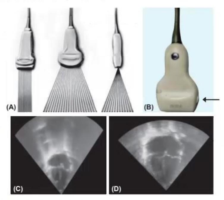
- What are name of 3 probes in Fig A?
- What 1s this arrow marked name in Fig. B?
- In Fig. C and D, which depth 1s appropriate when you are doing ECHO, how much depth is consider best?
Answer
- Linear, curved and array probes
- Probe marker
- Figure D is of appropriate depth (must be 75% of total screen).
MNJ03-002
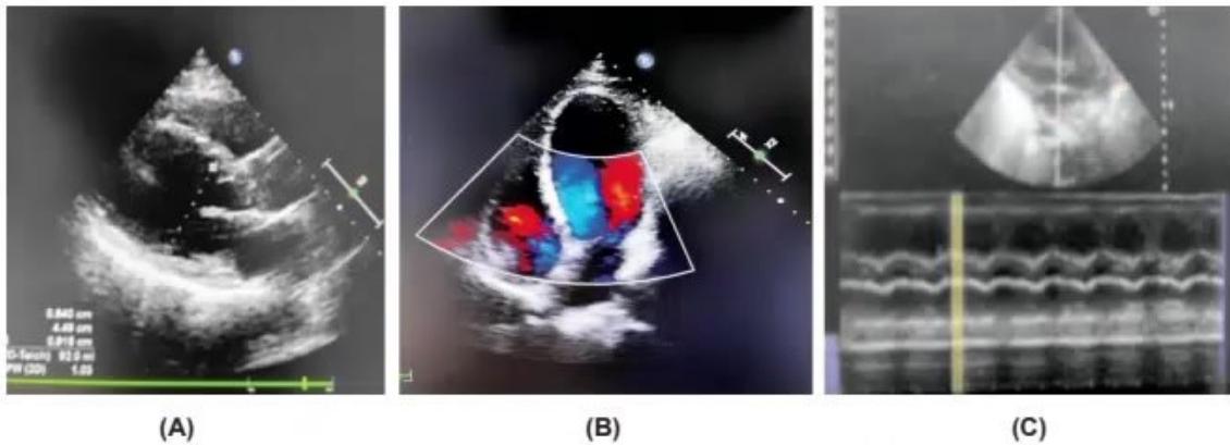
What are the modalities in given three images and write one use of each.
Answer
Grey scale to see structure and orientation HE ee we
- Doppler to see blood flow in vessel
- M MODE: to see motion in form of contractility.
- Hemodynamucally significant PDA Ans. 3.
- To see hypoperfusion, assessment and monitoring of PDA, assessment of neonatal shock
-
- As a marker of systemic hypoperfusion due to PDA: will see diastolic blood flow pattern.
MNJ03-003
A preterm neonate 26 weeks at day 3 became sick in form of tachycardia, desaturation, feed intolerance. You Observed baby has bounding pulses and oxygen need increased in last 12 hours. Your unit in-charge advised ECHO screening in all preterm babies around day 3. Kindly answer the following questions:
- What 1s possible diagnosis?
- Why ECHO screening important in preterm babies at day 3?
- What are importance of antenor cerebral artery Doppler in cases of PDA in neonate?
Answer
No model answer in source material.
MNJ03-004
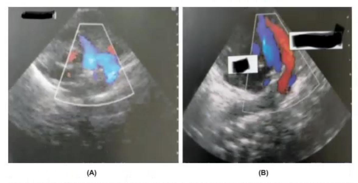
In given two images below differential normal image and abnormal duct and what is diagnosis in abnormal duct image? What are three important pathophysiological parameters of abnormal duct?
Answer
Image A: Closed duct (normal); Image B. Large patent duct (abnormal) and three pathophysiological parameters are:
- i. Pulmonary over circulation,
- ii. Systemuc hypoperfusion,
-
- High volume left to right shunt.
MNJ03-005
A neonate has issues related to his heart. A screening ECHO was planned by your unit head. While performing the ECHO this is the first image you saw on screen. Observe this image and answer the following question related to this.
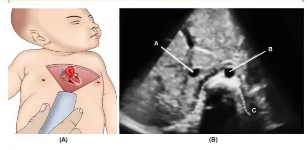
- Which 1s this view (Image A)
- What are the marked structures in (Image B)
- What is NEOPOCUS means
Answer
- Subcostal view
-
- A IVC, B: Aorta, C: Spine
-
- Neonatal point-of-care ultrasound.
MNJ03-006
A5 days old 26 weeks preterm baby started deteriorating in NICU. Baby has tachycardia, hyperactive precordium, increased pulse pressure. You diagnose this as ahemodynamically significant PDA and called the pediatric cardiologist. See the images below and answere the following questions:
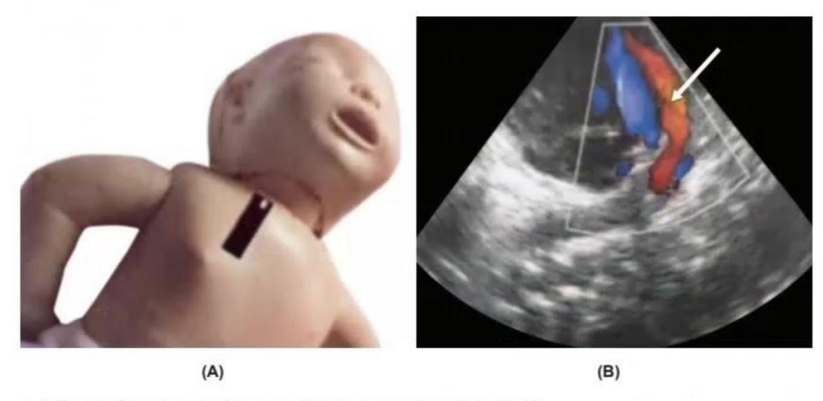

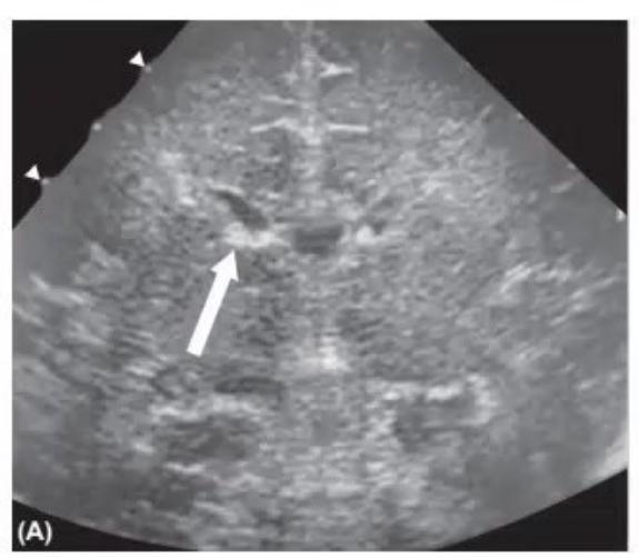
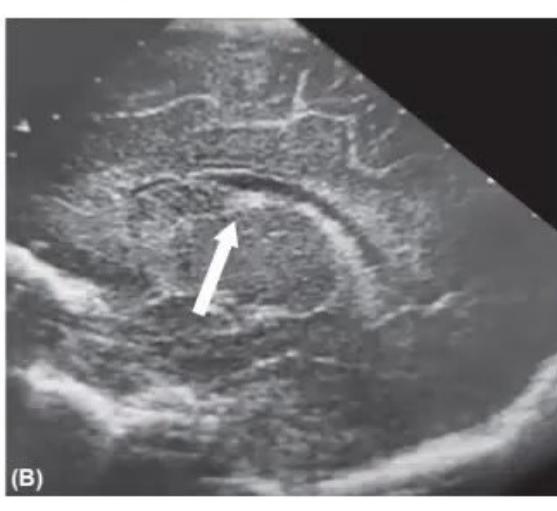

- What 1s this view to diagnosed PDA in neonate (Image A)
- What does the arrow marker show im image B
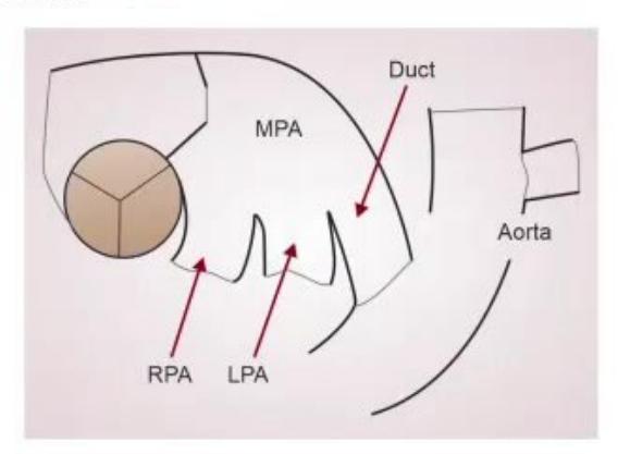
- Make the most important "triple leg view" line diagram indicating vessels.
- What are the name of two "views" showing IVH?
- Whats the grade of IVH?
- What 1s important structure shown in arrow in both images?
- Why this area 1s important for IVH in preterm for long-term outcome?
Answer
- High parasternal view (ductal view)
-
- Patent PDA
-
- See figure below.