MNJ06-001
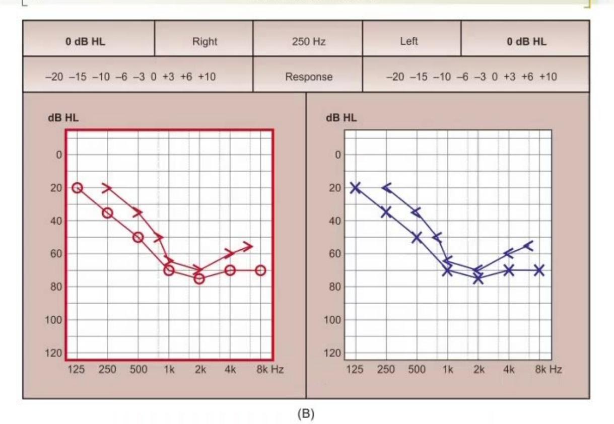
" Identify images A and B:
[ 62 OSCE Clinical Pediatrics »
Answer
No model answer in source material.
MNJ06-002
"
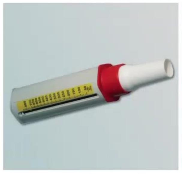
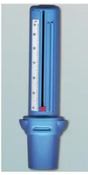
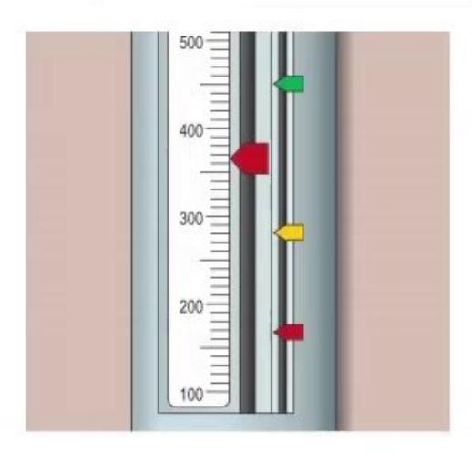
- Identify the instrument.
- Which illness 1s it used in?
- What are the three settings?
Answer
No model answer in source material.
MNJ06-003
*
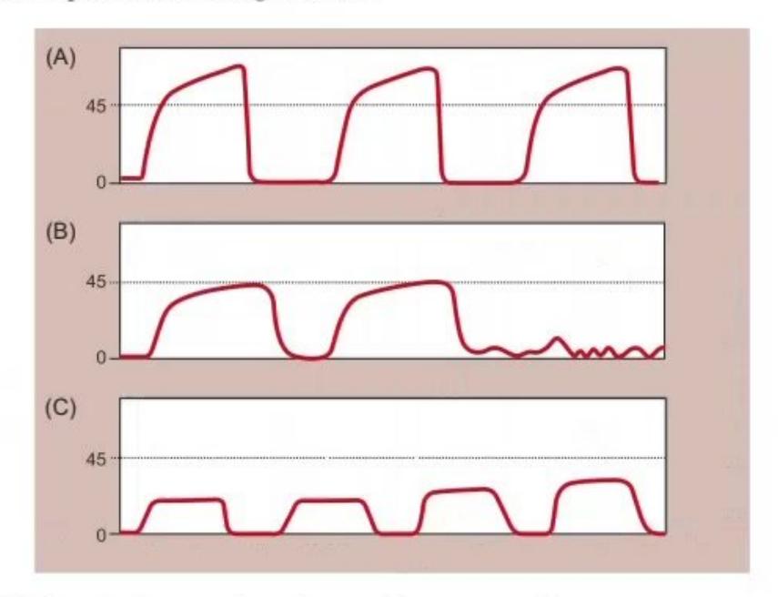
- What are these graphical techniques?
- What are interpretation for images A, B, C?
Answer
No model answer in source material.
MNJ06-004
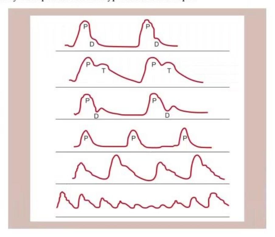
* Identify these pulse waveform types with one example.
Answer
No model answer in source material.
MNJ06-005
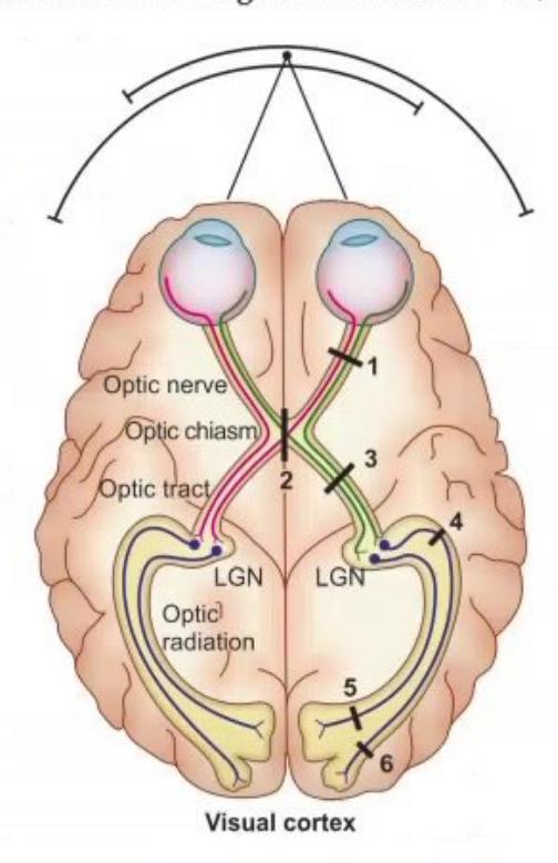
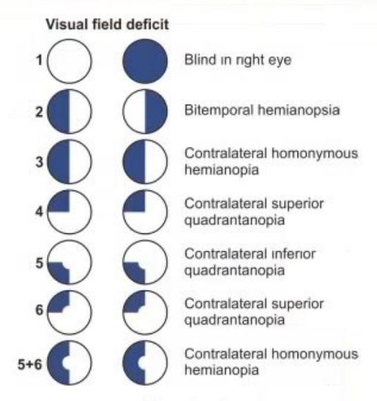
* Give the type of vision loss according to lesion at site 1-6. (2012 Question):
Answer
No model answer in source material.
MNJ06-006
*
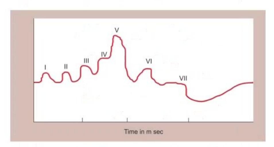
- Whats this test?
- Identify level I-VIL.
Answer
No model answer in source material.
MNJ06-007
*
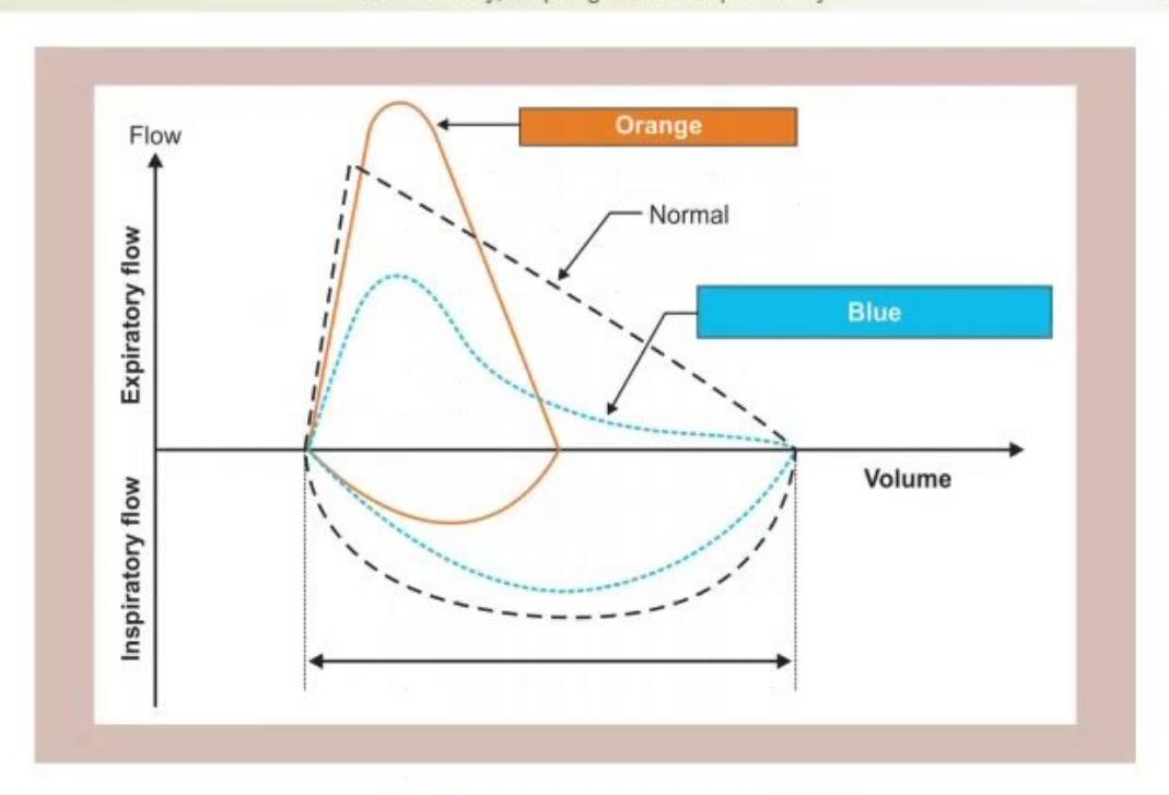
- Identify the graphical picture and give its use.
- Give one example of red and green flow.
Answer
No model answer in source material.
MNJ06-008
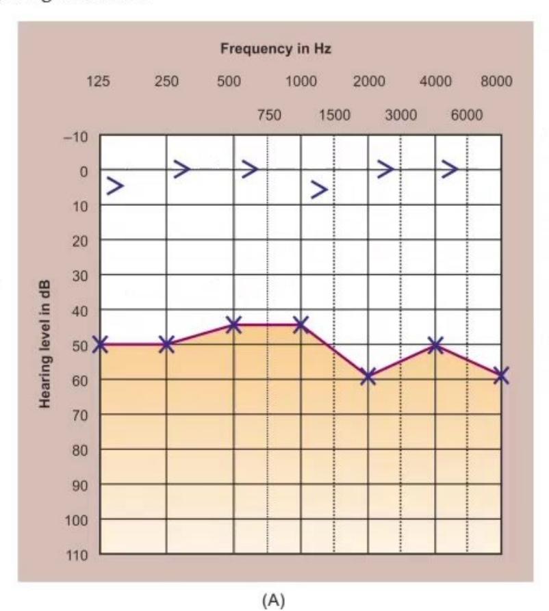
Audiogram from a7-year-old boy Ronit. His mother was worried by his hearing. He has passed his routine audiometry screen at 5 years and there was no family history of deafness. On examination, no abnormalities were found.
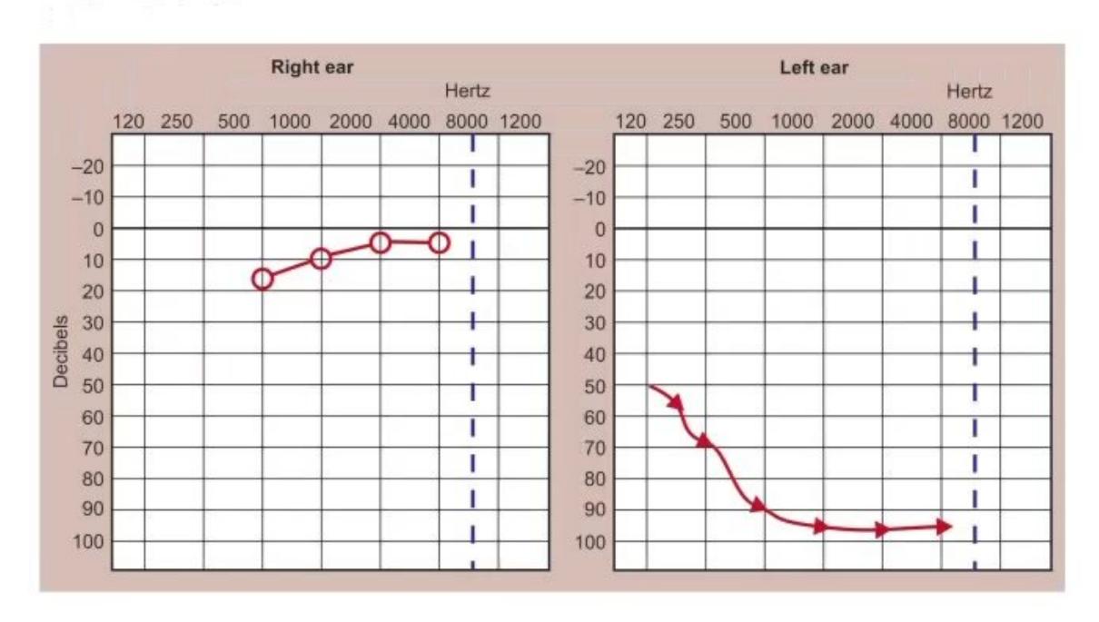
- Diagnose the given audiometry.
- Write down the causes.
Answer
No model answer in source material.
MNJ06-009
This is the audiogram of a4-year-old girl child who was referred because of severe speech delay:
ANSWERS
-
Ans. 1. 1. Conducting hearing loss-left ear
-
- Sensorineural hearing loss—B/L
-
-
Ans. 2. 1. Peak flow meter
-
- Bronchial asthma
-
- Green zone >80% peak expiratory flow
- Yellow zone 50–80% PEF
- Red zone <50% PEF
-
-
Ans. 3. 1. Capnograph
-
- a. Shark fin pattern—asthma
- b. Obstruction of ET
- c During CRP
-
-
Ans. 4. 1. Collapsing pulse: AR, PDA, artriovenous fistula
-
- Pulses bisferiens: AS, AR
-
- Dicrotic pulse: Typhoid fever
-
- Small volume collapsing pulse: MR
-
- Pulses alterans: AS
-
- Pulses paradox Severe asthma, SVC obstruction
-
-
Ans. 5. (See figure)
-
Ans. 6. 1. Bera
-
- Level (Pneumonic: C. coli)
- a. Cochlear nerves-waves I and II
- b. Cochlear nucleus-wave III
- c Superior olivary complex-wave IV
- d. Nulclei of lateral lemniscus-wave V
- e. Inferior colliculus-waves VI and VII
-
-
Ans. 7. 1. Flow volume curve, it is useful in differentiating type of respiratory failure
- Red—restrictive pattern Green—obstructive pattern
Visual axis
- Ans. 8. 1. Profound sensorineural deafness affecting the left ear
-
- Mumps infection after 5 years of age
- · SN deafness due to viral infection
- · Progressive herediatary deafness
- · Acoustin neuroma
-
- Ans. 9. 1. Bilateral partial sensorineural deafness
-
- Deafness
-
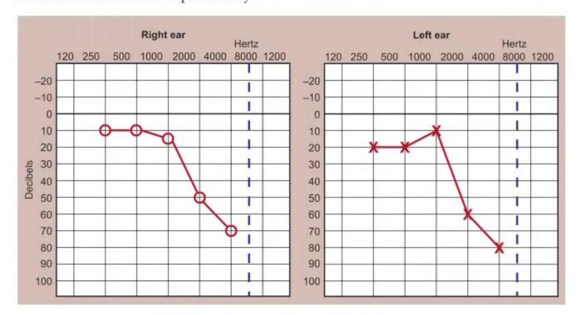

- What does this audiogram show?
- Whats the cause of her speech delay?
- Sensorineural hearing loss—B/L
- Bronchial asthma
- Green zone >80% peak expiratory flow
- a. Shark fin pattern—asthma
b. Obstruction of ET - Pulses bisferiens: AS, AR
- Dicrotic pulse: Typhoid fever
- Small volume collapsing pulse: MR
- Pulses alterans: AS
- Pulses paradox Severe asthma, SVC obstruction
- Level (Pneumonic: C. coli)
a. Cochlear nerves-waves I and II
b. Cochlear nucleus-wave III
d. Nulclei of lateral lemniscus-wave V
e. Inferior colliculus-waves VI and VII - Mumps infection after 5 years of age
- Deafness
Answer
No model answer in source material.