THG35-001
Answer the following questions from US cranium of a preterm infant born at 28 weeks gestational age (GA).
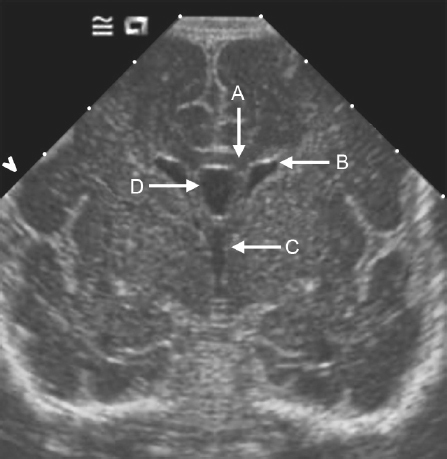
a. What is the section of scan?
b. Name the structures A, B, C, and D.
Answer
| a. | Coronal section |
| b. | A: Corpus callosum, B: Lateral ventricle, C: Third ventricle, and D: Cavum septum pellucidum |
THG35-002
Answer the following questions from US cranium of a preterm infant born at 30 weeks GA.
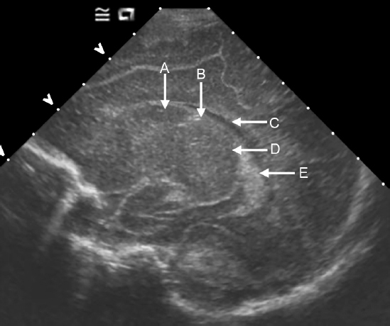
a. What is the section of scan?
b. Name the structures A, B, C, D, and E.
Answer
| a. | Sagittal section |
| b. | A: Caudate nucleus, B: Caudothalamic groove, C: Lateral ventricle, D: Thalamus, and E: Choroid plexus |
THG35-003
A 26-week-old GA preterm baby had a cranial US on day 2.
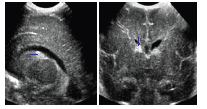
a. What does the highlighted area (blue arrow) show?
b. What grade is this?
Answer
| a. | Germinal matrix hemorrhage/intraventricular hemorrhage (GMH/IVH) |
| b. | Grade 1 |
THG35-004
A 27-week-old GA preterm baby was very sick at birth, needing ventilation, and been hypoxic for hours before recovering. She also suffered an Escherichia coli sepsis in the first week of life. She had a cranial US at 6 weeks of age (previous scans were normal).
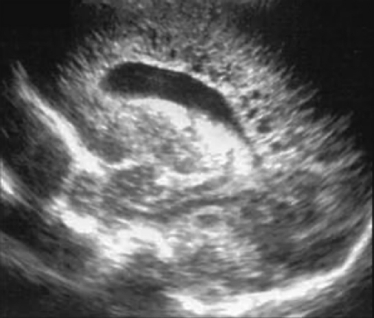
a. What does this scan show?
b. What grade is this?
c. What is the likely neurodevelopmental outcome of this baby if this finding is bilateral?
Answer
| a. | Extensive cystic PVL |
| b. | Severe: Grade 4 |
| c. | Abnormal neurodevelopmental outcome—spastic cerebral palsy (80–90% of babies with bilateral cystic PVL) |
THG35-005
A 24-week-old GA preterm baby was hypotensive for 3 days after birth, needed maximum support with ventilation requiring high frequency oscillation during the first week before recovering. He had a cranial US at 6 weeks of age.
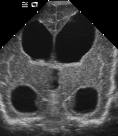
a. Bilateral cystic periventricular leukomalacia (PVL)
b. Bilateral porencephalic cyst
c. Bilateral ventriculomegaly following severe intraventricular hemorrhage (IVH) early on
d. Normal scan
Answer
c. Bilateral ventriculomegaly following severe IVH early on
THG35-006
Term baby had a cranial US at 24 hours of age. Answer the following questions:
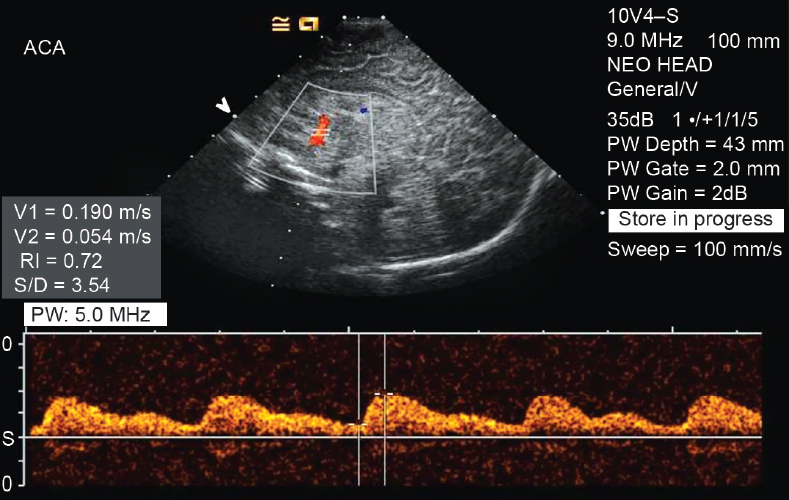
a. What type of US is this?
b. What is the expansion of RI?
c. What is the normal value?
d. In which condition, RI is useful to prognosticate the outcome?
Answer
| a. | Doppler ultrasound |
| b. | Resistive index |
| c. | 0.6–0.9 |
| d. | Hypoxic ischemic encephalopathy |
Figure Sources
All the figures are from author’s personal collection.