MNJ33-001
** See the X-ray image and answer the following questions.
aaa
- See the X-ray image and give the possible diagnosis.
- . What are the first two steps in treatment of hypoxic spell?
- In a cyanotic newborn, how can you distinguish pulmonary disease from cyanotic congenutal heart disease? Whe we
- Which cardiac conditions are associated with following: phe
- * Egg-shaped heart
- * Snowman silhouette
- * Rib notching
] Cardiology 459 |
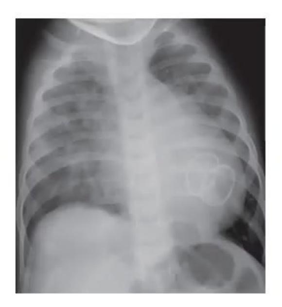
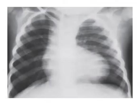
- What was the lesion?
- What was the procedure carried out?
- What are the complications?
Answer
No model answer in source material.
MNJ33-002
A 2-day-old neonate, birth weight: 3.1 kg, on examination, cyanosis +, soft murmur +, single 52. On investigation, hypoxia and acidosis present, chest X-ray shows normal cardiac size:
- What 1s most likely diagnosis?
- What is the emergency therapy?
- Whats the dosage and complications of the drug?
Answer
- Duct dependent cyanotic heart disease (most common: D-TGA with intact IVS/ pulmonary atresia with intact IVS)
- . IV prostaglandin E1 infusion.
- . 0.05 pg/kg and 0.1 pg/kg as continuous infusion, starting at the higher dose and tapering slowly.
Systenuc hypotension and apnea are the most common complications.
MNJ33-003
A term infant suddenly develops cardiac failure in the first week of life. The infant was pink, his pulses were collapsing but there is no cardiac murmur. His X-ray showed cardiomegaly. His echo showed normal heart and normal great vessels. His PDA was closed:
- What 1s the diagnosis?
- What other investigations will be indicated?
Answer
AV fistula
- Angiography
MNJ33-004
See the X-ray image below and answer the following questions.
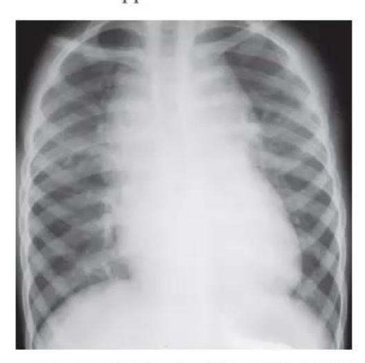
- What 1s the diagnosis?
- Whats this sign called?
- What are structures that cause this appearance in this condition?
Answer
oe Ber Total anomalous pulmonary venous draimage—supracardiac type
- Snowman appearance / figure of § appearance
- Cardiomegaly with mcreased vascular markings.
Dilatation of both the left and nght innominate veins and mght SVC producing the classical snowman of figure of 8 appearance.
Superior mediastinum is enlarged secondary to right SVC, innominate artery and ascending verhecal vein.
MNJ33-005
1-day-old infant appears dusky during feeding. O, is administered via nasal cannula, and 2 hours later she develops tachypnea. Findings include: HR—170 beats/min; RR—80/ min; R arm BP—48/30 mm Hg; R leg BP—52/32 mm Hg; and pulse oximetry, 90% on oxygen:
- Ifthe diagnosis is HLHS describe physical findings of pulse, murmur, precordium and 82.
- What was wrong in the management in the above scenario?
- Mention 5 CHD presenting in the first week of life
- Emergency management.
Answer
- HLHS Duct dependent lesion—signs of shock, poor pulses and metabolic acidosis, as systemic perfusion deteriorates, thereby depriving the systemic circulation of adequate perfusion. Hyperdynamic precordium and a loud, even palpable single loud P2 (52), no murmur
- . O, acts as a pulmonary vasodilator stealing systemic perfusion
- . Duct dependent SBF (Systemic blood flow)—AS, coarctation of aorta, [AA Duct dependent PBF (Pulmonary blood flow)—PA, HRHS. PA with intact IVS Duct dependent TGA with intact IVS Infradiaphragmate TAPVC
- . PGE] to keep PDA open.
MNJ33-006
A 5-day-old infant born at 31 weeks' gestation is on ventilatory support. He has a grade 2 holosystolic murmur that extends the second heart sound. Pulses are bounding. Precordial palpation is hyperdynamic. Echocardiography reveals a large patent ductus without other ductal-dependent congenital lesions:
- When would one suspect PDA in preterm clinically
- Mention 3 complications of PDA in neonate.
- Contraindications for indomethacin.
- Pharmacological closure of PDA—medication and dosage
Answer
- Tachypnea, tachycardia, RD, O, dependency, continuous murmur, child not gang weight, hyperdynamuc precordium, bounding pulses.
Cardiology 465
-
- Persistence of RD (BPD), NEC, ICH, O2/ventilator dependency, apnea
-
- Anemia, bleeding, electrolytes disturbances and renal dysfunction.
- IV/PO—indomethacin 0.2 mg/kg/dose × 3 doses 12 hr apart, PO—ibuprofen, PO—paracetamol.
MNJ33-007
When examining a healthy 3-year-old boy during a health supervision visit, you hear a grade 2 systolic ejection murmur most clearly at the upper left sternal border:
- Differential diagnosis of ESM in second left ICS.
- Auscultatory findings in ASD
- Types of ASD.
- Children with ASD and moderate to severe pulmonary hypertension which associated conditions should be ruled out.
Answer
- ASD, increased PBF, AS, PS and CoA
- ESM, soft, wide fixed split, low-pitched, audible diastolic murmur at the lower left sternal border with a rumbling quality due to increased tricuspid valve flow.
-
- Ostium primum, ostium secundum/PFO, sinus venosus ASD, coronary sinus ASD.
-
- Large ASD, TAPVC, PAPVC, cortriatriatrum, mitral valve abnormalities.
MNJ33-008
Following an uncomplicated delivery, a3.7 kg term infant develops cyanosis in the first hour of life. Findings at 3 hours of age include: Cyanosis; heart rate: 140 beats/min; respiratory rate: 56 breaths/min; no heart murmurs; pulse oximetry in room air: 70% saturation in the right hand and 75% in the foot; in 100% FiO, via head-hood oxygen—saturation increases to 90% in the foot; chest radiography is normal:
- Clinical differentials.
- Describe hyperoxia test.
- Causes of differential cyanosis/reversed differential.
Answer
- Pre- and post-ductal oxygen saturations
-
- If pre-ductal saturation higher than post-ductal saturation (differential cyanosis)
- Left heart abnormalities (such as aortic arch hypoplasia, critical aortic stenosis, interrupted aortic arch)
- · Persistent pulmonary hypertension
- If post-ductal saturation higher than pre-ductal saturation (reverse differential cyanosis)
- TGA with CoA or TGA with IAA
- TGA with supersystemic pulmonary vascular resistance.
Ans. 10.
| Causes of sudden cardiac death in children | ||||
|---|---|---|---|---|
| Structural/functional | Electrical | |||
| Hypertrophic cardiomyopathy | Long QT/short QT syndrome | |||
| Coronary artery anomalies | Wolff-Parkinson-White syndrome | |||
| Aortic rupture/Marfan's syndrome | Brugada syndrome | |||
| Dilated cardiomyopathy | Congenital heart block | |||
| Myocarditis/endocarditis | Catecholaminergic ventricular tachycardia | |||
| LVOT obstruction | Primary pulmonary hypertension | |||
| Mitral valve prolapse | Drugs/stimulants | |||
| Coronary artery atherosclerotic heart disease | Commotio cordis | |||
| Arrhythmogenic RV cardiomyopathy | ||||
| Postoperative congenital heart disease |
MNJ33-009
A 14-year-old girl falls during a race. She is unconscious, cyanotic, and has no pulse, but spontaneously revives within seconds. Both patient and family histories are benign. Results of physical examination:
- Causes of sudden cardiac death in children.
- Appropriate tests been excluded.
- Arrhythmias associated with syncope.
Answer
No model answer in source material.
MNJ33-010
A healthy 8-month-old boy has had intermittent episodes of intense cyanosis of the hands, lower arms, and feet since birth. The infant appears alert and playful during the episodes. At other times, his arms and legs are pink but havea "mottled" appearance. Physical examination reveals no remarkable findings other than mottling of the extremities:
- Causes of episodic acrocyanosis
- Diagnostic tests for this child
Answer
- Episodic acrocyanosis of the hands and feet, episodic arrhythmia, methemoglobinemia, cyanotic spells, seizures.
- Ambulatory ECG, ABG/MetHB to detect methemoglobinemia, echocardiography to detect congenital heart disease, EEG to detect a seizure disorder.
MNJ33-011
A 14-year-old boy in your practice is followed periodically for bicuspid aortic valve. He presents to you with 3 weeks history of fatigue and off and on fever of 101°F:
| 0.13. | ||
|---|---|---|
| 1 | At what age ECG resembles an adult ECG | |
| 2 | Tall P waves in lead II 1s defined as height more than | mm. |
| 3 | Maximum P wave duration in children 1s | seconds. |
| 4 | Maximum P wave duration in infants <12 months | seconds. |
| 5 | PR interval—lower limit in less than 3 years | seconds. |
| 0.14. |
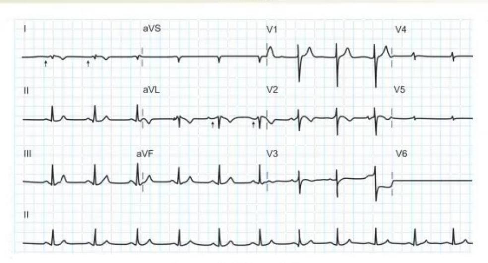
A 12-year-old boy presented with acute onset pain in left iliac fossa, fever, vomiting. Preoperative ECG is shown:
Cardiology 467 |
- What will you look for on physical examination?
- He has elevated WBC and ESR. What other test you will ask for?
- What organisms are likely to grow on culture?
- What treatment regime is ideal?
- What risk this boy has for sudden deterioration?
- Analyse the ECG.
- Mention likely malformations associated with this condition.
Answer
Infective endocarditis (rest answer see the Nelson).
MNJ33-012
- At what FiO; level, patient should be on continuous pulse oximetry?
- What are the causes of (external and internal) inaccurate SpO, readings?
- A child rescued from fire at school on admussion to PICU shows 100% SpO, on pulse oximeter Intensive care in charge still puts him on oxygen and orders ABG Why?
- Whats the oxygen carrying capacity of a child with SpO, of 97% and Hb of 10?
- What 1s the lowest "safe" level (normal Hb, etc.) of % saturation of oxygen, and the corresponding PaO,?
- What should be done when ambient light interferes with SpO, reading?
- When will you suspect "ductus dependent circulation" on day 1 of life (by use of pulse oxumeter)?
Answer
No model answer in source material.
MNJ33-013
- Principles of management of congestive cardiac failure
- What are the specific drugs used in the management of congestive cardiac failure?
Answer
No model answer in source material.
MNJ33-014
| 462 OSCE Clinical Pediatrics ]
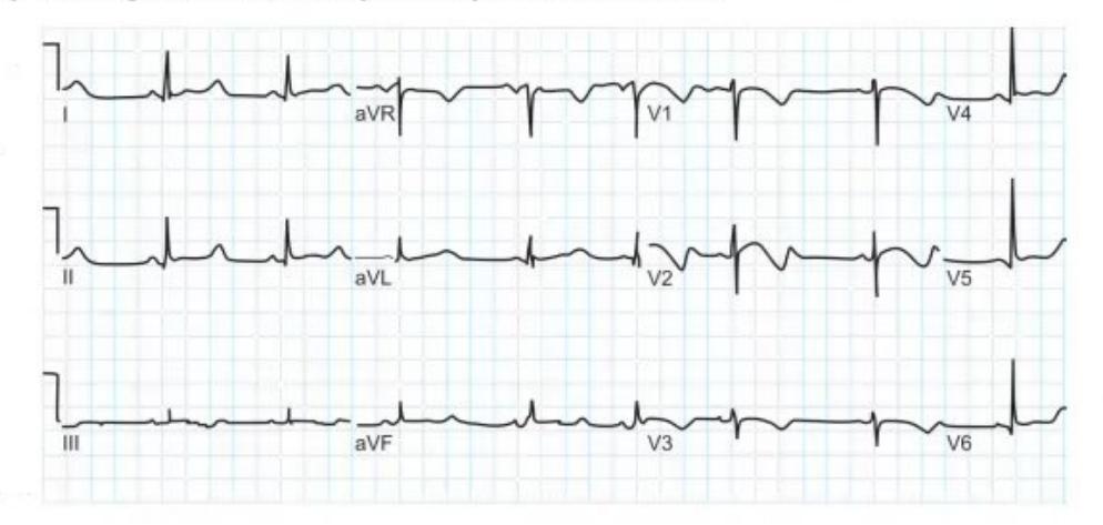
- Identify the abnormality in the ECG.
- What electrolyte abnormality can cause this?
- Why is it important to identify this rhythm disturbance?
Answer
- Prolonged QT interval.
-
- Hypocalcemia.
-
- As it can degenerate into dangerous rhythm—V Tac/VF and cardiac arrest
Ans. 18. 1.T,2.T,3 T,4 F,5.1T,6.T
*neonates had right axis deviation due to nght ventricle dommance
Cardiology 467
- *left axis deviation (superior axis) seen in-Tricuspid atresia, AVSD, Pulmonary atresia Noonan syndrome
- *RBBB-seen in COA, Ebstein anomaly, AVSD
- *Normal neonate have heart rate of 110–160/min, rising heart rate is always abnormal
Ans. 19. 1. F, 2 F, 3. F, 4. F
- *Oxygenated blood from placenta return to fetus via umbilical vein-50% enter in hepatic circulation and 50% bypass the liver through ductus venosus.
- *Most of blood from Inferior vena cava goes directly in right atrium through foramen ovale but blood from SVC goes in right ventricle.
- *85% of blood from right ventricle passes in to descending aorta via ductus arteriosus and 15 % enter in fetal lung.
MNJ33-015
True/False
- Heart block is associated with maternal SLE
- Right bundle branch block seen in COA. Ulm G2 he
- Romano ward syndrome is associated with prolong PR interval
- A rising heart rate 180 in a neonate is tachycardia.
- fh Superior axis 1s seen in tricuspid atresia.
- Neonates have right axis deviation.
Answer
No model answer in source material.
MNJ33-016
True/False about fetal circulation
- Umbilical vein carry deoxygenated blood in placenta from fetus
- Blood from superior vena cava preferentially flow across foramen ovale in to nght atrium
- 45% of right ventricular outflow enter enters the lung via pulmonary arteries
- Descending aorta connected to pulmonary artery via ductus venosus
Answer
No model answer in source material.
MNJ33-017
A 13 years old girl present with 3 months history of palpitation. She is fit and well grown without any recent illness and medication history. She is very good student in class also. She feel her heart rate rising and comes gradually down after 3—4 hours. No history of chest pain, collapse. Her symptoms usually present in school hours. She is good plays and not having any problem while playing. Mother tell no member in our family had heart disease. You found normal physical and systemic examination and her BP is also normal.
- What 1s possible diagnosis
- What test you will do before sending her home
- Cause of potental life threatening syncope 1n childrens(wmite any 3)
- When you will refer a child with syncope to cardiologist.
Answer
- Physiological tachycardia associated with situation related stress
-
- 12 leads ECG-always do this in any child with palpitation or syncope
-
- WPW syndrome, Prolong Q-T syndrome, HOCM
-
- If any abnormality in ECG and physical examination
MNJ33-018
From each of following clinical scenario pick the most likely diagnosis from the list.
- a Cardiomyopathy
- b Endocarditis
- A13 years old boy treated for rhabdomyosarcoma ay 4 years age and treated with full recovery. Now he has shortness of breath smce 6 month, on examination he looks tired with mcreased respiratory rate. He has gallop rhythm and raised JVP. X-ray chest showed cardiomegaly.
- A 15 years old boy with history of loss of appetite, off and on fever since 1 month. On examination you found muffed heart sound and low platelet count on blood test. His mother gave history recurrent rash over his face when she play cricket.
- A7 years old girl presented with swollen left knee after recovery form swollen ankle last week. She has fever and pink rash over his trunk. She gave history of sore throat last month. You ordered ECG and found first degree heart block.
c. Rheumatic fever
d. JIA
e. SLE
Answer
- a. Anthracycline induced cardiomyopathy-need lifelong annual ECHO
-
- e. Malar rash are photosensitive in SLE
-
- c prolong PR interval is called first degree heart block is a feature of RF
MNJ33-019
Cardiac investigations-choose the best investigation for given scenarios
- a 24 hours ECG
- b 12 leads ECG
- A3 years old boy came to you for URI, on examination you notices murmur which 1s soft systolic at right supraclavicular area and became louder on sitting.
- A12 years old boy with recurrent panic attacks with fast heart rate. On examination he has resting heart rate of 120/min. his systemic examination 1s normal.
- A6 years old boy who repaired for aortic stenosis at 18 month age, present with shortness of breath. On examination you found systolic murmur on aortic area.
c. Cardiac catheter
d. ECHO
e. MRI thorax
f. Stress test
Answer
- b. This patient has innocent murmur-venous hum only do ECG and reassure the parents.
-
- a. Resting heart rate 120 is high. May be SVT Do 24 hour ECG
- c Case of Aortic Valve Restenosis-ECHO can be done but cardiac catheter is gold standard to see the gradient and pressures across the valve.
MNJ33-020
Chest pain-choose the best diagnosis for given scenarios.
- b Pneumonia
- Al? years old girl referred to you for 11 month history of recurrent chest pain. The pain episode i is localized to left side and does not radiate. She recovered after few minutes. She is well grown, active girl without any clinical finding on examination.
- A115 years old boy rushed in causality for sudden collapse while playing football match. In causality he was awake and describe a severe chest pain with sweating. This 1s his first episode of this type of symptoms. On examination you found ejection systolic murmur at right upper sterna area that is radiating to carotid area No other finding in examination
- A110 years old girl present to you for 3 weeks history of left side chest pai that increase on deep breathing. There is no history of cough, fever, trauma and palpitation. Her cardiovascular, respiratory and GI examination is normal On examination you found tenderness over 2nd costochondral junction
a. Aortic stenosis
c. Pericardial catch syndrome
d. HOCM
e. Costochondritis
Answer
- c. Pericardial catch syndrome-this is classical presentation-recurrent chest pain for few minute that increase on deep breathing
-
- a. classical murmur of AS
-
- e. Costochondritis
MNJ33-021
An 8 years old boy is due for a minor procedure in OT today. Anesthetist cannot feel his femoral pulse. Child is referred to you and on examination he is nor cyanosed nor clubbed at finger. His femoral pulse was absent with forceful apex with out any thrill. A 3/6 systolic murmur is present at upper left sternum with radiation at back between the scapula. X-ray and angiogram are given below.
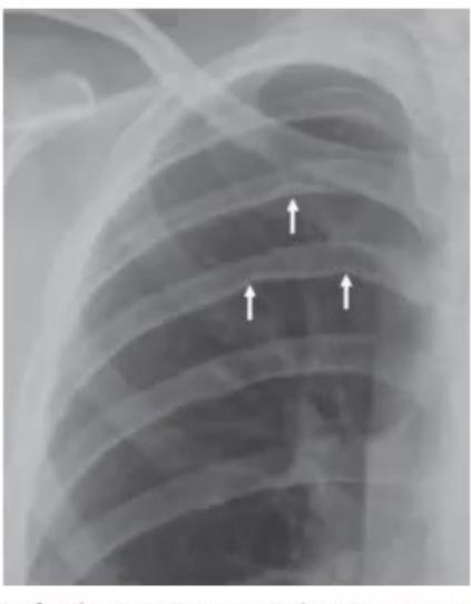
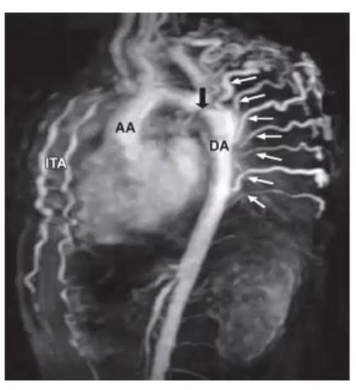
- What 1s finding in X-ray and MR angiogram?
- What is diagnosis on the base of chmical and inveshigation findings?
- What what 1s syndrome associated with this?
- Whatis plan of further treatment?
Answer
- Rib notching (X-ray) with coarctation just beyond the left subclavian artery (Angio)
-
- Coarctation of aorta
-
- Williams syndrome
-
- Surgical correction and refer to cardiology team