THG08-001
Regarding opioid analgesic in children
a. Which is the most potent opioid?
b. Why do we need special pain scales in pediatric intensive care unit (PICU)?
c. Name any sedation scores validated in children.
d. What is the drug of choice for opioid withdrawal? Which tool is used to assess withdrawal?
e. Which opioid works only with continuous infusion?
f. What is the antagonist drug for opioid overdose?
g. Which drug is used to reduce opioid dependence?
Answer
| a. | Sufentanil. Sufentanil is 5–10 times more potent than fentanyl and is the most potent opioid used in clinical practice. Fentanyl is 100 times more potent than morphine. |
| Relative drug potency is as follows: |
| Drug | Relative potency |
|---|---|
| Morphine | 1 |
| Meperidine | 0.1 |
| Methadone | 1 |
| Hydromorphone | 7 |
| Alfentanil | 10 |
| Fentanyl | 100 |
| Sufentanil | 500 |
| b. | Self-report measures are impossible in children intubated and sedated/paralyzed children. |
| c. | State behavior scale/COMFORT B score. |
| d. | Methadone, withdrawal assessment tool. |
| e. | Remifentanil. |
| f. | Naloxone (1–10 μcg/kg/dose). |
| g. | Buprenorphine is considered a partial agonist at the μ and κ receptors and may have a role in the prevention or treatment of opioid dependence. |
THG08-002
Nonopioid analgesic in children
a. What is the mechanism of action of most nonopioid analgesics?
b. What is the dose of rectal paracetamol?
c. Name one nonsteroidal anti-inflammatory drug (NSAID) that can be administrated through intravenous route.
d. Name two side effects of ibuprofen.
e. Which drug causes Reye syndrome and what are its features?
Answer
| a. | They act by inhibition of cyclooxygenase types I, II, and III and thus blocking peripheral and central prostaglandin production. |
| b. | 25–40 mg/kg, a maximum daily dose of 60 mg/kg, 80 mg/kg, and 90 mg/kg in preterm neonates, term neonates, and older children irrespective of route. |
| c. | Ketorolac. |
| d. | Gastrointestinal (GI) irritation, platelet dysfunction, hematuria, and bronchospasm. |
| e. | Aspirin. Reye syndrome occurs when aspirin is used in influenza like viral illness. It results in liver damage and cerebral edema leading to encephalopathy, seizures, decreased level of consciousness, irritability, vomiting, and diarrhea in smaller children. Laboratories show abnormal liver function with low glucose and high ammonia. |
THG08-003
Facts about vasoactive medications
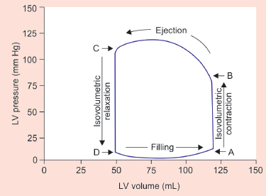
a. Aortic valve opens at which point?
b. Increase in afterload will be reflected mainly change in which segment?
c. At the same level of contractility, what strategies would restore the stroke volume for the patient with myocardial dysfunction and decreased contractility to his baseline?
d. How does nesiritide work?
e. What is the drug of choice with hypotension with normal cardiac output?
f. What are the receptor activities of norepinephrine?
g. What is the mechanism of action of milrinone?
Answer
| a. | Aortic valve opens at Point B. |
| b. | Increase in afterload will be reflected mainly change in BC segment. |
Loop ABCD depicts the pressure-volume relationship in a healthy heart. Contraction starts at point A, corresponding to the end-diastolic pressure and volume. Line AB signifies isovolumetric contraction. At point B, the aortic valve opens when the left ventricular (LV) pressure surpasses the pressure in the aorta. Ejection commences from point B until point C where LV pressure matches the maximal force generated by the ventricular wall at that particular end-systolic fiber length.
Isovolumetric relaxation commences at point C, coinciding with the closure of the aortic valve. The ventricular pressure declines along line CD. When LV pressure falls below left atrial pressure, mitral valve opens and blood flows into the LV. The difference between lines AB and CD represents the stroke volume. Point A signifies preload and point B signifies afterload.
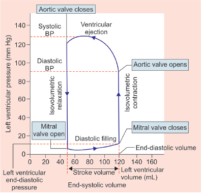
Cardiac cycle.
Courtesy: Dr Aishwarya Padmanaban.
| c. | Increase in preload and vasodilator therapy. |
Loop 1 normal, loop 2 poor contractility, loop 3 decreased afterload, and loop 4 increased preload. Stroke volume can be restored in situation with low contractility by increasing preload or decreasing afterload without changing the contractility like in loop 3 and loop 4.
| d. | Reducing preload and afterload. During cardiac failure, heart produces an endogenous B-type natriuretic peptide (BNP) and nesiritide is a synthetic peptide identical to the BNP. Nesiritide decreases preload and afterload, resulting in increased cardiac output/index without inducing reflex tachycardia or direct inotropic effect. It also promotes natriuresis and diuresis while inhibiting the renin–angiotensin axis and endogenous catecholamines. |
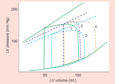
Relationship between LV pressure and volume.
Courtesy: Dr Aishwarya Padmanaban.
| e. | Norepinephrine is the drug of choice when the primary hemodynamic disturbance involves hypotension, a low systemic vascular resistance (SVR), and normal or high cardiac output following fluid resuscitation. |
| f. | α1 and β1. |
| g. | PDE3 inhibitor (phosphodiesterase), milrinone acts as an inotrope and vasodilator, producing a direct reduction in preload and afterload. PDE III inhibitors provide effective adjunctive therapy in the child with elevated pulmonary vascular resistance and reduced pulmonary blood flow by its lusitropic action of improving right ventricular (RV) relaxation in diastole. |
THG08-004
Ascites in children
a. What is the serum ascites albumin gradient (SAAG) ratio in portal hypertension?
b. What is the triglyceride cut off in chylous ascites?
c. What kind of cardiac procedures result in refractory ascites with or without chyle?
d. What is the neutrophil cut off in spontaneous bacterial peritonitis (SBP)?
e. What is the most common organism implicated in SBP in nephrotic syndrome?
f. What is the characteristic of secondary bacterial peritonitis?
Answer
| a. | SAAG ≥1.1 g/dL and total protein <2.5–3 g/dL. Transudate fluid has higher serum albumin to ascites gradient and hence SAAG is higher. In exudative or proteinaceous ascites due to causes other than portal hypertension SAAG is <1.1 g/dL. |
| b. | Milky ascites which increases with fat ingestion where triglycerides need to be minimum of >200 mg/dL. |
| c. | Fontan and bidirectional (BD) Glenn shunt and procedure which involve RV ventriculotomy like in tetralogy of Fallot (TOF) intracardiac repair. |
| d. | Neutrophils ≥250/uL. |
| e. | Streptococcus pneumoniae is seen specifically with SBP in nephrotic syndrome. Generally gram-negative bacilli like Escherichia coli are the most common cause of bacterial peritonitis in children. |
| f. | Multiple organisms are implicated in secondary bacterial peritonitis where there is a breach in the integrity of bowel like in cases of perforation. |
THG08-005
Acute peritoneal dialysis (PD)
a. What is the common site and size of acute PD catheter insertion in pediatric age group?
b. What are the mechanisms by which PD works?
c. What is done to increase ultrafiltration in PD?
d. What is done if PD is not draining well after working well for first 24 hours?
e. What is to be done to increase molecular clearance in PD?
f. PD drain effluent is cloudy. What is suspected?
Answer
| a. | 12 Fr 20 cm catheter is inserted usually in right or left iliac fossa infra umbilical area. |
| b. | Diffusion and convection. |
| Diffusion: Particles move from an area of higher concentration to an area of lower concentration across a semipermeable membrane. Smaller particles diffuse more freely, whereas larger particles have restricted movement. |
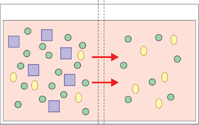
| Convection: Particles traversing across a semipermeable membrane, carried by ultrafiltered water, due to the effect of pressure (solvent drag). All particles up to the membrane’s cut off size move relatively equally. The concentration of effluent is equal to that of the original solution. |
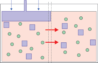
| c. | Increasing the concentration of dextrose in PD fluid makes it more hypertonic and increase the concentration gradient which will increase fluid removal. Also doing rapid cycles reducing dwell periods in each cycle to increase number of cycles in a given time will increase clearance. |
| d. | Problems with filling and draining dialysate through the PD catheter can be due to kinking of the catheter, fibrin plugs, omental obstruction, and catheter malposition. If the catheter fills but does not drain effectively, less ascites volume could be a reason and increasing dwell volume can help. Abdominal X-ray can verify correct positioning and identify any kinks in the catheter. If these maneuvers fail, revision or replacement of the catheter will be required. |
| e. | Increasing dwell volume (increase surface area) or dwell duration (increase exposure time). Example: Increase from 10 to 20 mL/kg to 20 to 30 mL/kg. Extended dwell periods between exchanges allows more time for the equilibration of dialyzable particles and ultrafiltration. |
| f. | Peritonitis. Dialysate should be sent for cell count, Gram stain, and bacterial culture if peritonitis is suspected. Empirical or specific antibiotics can be added in the dialysate for treating peritonitis via intraperitoneal route. |
THG08-006
Different types of shock and hemodynamic parameters
a. What is the formula for CaO2 (arterial oxygen content)?
b. What are the microcirculatory variables useful in shock monitoring?
c. Name any three dynamic parameters in fluid responsiveness.
d. What are the components of quick sequential organ failure assessment (SOFA) score?
e. What are the obstructive types of shock?
f. How does ventilation help in shock management?
Answer
| a. | CaO2 = (Hgb × 1.34 × SaO2) + (PaO2 × 0.003). |
| DO2 (oxygen delivery) = CO (cardiac output) × CaO2 | |
| Optimizing heart rate (HR), contractility, diastolic relaxation, preload, and afterload can improve cardiac output as stroke volume is dependent on these factors. | |
| Oxygen-carrying capacity can be improved by increasing hemoglobin levels and optimizing oxygen saturation. Oxygen delivery can be enhanced by manipulating these factors. | |
| b. | ABG, lactate, and superior vena cava oxygen saturation (ScvO2). They are useful markers of severity of illness and for prognosis. Systemic mixed venous oxygen saturation >70% suggests adequate cardiac output, but this assumes normal oxygen extraction. Various investigational methods, such as gastric tonometry, near-infrared spectroscopy, and muscle oxygenation, are used to evaluate regional circulation, but their clinical usefulness is yet to be proven. |
| c. | Dynamic fluid responsiveness parameters involve doing an intervention or monitoring difference in various phases of respiration and see the change in cardiac output. A fluid responsive patient has increase in cardiac output after giving fluid bolus. Stroke volume variation (SVV), pulse pressure variation (PPV), systolic pressure variation (SPV), inferior vena cava (IVC) collapsibility, passive leg raise (PLR), plethysmography variability index (PVI), and aortic outflow blood velocity (AOBV). There are prerequisites and limitations of all variables. In spontaneously breathing patients and patients with arrhythmia only passive leg rise test is possible. Aortic blood velocity and PLR are validated tools in children. |
| d. | SBP ≤100 mm Hg, respiratory rate (RR) >22 breaths/minute, and mental status [Glasgow Coma Scale (GCS) <15]. For rapid bed side identification of shock. |
| e. | Obstructive shock manifests as inadequate cardiac output because of physical restriction of forward blood flow, and the acute presentation may quickly progress to cardiac arrest e.g., cardiac tamponade, tension pneumothorax, pulmonary embolism, duct dependent congenital cardiac defects. |
| f. | Mechanical ventilation improves hemodynamic status in most shock states, especially in cardiogenic shock. Positive end-expiratory pressure (PEEP) reduces aortic transmural pressure gradient in systole thereby reducing afterload and during inspiration. Also mechanical ventilation reduces VO2 (oxygen consumption) by reducing WOB (energy spent on breathing which is disproportionately high in shock states). Mechanical ventilation can also reduce pulmonary vascular resistance provided optimal PEEP is given. |
THG08-007
Difficult airway management
a. What are the situations with physiologically difficult airway?
b. What are the assessment tools to identify difficult airway?
c. What is the immediate airway adjunct used in cannot intubate situations after giving neuromuscular blockade (NMB)?
d. What all factors make pediatric airway difficult?
e. Name some airway adjuncts to direct laryngoscopy used in difficult airway.
Answer
| a. | Severe hypoxia (ARDS), hypotension (shock), RV dysfunction, acidotic pH preintubation where airway is not anatomically difficult but patient may have major hemodynamic compromise peri-intubation, and so makes it a high-risk intubation mandating preparation for cardiac arrest. |
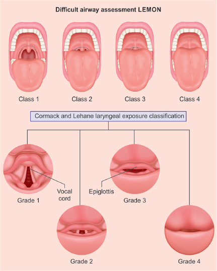
| b. | LEMON airway assessment and Cormack and Lehane classification of the laryngeal exposure. |
| c. | Laryngeal mask airway (LMA), a type of supraglottic airway. Patients with intact protective reflexes poorly tolerate the LMA, so its use is largely limited to those with severely depressed levels of consciousness or heavy sedation or anesthesia. |
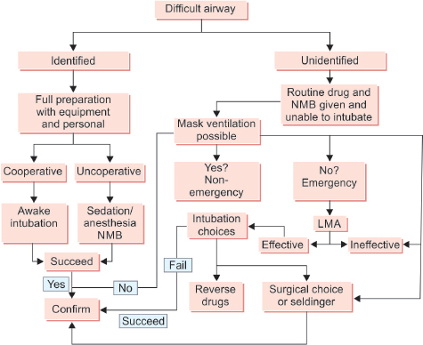
| d. | Relatively large tongue with less space, higher larynx (C1 in infancy, C3 to C4 at 7 years of age, and C4 to C5 in the adult), epiglottis relatively larger, and less tethered. Large occiput may cause of flexion of airway, cricoid ring is the narrowest portion of the child’s trachea making it difficult to pass the endotracheal (ET) tube. |
| e. | Gum elastic bougie, airway exchange catheter, lighted intubation stylet, flexible bronchoscopy, videolaryngoscopy, LMA, and Combitube. |
THG08-008
Fever with neutropenia
a. What is the cut off for neutropenia?
b. What is febrile neutropenia?
c. Which neutropenia granulocyte colony-stimulating factor (G-CSF) is used and at what dose?
d. Which combination of antibiotics is used in febrile neutropenia?
e. Enlist some causes of congenital neutropenia syndromes.
Answer
| a. | Neutropenia is defined as a decreased number of circulating neutrophils in the peripheral blood. An absolute neutrophil count <1,500/mm3 is defined as neutropenia, or <1,000/mm3 in children under 1 year of age. Neutropenia is classified as: |
| • | Mild if circulating white blood cell (WBC) counts are 1,000–1,500/mm3 |
| • | Moderate if 500–1,000/mm3 |
| • | Severe with increased risk for a life-threatening infection when the absolute neutrophil count is <500/mm3. |
| b. | Fever >100.4°F for >1 hour in a child with neutropenia (usually <500/mm3) with immunosuppression. Febrile neutropenia is the occurrence of fever during a period of significant neutropenia. |
| c. | Recombinant human granulocyte colony-stimulating factor (rh G-CSF or GCSF) stimulates the production of neutrophils from progenitor cells in the bone marrow. It is used for chemotherapy-related neutropenia at a dose 5–10 μg/kg administered subcutaneously. Absolute neutrophil counts improves (>1,500/mm3) within 10–14 days. |
| d. | Febrile neutropenia is a medical emergency and needs immediate admission with IV antibiotics covering gram-positive and gram-negative bacteria. Ceftazidime (or piperacillin/tazobactam) and vancomycin are often used as initial therapy [especially in patients with a central venous catheter (CVC)]. A carbapenem such as meropenem may also be considered, especially if extended-spectrum β-lactamase or AmpC producing organisms are a concern. Some centers use double coverage for gram-negative infections with an aminoglycoside and a β-lactam antibiotic in addition to vancomycin. |
| e. | Kostmann agranulocytosis, cyclic neutropenia, Shwachman syndrome, neutropenia with abnormal B or T lymphocytes (e.g., X-linked agammaglobulinemia), and severe congenital neutropenia. |
THG08-009
Hypertensive emergency
a. Define hypertensive emergency.
b. Discuss on organ involvement in hypertensive emergencies.
c. What are the goals in management of hypertensive emergencies?
d. What are the drugs used in hypertensive emergencies?
e. Which is the infusive β-blocker used in hypertensive emergency?
f. Define mechanism of action of labetalol.
Answer
| a. | Hypertensive crisis is defined as severe hypertension associated with end-organ damage (hypertensive emergency) or impending end-organ damage (hypertensive urgency). |
| b. | Brain, eyes, heart, and kidneys. Brain is commonly affected in children presents with hypertensive encephalopathy features such as headache, visual disturbances, nausea, and vomiting followed by altered sensorium, focal neurologic deficits, and coma. |
| • | Ocular manifestations—retinal hemorrhages, exudates, and papilledema. |
| • | Cardiac involvement—LV failure, pulmonary edema, or acute myocardial ischemia. |
| • | Renal involvement—hematuria, proteinuria, or azotemia. |
| c. | The primary goals of treatment are tightly controlled BP reduction and prevention of end-organ injury. The recommendation suggests reduction of the mean arterial pressure (MAP) by 20–25% within a period of 15 minutes to 2 hours. 25% reduction toward target BP is planned in first 8 hours followed by another 25% reduction in next 24 hours and slow reduction of next 50% over next 48–72 hours to achieve target BP. |
| d. | Sodium nitroprusside, labetalol, esmolol, nicardipine, fenoldopam, and enalaprilat. |
| e. | Esmolol. Esmolol is an intravenous, ultra-short-acting selective β1-adrenergic antagonist with no sympathomimetic action which may cause significant bradycardia. |
| f. | Labetalol is a competitive α1-adrenergic and β-adrenergic receptor antagonist. The action on α1 causes arterial smooth muscle relaxation and vasodilation, and that on β-receptors reduces BP by blocking reflex sympathetic stimulation of the heart. |
THG08-010
Tachyarrhythmias
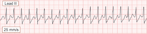
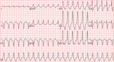
a. Identify the rhythm and how do we manage it.
b. What is the most common tachyarrhythmia in children?
c. What are the common mechanisms of arrhythmia?
d. Which are the shockable tachyarrhythmias?
e. Which are the tachyarrhythmias where synchronized cardioversion helps?
f. Identify the rhythm.
g. What is the drug of choice for ventricular tachycardia?
h. What is the drug of choice for Wolff–Parkinson–White (WPW) syndrome?
i. In long QT syndrome what is the time cut off?
Answer
| a. | Supraventricular tachycardia (SVT). Narrow QRS complex with HR >230 beats/minute for infants and >180 beats/minute for children with no identifiable normal P wave, no R-R variation, and no baseline variability. |
Adenosine is drug of choice. 0.1 mg/kg (first dose maximum 6 mg) via rapid push with three-way stopcock in a venous access close to heart. If not settling with second dose 0.2 mg/kg (maximum 12 mg) injection amiodarone is the next drug of choice. If patient becomes hemodynamically unstable synchronized cardioversion has to be done.
Long-term treatment for SVT includes medical antiarrhythmic therapy or catheter ablation.
| b. | Sinus tachycardia, commonly with fever. |
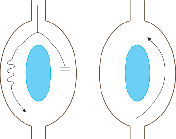
| c. | Re-entry, automaticity, and triggered activity. Re-entry most common as in SVT. |
| d. | Ventricular tachycardia with pulse, SVT with no pulse (unstable SVT). |
| e. | Ventricular tachycardia with pulse, SVT with no pulse (unstable SVT). |
| f. | Ventricular tachycardia—broad QRS complex tachycardia, atrioventricular (AV) dissociation, QRS and T wave different orientation due to automaticity. |
| g. | Injection amiodarone and lignocaine. |
| h. | Verapamil or diltiazem can be used in WPW syndrome. It is characterized by a short PR interval and delta wave (which represents ventricular preexcitation prior to normal activation of the AV node and His-Purkinje system) and episodes of tachycardia. Ebstein’s anomaly and L-transposition of the great arteries are the most common associated congenital anomalies. |
Digoxin or any AV node blocking agents should not be used. Adenosine should not be used in preexcited atrial fibrillation as it may lead to ventricular fibrillation by enhancing accessory pathway conduction to ventricles.
| i. | More than 500 ms is long QT definition and if >550 ms needs therapy. Although several genetic mutations have been recognized to cause long-QT syndrome, the most common being LQT1 (KCNQ1). |
THG08-011
Q8.11
a. What is COMFORT scoring used for?
b. What are the components and total scores in COMFORT score?
c. How do you interpret the score?
d. What are the other scores used for monitoring sedation in PICU?
Answer
a. • Sedation assessment tool, use to monitor sedation, and distress for ventilated patients which is validated for use in children.
| • | Helps in maintaining the level of sedation by titrating dose of ongoing sedative drug infusion. |
b. • COMFORT score has eight components (two are physiological parameters): Alertness, calmness, respiratory response, physical movement, BP, HR, muscle tone, and facial tension.
| • | Each component has score from 1 to 5, higher the score more distressed the child. |
c. • Each parameter is graded on a scale of one to five. Lowest score being eight and highest being 40.
| ◆ | Score ≥22: Inadequate sedation |
| ◆ | Score 11–22: Adequate sedation |
| ◆ | Score ≤11: Excessive sedation |
| d. | Bispectral index (BIS) and Richmond Agitation Sedation Scale (RASS). |
THG08-012
Q8.12
a. What is the mechanism of action of fentanyl?
b. Define metabolism, excretion, and half-life of fentanyl.
c. Mention any three indications for fentanyl.
d. What are the common route of administration (any four) and dose for intravenous administration?
e. What are the advantages of using fentanyl?
f. What are the adverse effects and antidote for fentanyl overdose?
Answer
| a. | Biochemically fentanyl is Mu (μ)-opioid receptor agonist, activation of Mu receptor produces analgesia and increase levels of dopamine. Less effect on delta and kappa receptor. |
| b. | Fentanyl is hepatically metabolized via the CYP450 enzyme system. Up to 75% of the drug metabolites are excreted in urine. Half-life of fentanyl is 3–7 hours, and 11–22 hours for continuous infusion. |
c. • Anesthesia induction and general anesthesia.
| • | Direct laryngoscopy for ET intubation. |
| • | Continuous infusion for sedation for ventilated patients. |
| • | Postoperative pain management/pain control analgesia (PCA). |
| • | Fentanyl patch for postoperative pain. |
| d. | Intravenous, intramuscular, intranasal, transdermal patch, and intrathecal. |
| e. | Lack of myocardial depression, no histamine release, and suppress stress response. |
| f. | Adverse effects: Chest tightening (rigidity) on rapid push, respiratory depression and unpredictable amnesia. |
| Monitoring: Respiratory rate, SpO2, HR and BP | |
| Antidote: Naloxone |
THG08-013
Q8.13
a. What is the mechanism of action of propofol?
b. Mention lipid content of propofol.
c. Mention any three indications for propofol.
d. What are the advantages of using propofol?
e. What are the contraindications for using propofol?
Answer
| a. | Activation of GABA receptor (prolonging the action of GABA), inhibition of NMDA receptor through sodium channel gating. |
| b. | Emulsion of soybean oil, egg lecithin, and glycerol. |
| c. | Induction and maintenance of anesthesia, short procedures, continuous sedation in ICU, and total intravenous anesthesia (TIVA). |
d. • Attenuation of bronchospasm, quick time of onset and shorter recovery time. antiemetic effect.
| • | Adverse events: Respiratory depression, profound hypotension, bradycardia, pain at injection site, propofol infusion syndrome. |
| e. | Hypersensitivity, shock/hypotension, generally not used in children <3 years of age, and hyperlipidemia. |
THG08-014
Q8.14
a. What is the mechanism of action of levosimendan?
b. Levosimendan comes under which group of vasoactive, mention one more from the same category.
c. What are the indications for levosimendan?
d. Mention advantages of using levosimendan over others of same group.
e. What are the adverse effects and contraindication for usage?
Answer
a. • Enhances calcium myofilament responsiveness by binding to cardiac troponin C (calcium sensitizer without increasing intracellular cAMP or calcium concentration)—this leads to increased contraction.
| • | It opens potassium ATP channels in myocytes and sarcolemma—effect seen as vasodilatation and cardioprotection. |
| b. | Levosimendan is a inodilator, i.e., increased contractility and vasodilatation. Other inodilators are dobutamine and milrinone. |
| c. | RV failure, chronic heart failure, perioperative cardiac surgeries with arrhythmias, and unstable hemodynamics. |
| d. | It increases inotropy without increasing cAMP and intracellular calcium levels. It does not increases myocardial oxygen demand— non-arrhythmogenic. |
| e. | Adverse effects: |
| • | Dose dependent, hypotension, and headaches |
| • | Should be avoided in case of torsades |
| • | Should be used with caution in case of renal and hepatic impairment |
Contraindicated in mechanical obstruction affecting ventricular filling and the outflow, severe hypotension, and tachycardia.
THG08-015
Q8.15
a. Which class of drug rituximab belongs to?
b. What are the mechanisms of action of rituximab?
c. Mention any three indications of rituximab.
d. Define administration and adverse effects of rituximab.
e. Mention contraindications of rituximab.
Answer
| a. | Rituximab is classified as monoclonal antibody. It is an anti-CD20 chimeric antibody. |
| b. | Rituximab binds to CD20 positive cells and causes cell death by antibody-dependent cell-mediated cytotoxicity (ADCC), complement-mediated cytotoxicity (CDC), antibody-dependent phagocytosis (ADP), and direct effect of binding of rituximab to CD20. |
| c. | Indications of rituximab: |
| • | Hematological conditions: Refractory ITP and HL. |
| • | Neurological: Autoimmune encephalitis (NMDA encephalitis), NMO spectrum disorders, multiple sclerosis, immune-related peripheral neuropathies, and refractory myasthenia gravis. |
| • | Rheumatological: Rheumatoid arthritis and systemic lupus erythematosus (SLE). |
| • | Renal: Nephrotic syndrome, renal transplant, and post-transplant graft-versus-host disease (GVHD). |
d. • Premedication with acetaminophen and antihistamine prior to administration. Should be used after diluting with 0.9% normal saline (NS)/D5%, and should be administered as slow infusion. No other drug should go along in the same line.
| • | Adverse effects: Most common is allergy and anaphylactic reaction, fever, chills, skin rash, hypotension, and arrhythmias. |
| • | Severe bacterial, viral or fungal infections, newer or reactivation of viruses like, herpes, cytomegalovirus (CMV), and varicella. |
| e. | Active infection, known hypersensitivity, heart failure, and ongoing arrhythmias. |
THG08-016
Q8.16
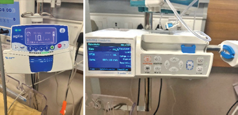
a. What are the equipment shown in the image called as? Mention any three types.
b. Mention the various usages of these pumps.
c. What are the advantages of these volumetric pumps?
d. What are the limitations of using volumetric pumps?
Answer
| a. | These are called volumetric pumps. Syringe pumps, infusion pumps, insulin infusion pumps, and patient-controlled anesthesia pumps. |
| b. | Administration of IV medications, fluid therapy, parenteral nutrition, blood transfusion, insulin delivery, chemotherapy, and pain management. |
| c. | Precise drug delivery, increased safety, audiovisual alarm in case of error, convenient to use, and mobility. |
| d. | Requires training, higher cost as compared to manual administration, and risk of malfunction. |
THG08-017
Q8.17
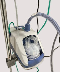
a. Identify this oxygen delivery device and mention various parts required for usage.
b. What are the general indications of this device in pediatric population?
c. Mention mechanism of action of the device and effect on the patient.
d. How to set flow rate and fraction of inspired oxygen (FiO2) at initiation?
e. What are the contraindications of using the device?
f. What should be monitored after initiation of the device?
g. What is the method of disinfection used for the device?
h. How frequently disinfection is required?
Answer
| a. | Heated humidified high-flow nasal cannula (HHHFNC) device. It consists of HFNC machine, humidification chamber, HFNC circuit, nasal interface, and distilled water for humidification. |
| b. | General indications: |
| • | Respiratory distress associated with infectious and noninfectious causes (bronchiolitis, bronchopneumonia, fluid overload, and congestive cardiac failure). |
| • | Postoperative respiratory support (i.e., tonsillectomy and adenoidectomy)/postextubation support. |
| • | Mild to moderate respiratory distress with hypoxemia (SpO2 <90%). |
| • | Weaning from NIV support. |
c. • Wash out of nasopharyngeal dead space.
| • | Reduce the inspiratory resistance. |
| • | Improves pulmonary compliance and recruitment by humidification and positive distending pressure. |
| • | Increases tidal volume and end-expiratory pressure. |
| • | Reduces respiratory rate and improves tolerance. |
| • | Improves mucociliary clearance. |
| d. | For children up to 10 kg—flow 2 L/kg/min, for each kg thereafter 0.5 L/kg/min (maximum up to 50 L/min) |
| e. | Main contraindications to start on HFNC are poor sensorium with non-maintainable airway, central apnea/hypopnea, upper GI surgery, facial trauma, severe upper airway obstruction, severe ARDS, and pneumothorax. |
| f. | While on HFNC we need to monitor HR, RR, WOB, and SpO2 till child get stabilized. Usual response is reduction in HR (by 20%) and RR (20%) within first 2–4 hours, with stabilization in SpO2 (should be able to maintain SpO2 with FiO2 <40%). |
| g. | HFNC machine needs to be disinfected by thermal disinfection method, using the disinfection tubing and air filter need to be replaced. |
| h. | It should be disinfected after each usage. |
THG08-018
Q8.18
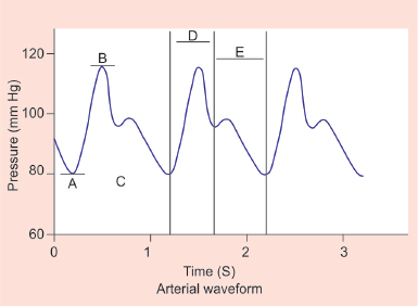
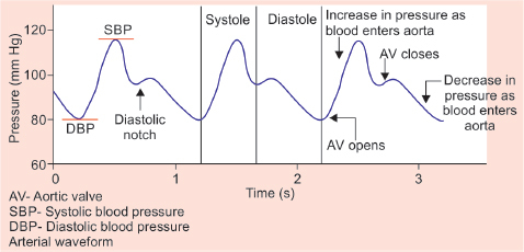
a. Identify the labels “A”, “B”, “C”, “D”, and “E” on the given arterial line waveform tracing.
b. Mention four preferred sites for arterial line insertion.
Answer
| a. | A—Diastolic BP, B—Systolic BP, C—Dicrotic notch, D—Systole, E—Diastole. |
| b. | Preferred sites are radial artery, femoral artery, posterior tibial, and dorsalis pedis |
THG08-019
Q8.19
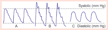
a. Identify the given trace.
b. Identify A, B and C in the following image.
c. Mention what is zeroing and leveling.
Answer
| a. | Arterial trace patterns. |
| b. | A—Normal, B—Underdamped, C—Overdamped. |
| Causes: |
| • | Underdamping—loose connectors, air bubbles in the tubing, blood clot in the circuit, or kinking of vascular catheter. |
| • | Overdamping—overly stiff circuit tubing or a defective transducer. |
| c. | “Zeroing” can be defined as the use of atmospheric pressure as a reference standard against which all other pressures are measured. “Leveling” can be defined as the selection of a position of interest at which the reference standard (zero) is set. |
THG08-020
Q8.20
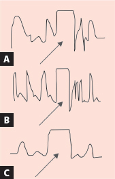
a. What is the test being performed below?
b. What is the interpretation of the test performed?
c. What is the influence of over- and underdamping on the measured blood pressure (BP)?
Answer
| a. | Test performed is square-wave test or flush test. |
| b. | After stopping of flush: |
| A. | 1.5–2 oscillations—adequate damping |
| B. | >2 oscillations—underdamping |
| C. | <1.5 oscillations—overdamping |
| c. | Underdamped system | Overdamped system |
| Overestimate SBP, underestimate SBP | Underestimate DBP, overestimate DBP | |
| MAP accurate | MAP accurate |
THG08-021
A child with meningitis on day 2 of the intensive care unit (ICU) stay shows electrolyte Na—122 mEq/L, K—3.9 mEq/L, Cl—102 mEq/L, HCO3—22 mEq/L.
a. Enumerate common causes with clinical parameters to differentiate them.
b. On evaluation of polyuria, three children shows the following findings. Identify 1, 2, and 3.
c. Name a test to differentiate these three conditions.
Answer
| a. | Common causes of hyponatremia in critically ill children are syndrome of inappropriate antidiuretic hormone (SIADH) and cerebral salt wasting syndrome. |
| SIADH | CSW |
|---|---|
| Low to normal urine | Polyuria |
| Euvolemia | Hypovolemia |
| Fluid restriction | Extra fluids and salt supplementation |
b. 1. Central diabetic insipidus
| 2. | Nephrogenic diabetic insipidus |
| 3. | Psychogenic polydipsia |
| c. | Water deprivation test: Psychogenic polydipsia will be able to produce concentrated urine in water deprivation test, whereas diabetes insipidus (DI) cannot produce concentrated urine in water deprivation test. Differentiation between central and nephrogenic DI can be done using desmopressin (DDAVP) administration. Concentrated urine is produced in central DI after DDAVP administration whereas no response is seen in nephrogenic DI. |
THG08-022
A 16-year-old male known wheezer went trekking at Mount Everest. After climbing 1,000 feet with an atmospheric pressure of 697 torr, he developed breathlessness and dizziness. He was taken to a nearby hospital where the arterial blood gas (ABG) result showed pH 7.1, PaCO2 48 mm Hg, and PaO2 66 mm Hg. Calculate the alveolar oxygen pressure (PAO2)
Answer
The partial pressure of oxygen in the alveolus (PAO2) can be predicted from the alveolar gas equation:
PAO2 = PiO2 − (PaCO2/R)
PB is barometric pressure
PH2O is the partial pressure due to water vapor
PAO2 is the alveolar partial pressure of oxygen
PiO2 is the inspired partial pressure of oxygen
R is the respiratory quotient
[R, is a measure of the metabolic rate of the tissues (CO2 production/O2 consumption)]
FiO2 is the inspired fraction of oxygen.
According to the above equation PAO2 is calculated as follow:
= 0.21(697 − 47) − 48/0.8
= 136 − 60
= 76 mm Hg
THG08-023
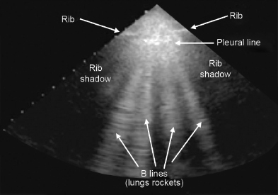
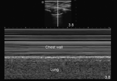
Ultrasound lung image is shown below. Describe the following:
a. What is bat sign in Lung ultrasound?
b. What are B lines and describe its significance?
c. What does the above image signifies?
Answer
| a. | Bat sign is seen in normal lung usg when the probe is placed longitudinally in chest. It represents the pleural line (which represents the parietal pleura) and the adjacent ribs. The ribs resemble the wings of the bat (black shadow), while the pleural line which lies below the ribs resembles the body of the bat. |
| b. | B lines, which are vertical artifacts produced by the pleural lines that move synchronously with lung sliding. They are hyperechoic in ultrasonography (USG) and laser shaped and are usually seen until the bottom of the screen, erasing the A lines. |
| Upto 2 B lines are seen in normal lung. B lines are absent in pneumothorax. Multiple B lines are seen in interstitial syndrome. | |
| c. | This image denotes the sea shore sign. In normal lung sliding images appears as the sea-shore in which the pleura and the overlying structures (chest wall) appear as horizontal echogenic lines (sea), while the underlying lung gives a grainy/sandy appearance. |
THG08-024
A 1-year-old female child with history of cough and cold for 5 days now presented with respiratory distress and hypoxia.
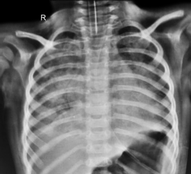
a. Identify the chest X-ray (CXR) findings.
b. She was initially started on noninvasive ventilation (NIV) with 50% FiO2, and after 4 hours the ABG showed pH 7.1, PCO2 46 mm Hg, PO2 60 mm Hg, and SpO2 88%. Classify severity according to the Pediatric Acute Lung Injury Consensus Conference (PALICC) criteria.
c. According to PALICC 2 guidelines what is the time period for trial NIV.
d. Define oxygenation criteria for possible acute respiratory distress syndrome (ARDS) and at-risk criteria according to the PALICC criteria.
Answer
| a. | Chest X-ray shows bilateral diffuse hazy infiltrates which is suggestive of ARDS. |
| b. | According to PALICC 2 guidelines severity grading should be done at least after a period of 4 hours of initiating respiratory support. |
Diagnosis of pediatric acute respiratory distress syndrome (PARDS) on NIV (NIV-PARDS) requires full facemask interface with continuous airway positive pressure/positive end-expiratory pressure ≥5 cmH2O.
| Mild/moderate NIV-PARDS: PaO2/FiO2 > 100 or SpO2/FiO2 > 150. | |
| Severe NIV-PARDS: PaO2/FiO2 ≤ 100 or SpO2/FiO2 ≤ 150 | |
| This child PF ratio = 60/0.5 = 120 (mild/moderate) | |
| c. | PALICC suggest that in patients with possible PARDS or at risk for PARDS on conventional oxygen therapy or HFNC who are showing signs of worsening respiratory failure, a time-limited trial of NIV [continuous positive airway pressure (CPAP) or bilevel positive airway pressure (BiPAP)] should be used if there are no clear indications for intubation. |
In patients on NIV who do not show clinical improvement within the first 6 hours of treatment or have signs and symptoms of worsening disease including increased respiratory/HR, increased WOB, and worsening gas exchange (SpO2/FiO2 ratio), intubation be used in comparison to continuing NIV.
d. Possible ARDS: Nasal CPAP/BiPAP or HFNC (≥1.5 L/kg/min or ≥30 L/min): PaO2/FiO2 ≤300 or SpO2/FiO2 ≤250
| At risk ARDS: Oxygen supplementation with any interface to maintain SpO2 ≥88% but not meeting definition for PARDS or possible PARDS. |
(Oxygen supplementation is defined as FiO2 >21% on invasive mechanical ventilation; or FiO2 >21% on NIV; or “oxygen flow” from a mask or cannula that exceeds these age-specific thresholds: ≥2 L/min (age <1 year), ≥4 L/min (age 1–5 years), ≥6 L/min (age 6–10 years), or ≥8 L/min (age >10 years).
For children on a mask or cannula, oxygen flow calculated as FiO2 × flow rate (L/min) (e.g., 6 L/min flow at 0.35 FiO2 = 2.1 L/min).
THG08-025
Pulse oximetry
a. What is the basic principle of pulse oximetry?
b. What factors affect the pulse oximetry readings?
c. What are the wavelengths used in the pulse oximeter to calculate oxyhemoglobin and deoxyhemoglobin ratios?
d. What is the saturation above which pulse oximetry will be accurate?
Answer
| a. | Pulse oximetry works according to the principle of Beer–Lambert law, which states that the concentration of a solute in a solvent can be determined by light absorption. |
Oxygenated hemoglobin and deoxygenated hemoglobin have different optical absorption spectra.
Pulsatile optical absorbance in biological tissue is primarily due to arterial blood.
The attenuation of light passing through tissue is pulsatile in nature due to arterial pulsation, and this altering attenuated light is analyzed to determine the saturation.
Nonpulsatile portions (capillary/venous blood) are ignored in the analysis.

b. • Poor cardiac output/low-perfusion states
| • | Motion artifact |
| • | Increased venous pulsations |
| • | Optical interference from environment |
| • | Dyshemoglobinemias: Carbon monoxide, methemoglobinemia, fetal hemoglobin |
| • | Dyes and pigments: Methylene blue and indocyanine green |
| c. | Wavelengths of 660 nm (red) and 940 nm (infrared) are used because the absorption characteristics of these oxygenated and deoxygenated hemoglobins are significantly different at these two wavelengths. |
Deoxygenated hemoglobin absorbs more red light while oxygenated absorbs more infrared light.
| d. | Mostly confidence limit of ±2–4% for pulse oximetry readings if saturation is >70%. Limited amount of calibration exists for saturation <70% making the reading less precise and variable. |
THG08-026
Following is the picture of capnogram.
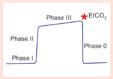
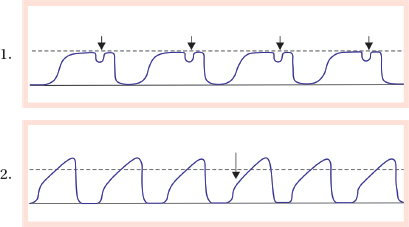
a. Explain the phases of capnogram.
b. Identify the following capnograms:
c. Enumerate few differential diagnoses of absent capnogram.
d. What are the types of capnography? Mention few advantages and disadvantages of each.
Answer
| a. | Phase I—inspiratory baseline, phase II—expiratory beginning (mixture of anatomical and physiological dead space) phase III—alveolar expiration plateau, phase 0—inspiration beginning. |
| b. | 1. Curare effect (weaning of neuromuscular blockade) |
| 2. Shark fin appearance (loss of plateau in expiration due to bronchospasm) | |
| c. | Cardiac arrest, tube displacement, pulmonary embolism, and equipment malfunction |
| d. | Main stream: |
| • | Advantages: No delay in sampling, no obstruction, etc. |
| • | Disadvantages: Older version are heavyweight, secretions may clog sensors |
Side stream:
| • | Advantages: Easy to connect, can be used in abnormal position (prone) |
| • | Disadvantages: Sampling tube obstruction, water vapor pressure change affects PCO2 |
THG08-027
Q8.27
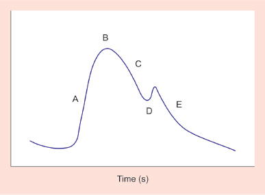
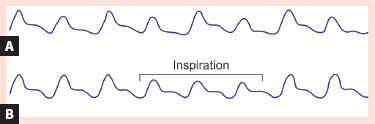
a. Identify the following waveform with its parts.
b. What are the common sites for tracing this waveform?
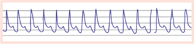
c. A 3-year-old male child admitted with septic shock shows the following distorted tracings on monitor after few hours of resuscitation. Identify the waveform findings and test to confirm the findings.
d. Identify the below waveforms and the condition associated with them.
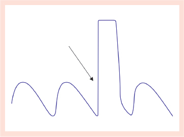
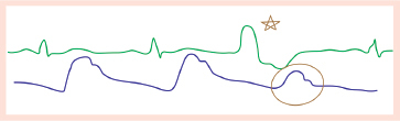
e. A 4-month-old infant with hypotensive shock and on adrenaline infusion. Identify the following condition and justify the waves.
Answer
| a. | Arterial wave form: |
| A—systolic upstroke | |
| B—peak systolic pressure | |
| C—systolic downstroke | |
| D—dicrotic notch | |
| E—diastolic run off | |
| b. | Radial artery, ulnar artery, axillary artery, brachial artery, dorsalis pedis, posterior tibial, femoral artery, superficial temporal artery, and umbilical artery |
| c. | Over dampened wave form. Fast flush test/square test shows <1.5 oscillations/absent dicrotic notch. |
| d. | A—pulsus alternans seen commonly in LV failure |
| B—pulses paradoxus seen in asthma and pericardial effusion | |
| e. | Green wave tracing shows ECG and blue line shows arterial tracing. The arterial waveform shows reduced systolic peak as stroke volume is less in premature ventricular contraction. |
THG08-028
Answer the following questions regarding monitoring of saturation.
a. What is mixed venous oxygen saturation?
b. Difference between ScvO2 and SvO2 levels.
c. What is the normal range and what causes low SvO2 levels?
d. What causes normal or high SvO2 levels with lactic acidosis?
Answer
| a. | Mixed venous oxygen saturation (SvO2) refers to the oxygen content of the blood that returns to the heart after meeting tissue needs. SvO2 is commonly used as a measure of the balance between O2 demand and supply. |
| b. | 0 | SvO2 | ScvO2 |
|---|---|---|---|
| Sampling site | Tip of pulmonary artery | Central vein (IJV/subclavian/SVC/IVC) | |
| Representation | Represents overall oxygen extraction of body | Represents either upper body and brain or lower body based on the sampling site | |
| Normal value | 75% (60-80%) | >70% |
| c. | The O2 saturation of Hb reaches stability in the RV outflow tract after the differences in saturation from the IVC, superior vena cava, coronary sinus, and Thebesian veins equilibrate. The normal saturation of Hb in the pulmonary artery is 75% (60–80%). |
| d. | Reduction in SvO2 : |
| • | Decreased O2 delivery |
| • | Decreased cardiac output |
| • | Decreased arterial O2 saturation |
| • | Decreased hemoglobin concentration/increased O2 consumption |
It can be seen in late septic shock, or in cell poisoning such as cyanide, where even when oxygen delivery is normal or near normal inability of tissue to utilize oxygen leads resort to anaerobic metabolism which cause rise in lactic acid.
Other causes of high SvO2:
| • | Decreased O2 consumption |
| • | Decreased O2 extraction |
| • | Left-to-right intracardiac shunt |
THG08-029
An 1-year-old male child developed cyanosis and hypoxia SpO2 82% under oxygen.
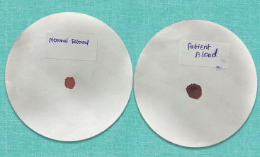
a. Look at the blood blot given in the image. What is the probable diagnosis and investigation of choice?
b. What are the hemoglobin forms measured by the above investigation?
c. What is the principle of the above investigation?
d. What is the treatment of choice in toxic methemoglobinemia?
Answer
| a. | The characteristic chocolate brown appearance of the blood sample on the right denotes methemoglobinemia. Co-oximetry will be the test of choice. |
| b. | It measures the levels of oxyhemoglobin, carboxyhemoglobin, methemoglobin, and reduced hemoglobin. |
| c. | Co-oximetry works on Beer–Lambert law. Multi-wavelength co-oximeters use several, rather than two, wavelengths of light (e.g., four to eight) to detect oxyhemoglobin, deoxyhemoglobin, carboxyhemoglobin, and methemoglobin (e.g., methemoglobin at 630 nm). |
| d. | Methylene blue administered iv (1–2 mg/kg) is used to treat toxic methemoglobinemia. Maintenance dose: 100–300 mg/day oral. |
THG08-030
A 9-year-old male child with fever and cough for 10 days, presented with respiratory distress and severe left side chest pain and scoliosis.
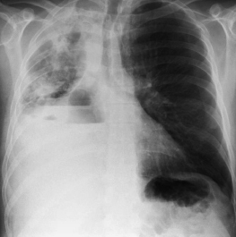
a. Identify the condition.
b. Mention the most common organisms causing the above condition.
c. What are the stages of the above condition?
d. What are the indications of surgery?
Answer
| a. | Empyema thoracis. |
| b. | Staphylococcus aureus, Streptococcus pneumoniae, and Streptococcus pyogenes are the organisms most implicated in empyema thoracis. |
| c. | (i) Exudative stage (1–3 days): Fluid is thin and lungs can readily re-expand. At this point fluid is considered a simple parapneumonic effusion characterized by normal pH and glucose levels. Pleural fluid analysis shows pH >7.30, glucose >60 mg/dL, pleural fluid/serum glucose ratio >0.5, lactate dehydrogenase (LDH) <1,000 IU/L while Gram stain and culture is negative for microorganism. |
(ii) Fibrinopurulent stage (4–14 days): A significant number of polymorphonuclear leukocytes and fibrin accumulate in the effusion. Pleural fluid pH and glucose level fall while LDH rises. Effusion becomes purulent and viscous leading to development of empyema. Pleural fluid analysis shows purulent fluid or pH <7.10, glucose <40 mg/dL, and LDH >1,000 IU/L. Gram stain and culture reports show microorganisms.
(iii) Organizing stage (after 14 days): Fibroblasts grow into exudates on both the visceral and parietal pleural surfaces, producing an inelastic membrane “the peel”. Thickened pleural peel may prevent the entry of antimicrobial drugs in the pleural space and in some cases can lead to drug resistance. A thickened pleural peel can restrict lung movement and it is commonly termed as trapped lung.
d. • Sepsis or infected fluid not effectively controlled with antibiotics or implantable cardioverter defibrillator (ICD).
| • | Presence of significant distress due to thickened pleura. |
| • | ICD output decreases but with clinical deterioration with persistent effusion in imaging. |