THG27-001
Q27.1
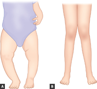
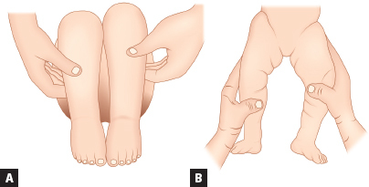
a. What is the diagnosis in images A and B?
b. Presence of this condition beyond ________ years of age is pathological (in image A).
c. Presence of this condition beyond ________ years of age is pathological (in image B).
d. Mention (at least four) pathological causes in image A.
e. Mention (at least four) pathological causes in image B.
Answer
| a. | Image A: Genu varum (bowlegs) |
| Image B: Genu valgum (knock-knees) | |
| b. | 2 years in image A |
| c. | 4–6 years in image B |
| d. | Image A: Metabolic bone disease (vitamin D deficiency rickets, vitamin D-resistant rickets, and hypophosphatasia), asymmetric growth arrest (trauma, infection, tumor, and Blount), bone dysplasia (achondroplasia and metaphyseal dysplasia) |
| e. | Image B: Metabolic bone disease (rickets and renal osteodystrophy), skeletal dysplasia, post-traumatic physeal arrest, tumors, and infection. |
THG27-002
Q27.2
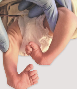
a. What is this condition?
b. What are the components of this condition?
c. What are the types?
d. What are the commonly associated conditions?
e. Name the standard method of initial treatment.
Answer
| a. | Talipes equinovarus |
| b. | Cavus (plantar flexion): Adductus (adduction of the forefoot/midfoot over hindfoot); Varus (hindfoot is in varus); Equinus (hindfoot is in equinus) |
| c. | Positional, congenital, syndromic, or neuromuscular |
| d. | Myelodysplasia, arthrogryposis, developmental dysplasia of the hip (DDH), chromosomal syndromes—trisomy 18, 22q11 deletion |
| e. | Ponseti method |
THG27-003
A 11-year-old boy with complaints of (c/o) foot pain on walking, difficulty in shoe wear, and frequent ankle sprains.
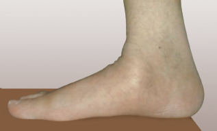
a. What is the diagnosis?
b. Name the different types of this condition.
c. What are the associated neurological conditions you will look for?
d. What is the treatment option for this child?
Answer
| a. | Pes planus (flat foot) |
| b. | Three types: Flexible flatfoot; a flexible flatfoot with a contracture of the Achilles tendon, and a rigid flatfoot |
| c. | Hereditary motor sensory neuropathies, Friedreich ataxia, and spinal dysraphism |
| d. | Orthosis such as a medial arch support |
THG27-004
X-ray of a 25-day-old neonate with history of (h/o) not moving right lower limb and fever of 8-day duration.
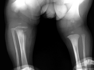
a. What are the X-ray findings?
b. What is the diagnosis?
c. What are the common organisms likely in this child?
d. What are the imaging modalities (other than X-ray) available for diagnosing this condition and which is most sensitive?
e. What is the initial empirical therapy for this child?
Answer
| a. | Right upper medial tibial metaphyseal lytic lesion; thick periostitis and interrupted soft tissues around. |
| b. | Acute osteomyelitis |
| c. | Staphylococcus aureus; Group B Streptococcus and gram-negative enteric bacilli |
| d. | Computed tomography, magnetic resonance imaging (MRI), radionuclide studies; MRI most sensitive |
| e. | Antistaphylococcal penicillin, such as nafcillin or oxacillin or cloxacillin (150–200 mg/kg/24 hour divided q6h IV), and a broad-spectrum cephalosporin, such as cefotaxime (150–225 mg/kg/24 hour divided q8h IV). If methicillin-resistant Staphylococcus is suspected, vancomycin is substituted for nafcillin. |
THG27-005
Clinical photographs of two children with limited abduction of hip.
a. Describe the clinical sign seen in images A and B?
b. What is the diagnosis?
c. Name three risk factors.
d. Name the specific maneuvers performed in newborn period for diagnosis of this condition.
e. Name the preferred modality for diagnosis in: (i) Newborn 0–1 month, (ii) 1–4/6 months, (iii) beyond 4/6 months to 1.5 year
Answer
| a. | Image A: Shortening of the thigh—placing both hips in 90° of flexion and comparing the height of the knees (the Galeazzi sign) |
| Image B: Asymmetry of thigh folds | |
| b. | Developmental dysplasia of the hip |
| c. | Breech presentation, oligohydramnios, large birth weight, first pregnancy |
| d. | Barlow provocative maneuver; Ortolani test |
| e. | (i) Clinical examination, (ii) USG, and (iii) X-ray hip |
THG27-006
Q27.6
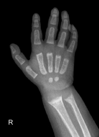
a. Describe three findings in this X-ray.
b. What is the diagnosis?
c. What is the most important radiological clue for improvement when this is treated?
d. List five biochemical tests for confirmation of diagnosis.
Answer
| a. | (i) Widening, cupping, fraying of distal ends of radius and ulna; (ii) decrease in bone density; (iii) increase in the distance between distal ends of radius and ulna and metacarpals |
| b. | Rickets |
| c. | Appearance of provisional zone of calcification |
| d. | Ca, PO4, alkaline phosphatase (ALP), parathyroid hormone (PTH), and 25(OH) vitamin D |
THG27-007
An infant presents with pain, hard swelling in bilateral thigh, no erythema and no warmth. X-ray done showed the following finding:
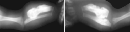
a. What is seen in the X-ray?
b. What is the diagnosis?
c. Which is the most common bone involved?
d. Which bones are not involved?
e. Name the differential diagnosis for this condition (any three).
Answer
| a. | Cortical thickening—right femur |
| b. | Caffey disease |
| c. | Mandible |
| d. | Phalanges and vertebral bodies |
| e. | Differential diagnosis: Chronic vitamin A intoxication, prolonged prostaglandin E infusion, scurvy, and primary bone tumors. |
THG27-008
A 10-year-old child presents with limping gait of 1-month duration and X-ray hip done showed the following finding:
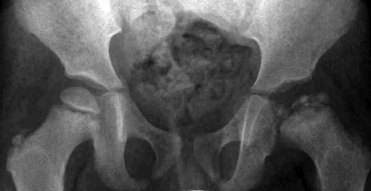
a. What are the X-ray findings?
b. What is the likely diagnosis?
c. Mention probable etiological/causative factors (at least four).
d. Investigation of choice in early stage of disease when X-ray is noncontributory.
e. What is the Prognosis?
Answer
| a. | Epiphyseal fragmentation in left hip in anteroposterior (AP) view |
| b. | Legg–Calvé–Perthes disease |
| c. | Factor V Leiden mutation; protein C deficiency; protein S deficiency; lupus anticoagulant; anticardiolipin antibodies; antitrypsin and plasminogen activator |
| d. | MRI; radionuclide bone scanning (with technetium-99m) |
| e. | Poor (onset after 9 years poor prognosis) |
THG27-009
A child presents with anemia, hepatosplenomegaly. X-ray done showed the following finding:
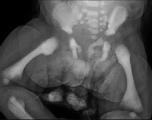
a. Describe the common findings in X-rays shown in above image.
b. Name two conditions with similar radiological findings.
c. Mention two radiological and two clinical differences in these two conditions.
d. What is this condition?
e. How many types are there?
f. What is the probable curative option for this condition?
Answer
| a. | (i) Dense bones and (ii) Cortex not made out |
| b. | (i) Osteopetrosis and (ii) Pyknodysostosis |
| c. | Clinical: (i) Pyknodysostosis—good prognosis and (ii) Osteopetrosis— anemia |
| Radiological: (i) Small mandible and (ii) Hypoplasia of distal phalanx in pyknodysostosis | |
| d. | Osteopetrosis |
| e. | Three types: (i) Autosomal recessive (AR)—infantile, (ii) AR—intermediate, and (iii) autosomal dominant (AD)—adult |
| f. | Bone marrow transplantation |
THG27-010
Q27.10
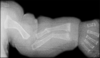
a. Describe the X-ray shown in above image.
b. What is the diagnosis?
c. What is the triad of this condition?
d. Which is the most severe form?
e. What is the underlying defect?
f. Mention two pulmonary complications.
g. What are the medical treatment options of this condition?
Answer
| a. | Generalized osteoporosis; humerus deformity; radius and ulna fractures with callus formation |
| b. | Osteogenesis imperfecta |
c. • Fragile bones
| • | Blue sclera |
| • | Early deafness |
| d. | Type 2 (perinatal lethal) |
| e. | Structural or quantitative defects in Type I collagen |
| f. | Recurrent pneumonias and declining pulmonary function |
| g. | Growth hormone; bisphosphonates |
THG27-011
A 6-year-old girl has short stature, large head and bowing of legs. Following are the X-rays. Answer the following questions:
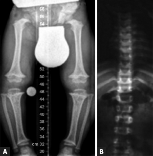
a. What are the X-ray findings in images A and B?
b. What is the diagnosis?
c. What is the mode of inheritance?
d. Explain problems/complications associated with this condition (any six).
Answer
| a. | Image A: All the tubular bones are short; fibula is longer than tibia. |
| Image B: Interpedicular narrowing of vertebrae | |
| b. | Achondroplasia |
| c. | Autosomal dominant |
| d. | Hydrocephalus; Spinal cord compression at foramen magnum = hypotonia, failure to thrive, quadriparesis, central and obstructive apnea, and sudden death; spinal cord compression at lumbar spine = paresthesia, numbness, and claudication in the legs, loss of bladder and bowel control; Bowing of the legs; Other common problems include dental crowding, articulation difficulties, obesity, and frequent episodes of otitis media, which can contribute to hearing loss. |
THG27-012
A 2-year-old child was brought with convulsions and chest X-ray done showed the following:
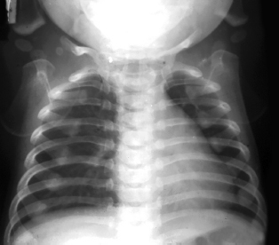
a. What is the finding in the X-ray and what is the most likely diagnosis?
b. What are the four common presenting features of this condition?
c. What is the bedside laboratory test that will confirm the diagnosis?
d. What is the confirmatory test?
e. What treatment is instituted?
f. What are the other measures to be undertaken?
Answer
| a. | X-ray chest shows bands of increased density at the metaphysis of upper end of humerus bilaterally, increased density of costochondral junctions, medial border of both scapula and manubrium sternum, indicating heavy metal deposition. |
| Diagnosis: Lead poisoning | |
| b. | Pain abdomen, constipation, pica, vomiting, seizures, and anemia |
| c. | Peripheral smear—basophilic stippling |
| d. | Elevated levels of serum lead, free erythrocyte protoporphyrin |
| e. | Chelation therapy with intravenous calcium sodium edetate for 5 days |
| f. | Health education to the family, avoid lead containing products |
THG27-013
A 10-year-old boy brought for evaluation of tall stature—height for age >3 standard deviation (SD) above mean for age, upper segment:lower segment (US:LS) ratio 0.8, and arm span to height ratio 1.5. Child has pectus carinatum, arachnodactyly, and joint laxity. There is positive family h/o similar condition across generations.
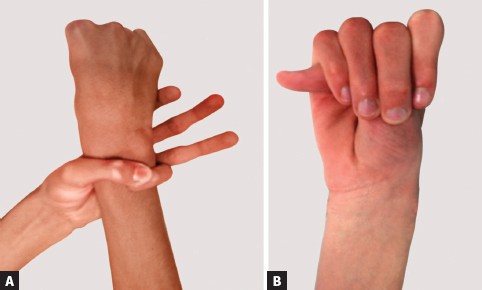
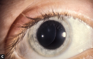
a. Name and describe the two clinical signs shown in above image?
b. What is the ocular finding?
c. What is the diagnosis?
d. Name four cardiovascular manifestations of this condition.
e. What are the pulmonary manifestations?
Answer
a. i. Wrist sign: When the wrist is grasped by the contralateral hand, the thumb overlaps the terminal phalanx of the 5th digit.
| ii. | Thumb sign: When the hand is clenched without assistance, the entire thumbnail projects beyond the border of the hand. |
| b. | Ectopia lentis (dislocation of the ocular lens) |
| c. | Marfan syndrome |
| d. | Mitral valve/tricuspid valve—prolapse/insufficiency; aortic valve insufficiency; supraventricular arrhythmias—atrial fibrillation/supraventricular tachycardia (AF/SVT); prolonged QT; ventricular dysrhythmias. Aortic aneurysm/dissection |
| e. | Restrictive lung disease; spontaneous pneumothorax |
Figure Sources
Figures of Q27.2, Q27.3, Q27.4, Q27.6, Q27.7, Q27.9, Q27.10, Q27.11 and Q27.13 are from open source.
Figures of Q27.1 and Q27.5 are redrawn by artist.
Figures of Q27.8 and Q27.13C are from author’s personal collection.