VVK18-001
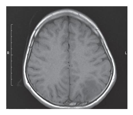
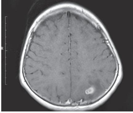
a. What is the radiological finding?
b. What is the diagnosis?
c. Enlist 2 commonest clinical conditions leading into above abnormalities.
d. What is cause of difference in radiological finding between 2 images?
Answer
- a. Hypointense lesion in posterior occipital lobe
- b. Ring enhancing lesion
- c. Inflammatory granuloma
- d. TBM, neurocysticercosis
- e. Image B is post contrast.
VVK18-002
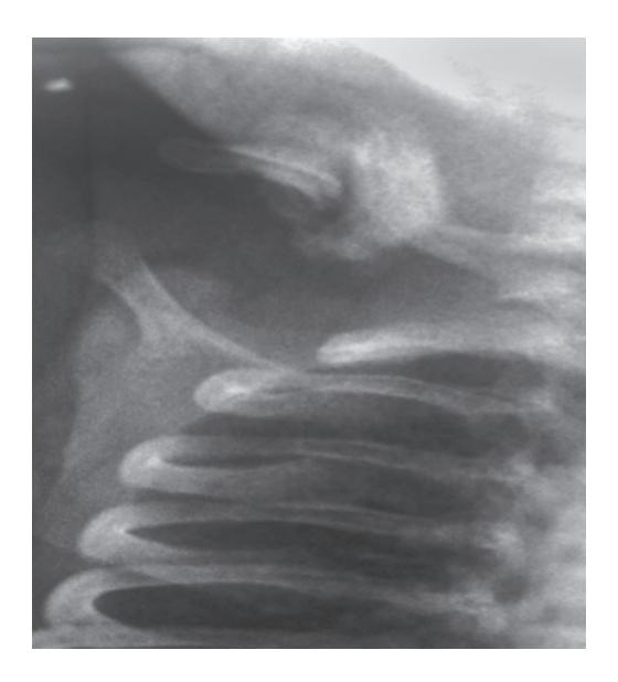
a. What is the diagnosis?
b. What is the commonest cause of abnormality in neonates and older child?
c. What is the treatment?
Answer
- a. Clavicular fracture with callus formation
- b. Neonate—birth trauma Older child—direct trauma to the clavicle
- c. Conservative in most cases rarely needing surgical correction.
VVK18-003
A 12-year-old boy presented with first episode of nonfebrile left focal seizures.
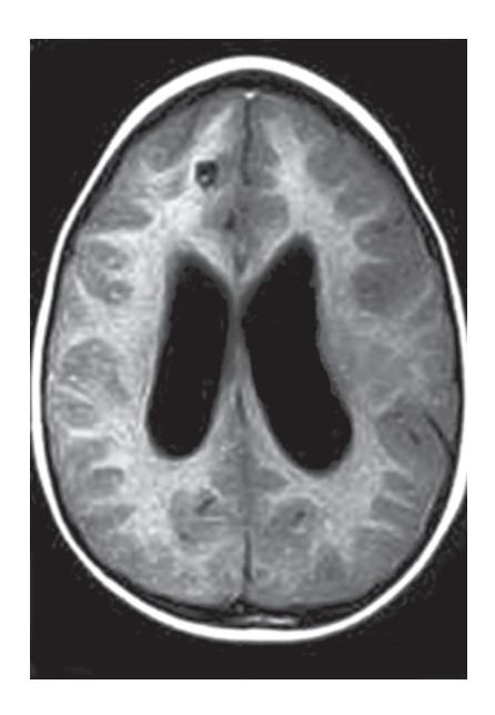
a. What is the most likely diagnosis?
b. What is the etiology?
c. What is the treatment?
Answer
- a. Neurocysticercosis (with mild ventricular dilatation).
- b. Taenia solium or pork tape worm.
- c. Albendazole (15 mg/kg/day, BID) for 28 days with steroids for 2–3 days before and immediately after initiating therapy. Anticonvulsant—carbamazepine or phenytoin.
VVK18-004
A 12-year-old boy presented with history of fever, headache and generalized tonic clonic seizures. His MRI shows hypertense lesion in the right temporal lobe.
a. What is the most likely diagnosis?
b. How can you confirm the diagnosis?
c. What is the drug of choice?
Answer
- a. HSV encephalitis.
- b. CSF viral studies for HSV-PCR.
- c. IV Acyclovir (5–10 mg/kg or 250 mg/m2 every 8 hour).
VVK18-005
Answer following questions based on X-ray seen.
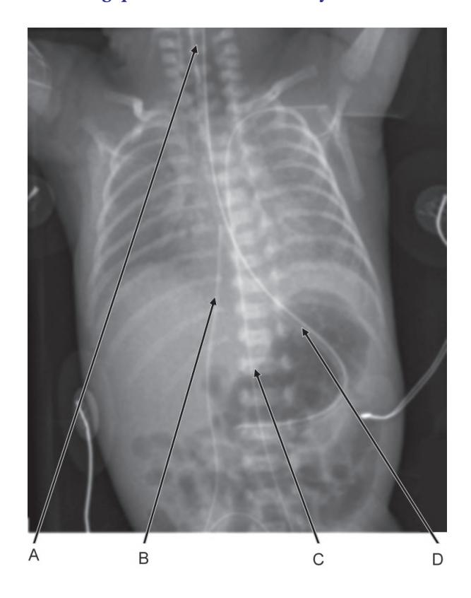
a. What are A, B, C and D?
b. What is the ideal position of placement of A, B, C and D on X-ray?
c. Which of the above is incorrectly placed?
Answer
- a. A-Endotracheal tube.
- B and C-Umbilical venous cannula and umbilical arterial cannula.
- D-Nasogastric tube.
- b. A-Endotracheal tube: T2-T3.
- B and C-Umbilical venous cannula—Just above the diaphragm and umbilical arterial cannula, high:T7-T10; low: L2-L3.
- D-Nasogastric tube—In the stomach.
- c. All of the above.
VVK18-006
A 10-year-old girl was admitted in PICU for acute gastroenteritis with severe dehydration. 24 hours after the admission she had an episode of generalized seizures. On examination, her Glasgow coma scale score is 7, heart rate 60/min, respirations are irregular and blood pressure is normal. Review the CT given below and answer the questions.
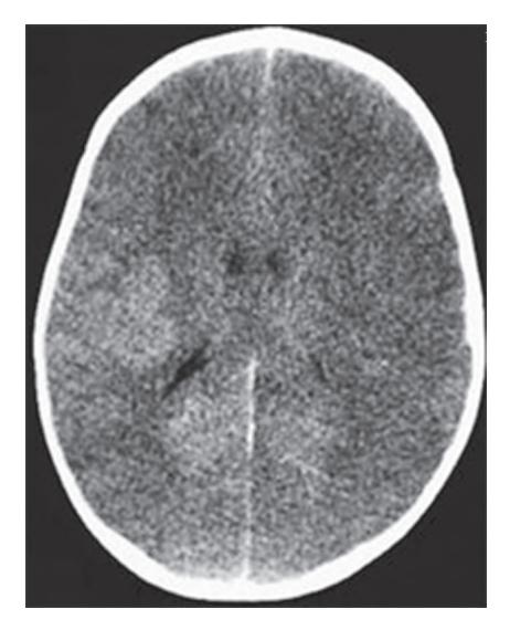
a. What are your findings on the CT scan?
b. What is the immediate management after the "ABC"?
c. What is the most common electrolyte disturbance which can be the cause of above problem in this patient?
Answer
- a. Cerebral edema in view of loss distinctness of the sulci and gyri. Due to increased ICP, the cortex is compressed up against the skull. The space between the cortex and the skull is obliterated.
- b. Hyperventilation, mannitol.
- c. Hyponatremia.
VVK18-007
Given below is the chest X-ray of a 6-week-old male infant. The infant has been having persistent tachypnea since birth. He has been treated with multiple courses of antibiotics for presumed pneumonia. Study the X-ray and answer the questions.
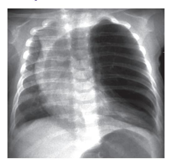
a. List 3 positive findings on chest X-ray.
b. What is the diagnosis in this X-ray?
c. What is the most common site of involvement in the above diagnosis?
Answer
a. Hyperinflation of the left upper lobe. Paucity of vascular markings of the left upper lobe. Mediastinal shift to the right.
Atelectasis of the left lower lobe.
Flattening of the left hemidiaphragm.
- b. Congenital lobar emphysema.
- c. Left upper lobe.
VVK18-008
Given below is the X-ray of a term male infant who presented with abdominal distention.
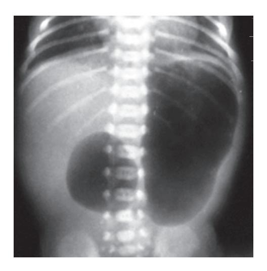
a. Name the abnormality seen.
b. What is the likely diagnosis?
c. What is the commonest chromosomal aberration associated with this abnormality?
d. What is the commonest upper gastrointestinal abnormality associated with this abnormality?
Answer
- a. Double bubble appearance.
- b. Duodenal atresia.
- c. Down's syndrome.
- d. Annular pancreas.
VVK18-009
A term female newborn was noted to have significant respiratory distress soon after birth. Study the X-ray and answer the questions.
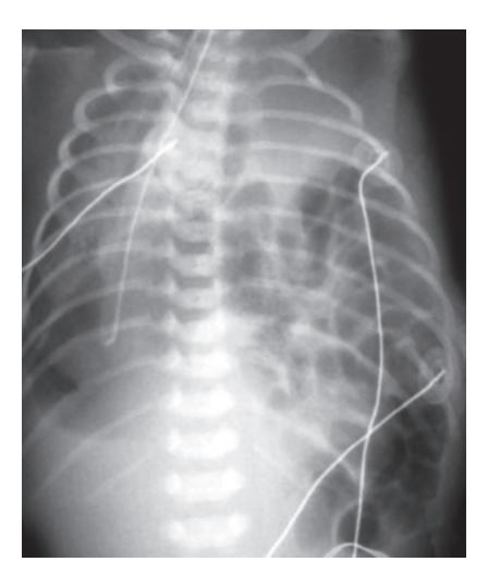
a. Describe the X-ray findings.
b. What is the diagnosis?
c. List 2 precautions to be taken during resuscitation to avoid any further deterioration in the clinical condition.
d. What other anomalies can be associated with this condition?
Answer
- a. There is a cystic mass (bowel loops) on the left side displacing the heart to the opposite side.
- b. Congenital diaphragmatic hernia.
- c. Place an OG/NG tube.
Not to give IPPV via bag and mask.
d. Pulmonary hypoplasia/aplasia with congenital heart disease, Cantrell's pentalogy.
VVK18-010
Given below is an X-ray of a 24-hour-old neonate.
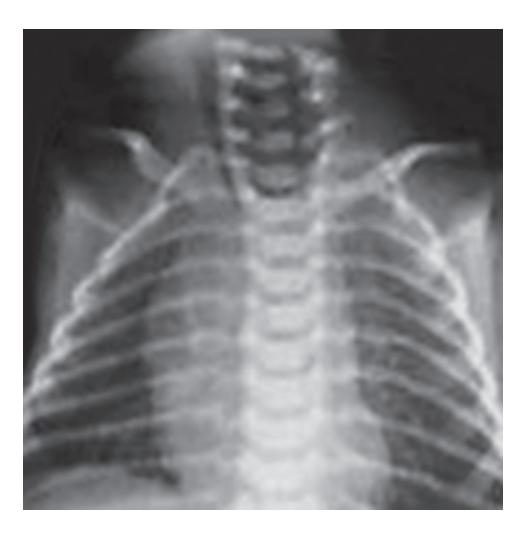
a. What is the most likely diagnosis?
b. Which is the most common subtype?
c. Give one common association with this condition.
Answer
- a. Esophageal atresia.
- b. Esophageal atresia with distal TEF (85%).
- c. VATER/VACTREL.
VVK18-011
Given below is X-ray of a 12-year-old seen by dentist with multiple dental abnormalities. This child has a history of delayed closure of fontanel and droopy shoulders.
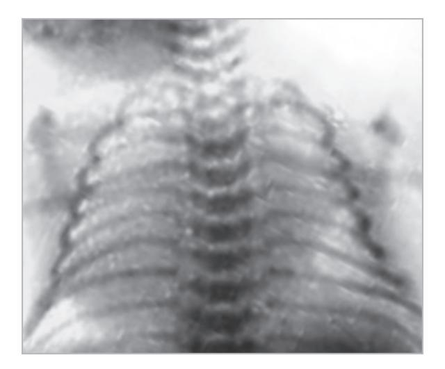
a. What is the abnormality on the X-ray?
b. What is the likely diagnosis?
c. List 3 abnormalities which are commonly seen with this diagnosis.
d. What is the clinical sign supporting the pathology seen in the X-ray above?
Answer
- a. Absent clavicles.
- b. Cleidocranial dysplasia.
- c. i. Drooping shoulders.
- ii. Open fontanelles, prominent forehead, mild short stature—because of delayed ossification of the cranial bones with multiple ossification centers (wormian bones), and delayed ossification of pelvic bones.
- iii. Dental anomalies (numerous teeth abnormalities including delayed eruption of teeth).
- d. Both shoulders can be approximated together anteriorly.
VVK18-012
Given below is the HIDA scan of a 2 months old 6 hours post dye injection.
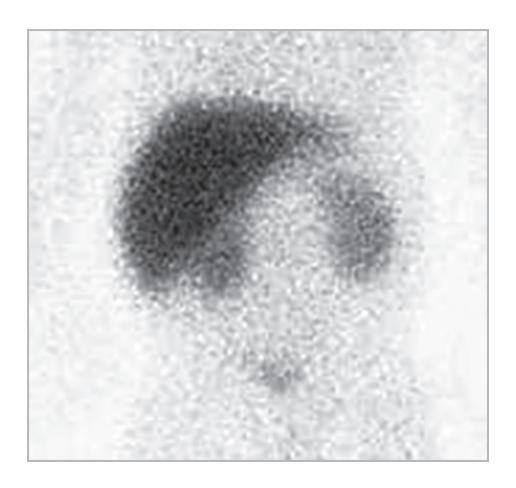
a. What are the findings on the scan?
b. What is the most likely diagnosis?
c. Which dye is used in this scan?
d. List any 6 supplementary medications required for this infant (including the dosing of any 4).
Answer
- a. No dye reaching the intestine.
- b. Biliary atresia.
- c. Iminodiacetic acid (Tc iminodiacetic) compound.
- d. Aquasol A—10,000-15,000 IU/day.
Oral α-tocopherol or TPGS 50–400 IU/day.
25-hydroxycholecalciferol 3-5 µg/kg/day or D2 5,000–8,000 IU/day.
Water-soluble derivative of menadione 2.5—5.0 mg every other day.
Calcium, phosphate, or zinc supplementation.
Choleretic bile acids and ursodeoxycholic acid, 15-20 mg/kg/day.
Dietary formula or supplements containing medium-chain triglycerides.
VVK18-013
A 15-month-old child was brought to pediatric emergency room with history of sudden onset of cough and choking. While coming to the Hospital he became asymptomatic. A chest X-ray performed is given below.
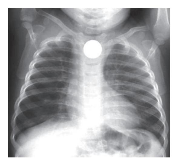
a. What is the diagnosis?
b. List 2 possible complications.
c. What is the definitive treatment for this child? This child has a sudden cardiorespiratory deterioration while in emergency.
d. What is the next step in management?
Answer
- a. Esophageal foreign body.
- b. Esophageal perforation.
Oesophageal obstruction.
Tracheal compression.
- c. Endoscopy—diagnostic and therapeutic.
- d. 5 abdominal thrust (Hemlich manouver) with the child sitting or lying down.
VVK18-014
This 12-year-old was intubated on arrival at emergency room following a road accident. Her intubation was difficult needing multiple attempts. The X-ray provided below was performed soon after admission.
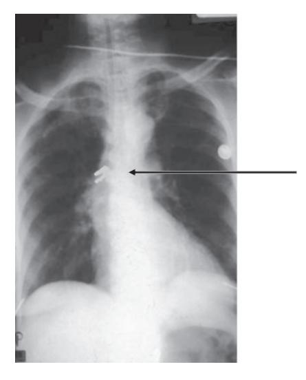
a. What is foreign body pointed in the X-ray?
b. Where is the location of this foreign body?
c. How would you remove the foreign body?
Answer
- a. Broken tooth.
- b. Right main bronchus.
- c. Rigid bronchoscopy.
VVK18-015
A 11-month-old male infant presented to pediatric OPD with highgrade fever and labored breathing. On auscultation, breath sounds are barely audible on right side. The X-ray performed is shown below.
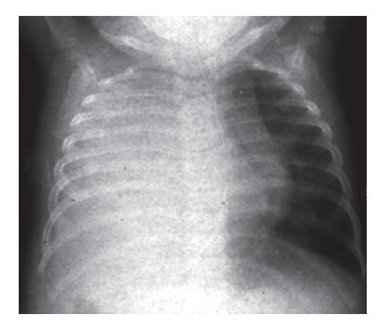
a. List 3 positive findings on chest X-ray.
b. What is the likely diagnosis?
Answer
a. Diffuse homogenous opacity in entire right lung field.
Mediastinal shift to left side.
Obliterated right CP angle.
b. Right pleural effusion.
VVK18-016
This is an X-ray of the abdomen in a 12-year-old girl who was brought to the pediatric OPD with chief complaint of recurrent abdominal pain. She has been bedridden since age of 7 years due to paraplegia following transverse myelitis.
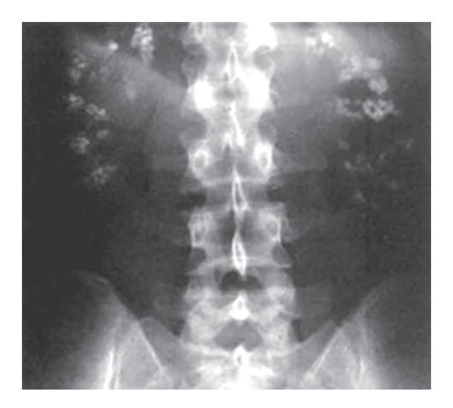
a. What abnormality can be seen on the plain abdominal film?
b. What is the diagnosis?
c. What are the definitive treatment options? (List any 2)
d. List 4 causes of this radiological finding.
Answer
- a. Bilateral radiopacities seen in the X-rays—calcification of kidneys.
- b. Nephrocalcinosis.
- c. Shock wave lithotripsy
Ureteroscopy.
Percutaneous nephrolithotomy.
d. Drug—Frusemide therapy
Medullary sponge kidney
Hyperoxalemia
Renal tubular acidosis
Hypoparathyroidism
Hypophosphatemic rickets
Sarcoidosis
Prolonged immobilization.
VVK18-017
A 3-year-old presented with an abdominal mass and hypertension. On investigation, she was noted to have microscopic hematuria. The CT scan performed show a large nonoccupying more than 70% of the abdomen.
a. What is the likely diagnosis?
b. List 2 syndromes associated with this condition.
Answer
- a. Wilms' tumor.
- b. WAGR syndrome is a contiguous gene deletion syndrome that consists of Wilms tumor, aniridia, genitourinary abnormalities
Beckwith-Wiedemann syndrome
Denys-Drash—Early onset renal failure with renal mesangial sclerosis, male pseudohermaphrodism, increased risk of Wilms tumor.
VVK18-018
A 5-year-old is diagnosed to have unilateral Wilms tumour. As a part of further investigation and staging of the disease, a CT scan of chest was performed.
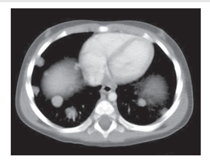
a. What is the finding on the CT scan?
b. What is the staging of his disease?
c. What other investigation will guide the treatment?
d. What is the treatment?
Answer
- a. Bilateral pulmonary metastases.
- b. Stage IV.
- c. Histopathology—Favorable/anaplastic.
- d. Surgical extirpation, chemotherapy and radiotherapy.
For stage IV tumors with favorable histology, vincristine, actinomycinD, and doxorubicin are administered, and radiation therapy is also administered to all the sites of known disease, particularly the lungs.
For tumors with unfavorable histology, vincristine, actinomycin D, doxorubicin, and cyclophosphamide are administered, and radiation therapy is also administered to all the known sites of disease.
VVK18-019
Given below is the CT scan of a 2-month-old. This child was born at term and had a stormy neonatal course. He was diagnosed to have neonatal sepsis and seizures on day 3 of life and was treated with antibiotics for 7 days.
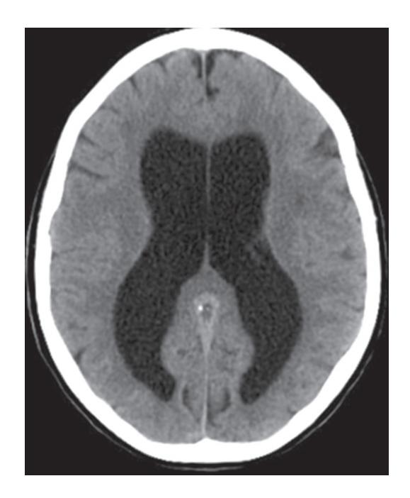
a. What is the radiological diagnosis?
b. What is the most likely cause in this infant?
c. How do you calculate VP ratio?
d. What is the medical management of the condition?
Answer
- a. Hydrocephalous.
- b. Neonatal meningitis.
- c. Divide the maximum ventricular measurement by maximum parenchymal measurement at the level of third ventricle.
- d. Acetazolamide and furosemide to decrease secretion by choroid plexus.
VVK18-020
A 8-year-old child presents to pediatric OPD with history of drooling of saliva and high-grade fever.
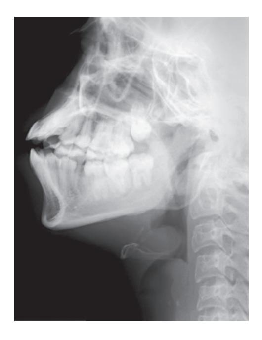
a. What is the most likely diagnosis?
b. What is the commonest organism causing this problem?
c. What is the treatment after stabilization of the child?
Answer
- a. Epiglottitis.
- b. H. influenzae.
- c. IV antibiotics—Ceftriaxone, cefotaxime, or a combination of ampicillin and sulbactam.
VVK18-021
An incidental finding was noted on reviewing the abdominal CT of a 12-year-old male for abdominal pain. The finding was later confirmed on history and physical examination.
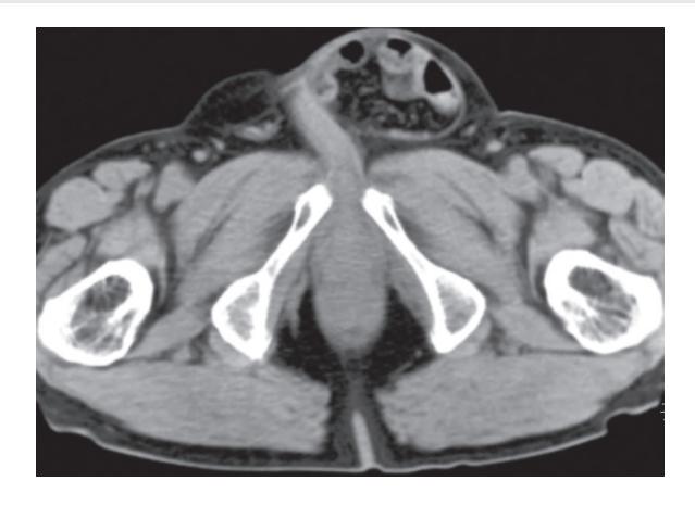
b. What is the diagnosis?
c. What is the treatment?
Answer
- a. Gas—containing bowel loops as well as mesenteric fat and vessels in the left inguinal canal.
- b. Inguinal hernia.
- c. Surgical repair.
VVK18-022
Given below is an X-ray of a 3-year-old female child with frontal bossing, bowing of legs and a large abdomen.
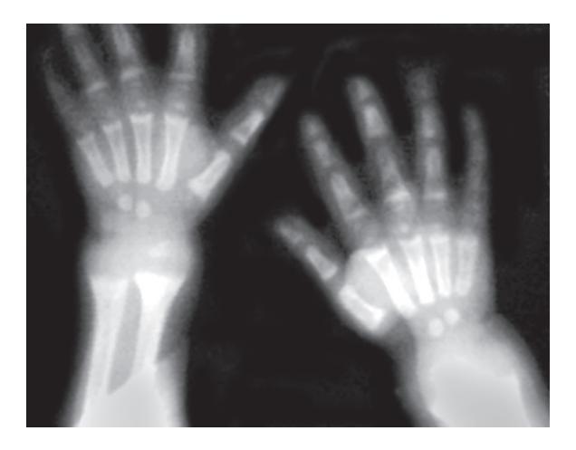
a. What are the findings seen on the X-ray?
b. What is the most likely diagnosis?
c. Enlist 3 common skeletal deformities seen in association with this disease.
d. What is the biochemical abnormality seen in this disease?
Answer
a. Fraying of metaphysis
Cupping of distal end of radius and ulna Widening of distal end of metaphysis
Generalized rarefaction.
- b. Rickets
- c. Head: Craniotabes, frontal bossing, delayed fontanelle closure, craniosynostosis
Back: Scoliosis, kyphosis, lordosis
Chest: Rachitic rosary, harrison groove
Extremities: Enlargement of wrists and ankles
Valgus or varus deformities
Windswept deformity (combination of valgus deformity of 1 leg with varus deformity of the other leg)
Anterior bowing of the tibia and femur
Coxa vara.
d. Serum calcium—normal or decreased
Serum phosphorus—decreased (except in CRF)
Serum alkaline phosphatase—increased.
VVK18-023
Identify the structures shown in cranial USG shown below.
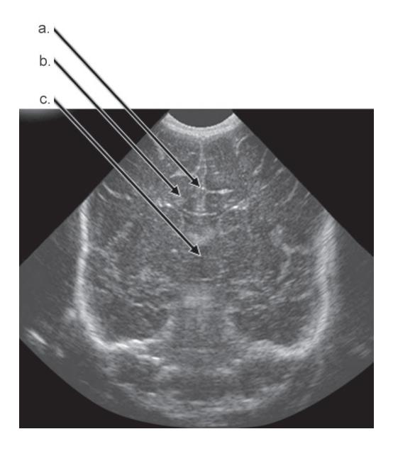
Answer
- a. Corpus callosum.
- b. Lateral ventricle.
- c. Third ventricle.
VVK18-024
A 28-week-old male infant is admitted to NICU. Given below is the cranial USG of this neonate at 48 hour age.
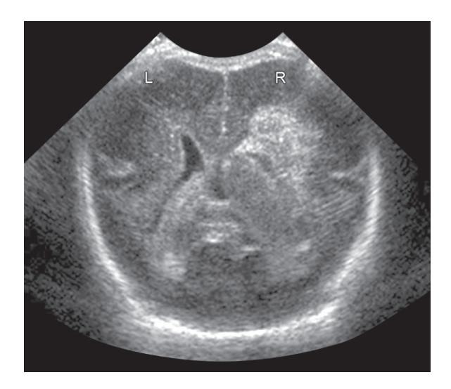
a. What are the cranial ultrasound findings?
b. What is the complete diagnosis?
c. How do you stage the disease based on cranial USG?
Answer
- a. Right intraventricular opacity Right periventricular echogenicity.
- b. Right sided grade IV intraventricular hemorrhage.
- c. Grade I—Confined to germinal matrix Grade II—Intraventricular without ventricular dilatation Grade III—Intraventricular with ventricular dilatation Grade IV—Periventricular hemorrhagic infarction.
VVK18-025
Given below is the cranial USG of a 27-week-old female neonate.
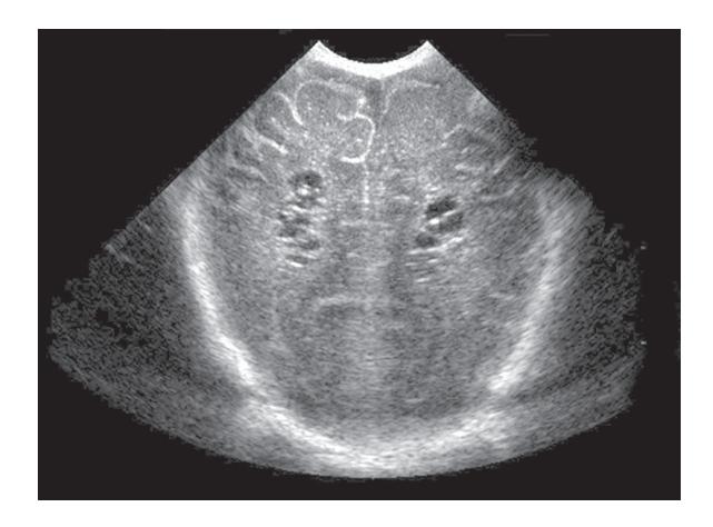
a. What are the cranial ultrasound findings?
b. What is the diagnosis?
c. What is the most likely cause of the diagnosis?
Answer
- a. Bilateral parenchymal cysts.
- b. Cystic PVL.
- c. Intraventricular hemorrhage/ischemia.
VVK18-026
Given below is the X-ray of 14-year-old male following a road traffic accident.
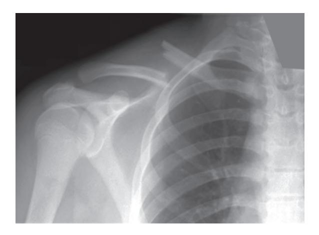
a. What is the abnormality seen on the X-ray above?
b. List 1 possible complication.
Answer
- a. Right clavicular fracture.
- b. Brachial plexus injury.
VVK18-027
A 32-week-old male infant is born via spontaneous vaginal delivery. He was born in good condition and did not require any resuscitation. Mother had no antenatal concerns and did not receive any medications
prior to delivery. Baby developed respiratory distress soon after birth and was intubated and ventilated.
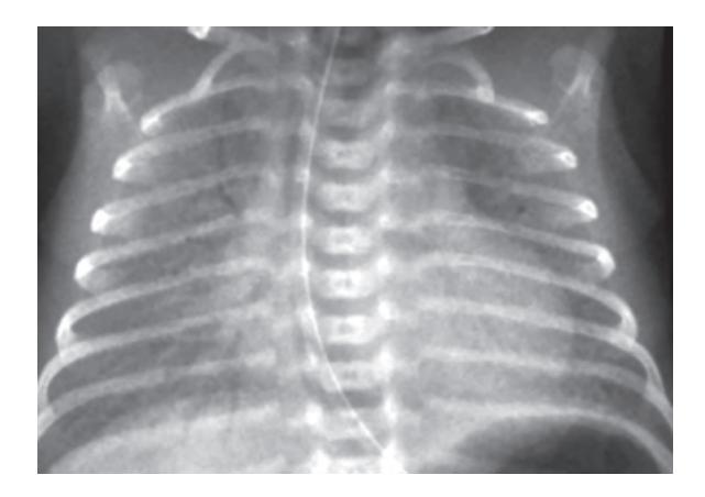
a. What is the most likely diagnosis on chest X-ray?
b. List two most important findings suggesting the diagnosis on chest X-ray.
c. What maternal intervention could have decreased the incidence of the likely diagnosis?
Answer
- a. Respiratory distress syndrome.
- b. Fine reticulogranular opacities Air bronchogram.
- c. Antenatal steroids.
VVK18-028
A 35-week-old female is born via elective LSCS. She cried soon after birth and did not require any resuscitation. She is born to a primigravida mother, who had no risk factors for sepsis. She developed tachypnea soon after birth with mild to moderate subcostal recessions.
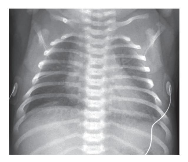
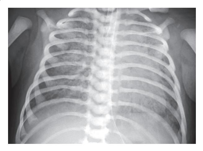
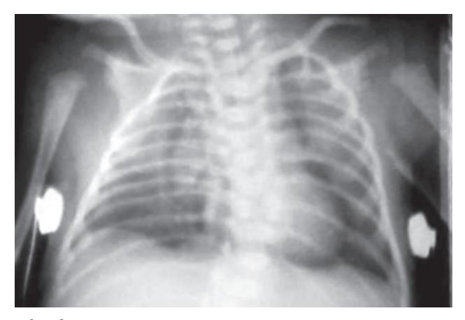
a. List two positive findings on chest X-ray.
b. What is the likely diagnosis?
c. What is prognosis?
d. Is there a role of diuretics in this condition?
a. List the positive findings on chest X-ray.
b. What is the likely diagnosis?
c. List 3 most important steps in management of this neonate.
a. What is the diagnosis?
b. List two bedside procedures, which could have helped in confirming the diagnosis before performing the X-ray.
c. What is the definitive treatment in this neonate?
d. What is the treatment option if the infant was asymptomatic?
Answer
- a. Hyperinflated lung fields Fluid in horizontal fissure.
- b. Transient tachypnea of newborn
- c. Good
- d. No.
VVK18-029
This term neonate was born via normal vaginal delivery. He was noted to have significant respiratory distress soon after birth.
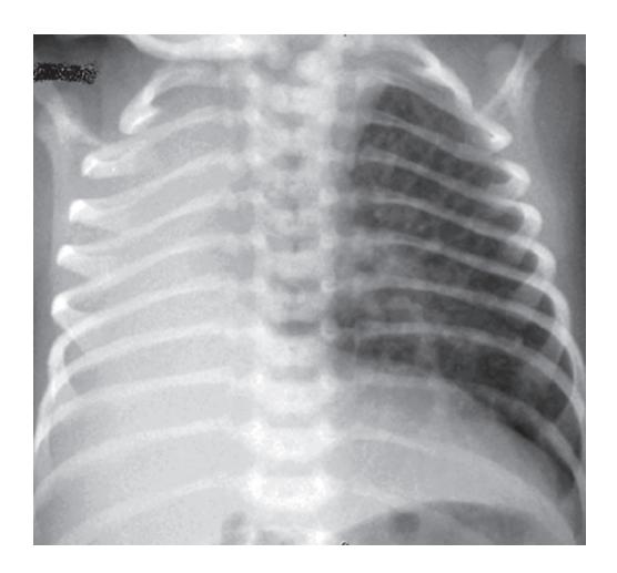
a. What are the findings on the chest X-ray?
b. What is the most likely diagnosis?
c. Give one congenital abnormality, which can be associated with this condition.
Answer
- a. No pulmonary tissue or bronchial markings seen on right side Mediastinal shift to the right Crowding of ribs on right side.
- b. Agenesis of right lung.
- c. Vacterl sequence
Ipsilateral facial and skeletal malformations
Central nervous system malformations
Cardiac malformations.
VVK18-030
This 12-year-old presents in pediatric OPD with respiratory distress of one day duration. He was reviewed in the OPD two days back with complaint of cough and cold and was diagnosed to have viral URI.
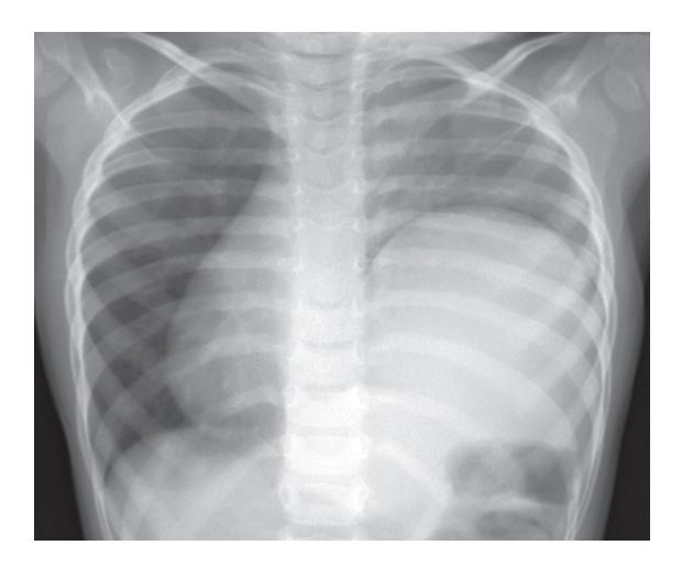
a. What is the most likely diagnosis?
b. How would you confirm the diagnosis?
Answer
- a. Eventration of the diaphragm.
- b. CT Chest.
VVK18-031
Given below is the X-ray of a 12-year-old female who presented to pediatric OPD with complaint of back pain.
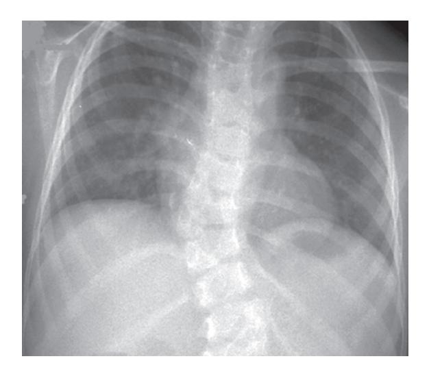
a. What is the diagnosis?
b. What extent of above disease causes cardiopulmonary compromise?
c. What is the definitive indication of surgery in this diagnosis?
d. List four conditions which can be associated with this diagnosis.
Answer
- a. Scoliosis.
- b. Idiopathic thoracic curvatures may not create significant alterations in cardiopulmonary function until they have reached 80–90°.
- c. Cardiopulmonary compromise
Difficulty in walking
(Most common indication is cosmetic deformity).
d. Congenital, e.g. Failure of formation—Wedge vertebrae, hemivertebrae Neuromuscular Neuropathic diseases
Poliomyelitis
Spinocerebellar degeneration (Friedreich ataxia, Charcot Marie—Tooth disease)
Syringomyelia
Spinal cord tumor
Spinal cord trauma
Spinal muscular atrophy
Duchenne muscular dystrophy
Arthrogryposis.
Syndromes e.g. Neurofibromatosis, Marfan syndrome.
Idiopathic.
VVK18-032
Identify the cardiac disease on the basis of the chest X-ray in this neonate.
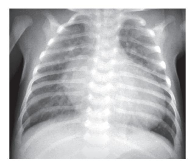
a. What is the diagnosis?
b. What is the name given to this typical chest X-ray presentation?
c. Name the drug used in this neonate soon after birth. The infant's condition fails to improve despite of above drug.
d. Name the procedure which can be performed prior to the definitive surgery.
e. Name the surgical procedures used to correct this anomaly and what age is this procedure performed?
Answer
- a. Transposition of great arteries.
- b. Egg on side appearance.
- c. Prostaglandin E1.
- d. Rashkind balloon atrial septostomy.
- e. Jantene: Arterial switch procedure—usually within first 2 to 4 weeks of life Mustard procedure.
VVK18-033
Identify the cardiac disease on the basis of the chest X-ray.
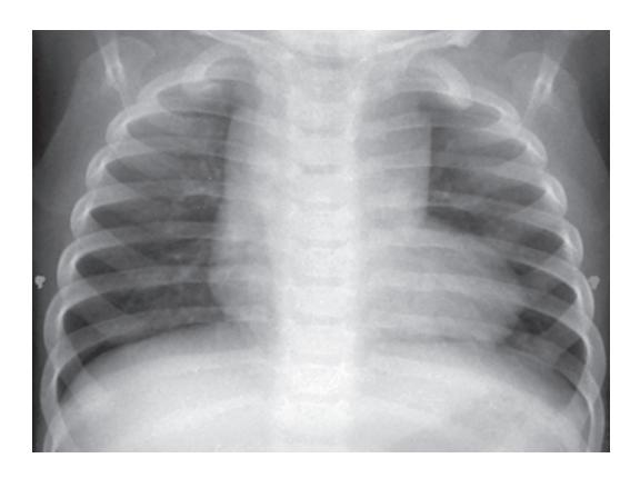
a. What is the diagnosis?
b. What is the name given to this typical chest X-ray presentation?
c. What are findings on cardiac examination?
d. List two most common neurological complications associated with the above diagnosis.
Answer
- a. Tetrology of Fallot.
- b. "Coeur en sabot"- boot shaped heart.
- c. Ejection systolic murmur heard best in left upper sternal edge. Single second heart sound.
- d. Cerebral thrombosis Brain abscess.
VVK18-034
Identify the cardiac disease on the basis of the chest X-ray.
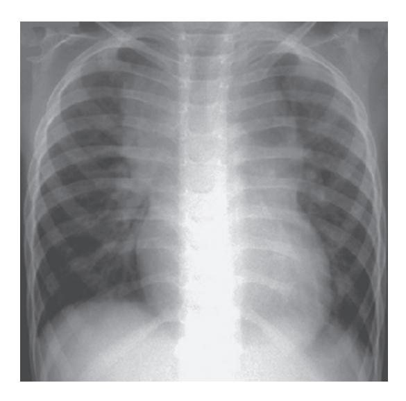
a. What is the diagnosis?
b. What is the name given to this typical chest X-ray presentation?
c. What is the clinical presentation of the infant with this diagnosis?
Answer
- a. Supracardiac TAPVD.
- b. "Snowman sign".
- c. Mild cyanosis Cardiac failure Recurrent chest infection Pulmonary hypertension.