THG01-001
A 5-day-old infant presents with respiratory distress and sacral edema. Antenatal history was normal and the delivery was uneventful. Baby cried immediately after birth, took feeds well initially, and was discharged on day 2 of life.
a. What are the differential diagnoses you will consider?
b. What other history will you elicit?
c. Mention five signs of congestive cardiac failure (CCF).
Answer
| a. | Late onset sepsis and CCF. |
| b. | Any bad child rearing practices, any acute infections in caretakers, feeding difficulty—frequent short feeds, forehead sweating, and suck-rest-suck cycle. |
| c. | Tachycardia, respiratory distress, basal crackles, hepatomegaly, and sacral edema. |
THG01-002
Regarding the examination findings in cardiovascular system.
a. How do you clinically examine for apex beat?
b. How to identify a heaving apex? What does it indicate?
c. What is the clinical finding in right ventricular hypertrophy (RVH)? How do you grade it?
d. How do you look for precordial bulge?
Answer
| a. | Child in sitting position and leaning forward. Use palmar surface of both hands to check both the sides of chest. It is the outermost and lowermost point of palpable cardiac impulse. |
| b. | It is broad, forced, and sustained cardiac impulse. It indicates left ventricular (LV) pressure overload hypertrophy. |
| c. | Parasternal heave |
| Grade 1—visible, not palpable |
| Grade 2—visible, palpable, and obliterable | |
| Grade 3—visible, palpable, but not obliterable | |
| d. | Make the child lay supine and look up from the child’s feet. |
THG01-003
Q1.3
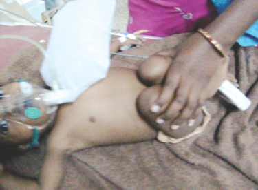
a. What is this position? When is it used for?
b. What relevant history will you ask?
c. Mention the cardiovascular system (CVS) findings you expect.
d. What are the mechanisms by which this maneuver helps the child?
Answer
| a. | Knee–chest position. To abort a cyanotic spell in children with congenital cyanotic heart disease. |
| b. | History of recurrent lower respiratory tract infections (LRTIs), history of failure to gain weight, bluish discoloration of lips, and history of assuming similar position prior. |
| c. | Central cyanosis, clubbing, chest deformities, precordial bulge, parasternal heave, and heart murmur. |
| d. | Squatting or knee chest position increases the systemic vascular resistance, reduces the venous return, and thereby reduces the right to left shunting of the blood. This increases the pulmonary blood flow and temporarily improves the cyanosis and hypoxemia. |
THG01-004
Regarding blood pressure.
a. What are the different methods of measuring blood pressure (BP) in a clinical setting?
b. How do you select the appropriate cuff size? How does it affect BP?
c. How do you calculate the minimal systolic blood pressure (SBP) for a child?
Answer
| a. | Palpatory, auscultatory, and oscillatory. |
| b. | The cuff length should cover two-thirds of the arm circumference and width should cover 40% of the arm circumference. A smaller cuff records falsely high reading and vice versa. |
| c. | Hypotension is considered if SBP is < (age × 2) + 70 mm Hg. |
THG01-005
Q1.5
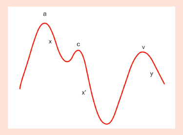
a. What do these waves represent?
b. How do you clinically measure it?
c. What is the normal value?
Answer
| a. | Jugular venous pressure waves. a—atrial contraction, x—atrial relaxation, c—tricuspid valve closure, x’—passive atrial filling, v—maximal atrial filling, and y—atrial emptying. |
| b. | Position the patient supine in bed, elevate the patient’s head to approximately 30–45°, and measure or estimate the vertical height of the meniscus of the right internal or external jugular vein above the sternal angle (angle of Louis) which is approximately 5 cm above the mid right atrium. |
| c. | Normal pressure is 6–8 cmH2 O. |
THG01-006
Regarding ascites.
a. Define ascites.
b. What is puddle sign? How to demonstrate?
c. When can a “fluid thrill” be elicited?
d. Name two causes of exudative ascites.
e. Name two causes of transudative ascites.
f. What is chylous ascites? Name one condition.
Answer
| a. | Ascites—presence of free fluid in peritoneal cavity. |
| b. | Puddle sign—sign used to demonstrate minimal ascites (120 mL). |
| Demonstration: To demonstrate puddle sign—patient lies prone for 5 minutes and then placed on hands and knees. Then, flick the flanks lightly while moving the stethoscope from the most dependent part of the abdomen to the flanks. Positive sign—change in intensity and character of the percussion note. | |
| c. | Fluid thrill—1.5 liters. |
| d. | Exudative ascites—tuberculous ascites and malignancy. |
| e. | Transudative ascites—cirrhosis and nephrotic syndrome. |
| f. | Chylous ascites—lipid-rich lymph in peritoneal cavity due to lymphatic obstruction, e.g., lymphatic duct obstruction. |
THG01-007
Regarding clinical examination of abdomen.
a. Name one condition where scaphoid abdomen is seen.
b. What is the surface marking of transpyloric line?
c. What is the surface marking of interiliac line?
d. What is borborygmus?
e. What is Terry's nail?
Answer
| a. | Congenital diaphragmatic hernia. |
| b. | Transpyloric line: Lies at level of first lumbar vertebra and midway between suprasternal notch and symphysis pubis. |
| c. | Interiliac line: Connects the two tubercles of iliac crest. |
| d. | Loud, rumbling, high-pitched gurgling sounds resulting from gush of gas or fluid through lumen of bowel in small bowel obstruction. |
| e. | In cirrhosis, 80% proximal nail is opaque and remaining 20% remains normal pink. |
THG01-008
On clinically examining the eyes, the following findings are noted.
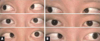
a. List out the cranial nerves supplying intraocular muscles and their muscle supply.
b. Explain the clinical findings in the image A. Name the nerve affected, muscle supplied by the nerve, and area of the brain where the nerve arises from.
c. Mention four causes of the affected nerve palsy.
d. How will you examine eye movements in infants and in a comatose patient?
e. Differentiate clinical findings of internal and external ophthalmoplegia.
Answer
a.
| Oculomotor nerve (3rd cranial nerve) | Superior, inferior, and medial recti, inferior oblique, and levator palpebrae superioris |
| Trochlear nerve (4th cranial nerve) | Superior oblique |
| Abducens nerve (6th cranial nerve) | Lateral rectus |
| b. | Image B shows normal eye movements. In image A, right eye is able to abduct normally, left eye abduction is absent, left abducens nerve palsy, and causing left lateral rectus palsy. It arises from the pons. |
| c. | Cavernous sinus thrombosis, complicated otitis media or mastoiditis, internal carotid aneurysm, ischemic stroke, metabolic diseases such as Wernicke disease and demyelinating lesions, aneurysm, carcinomatous meningitis, procedure-related injury (e.g., spinal anesthesia and post-lumbar puncture), inflammatory lesions (e.g., sarcoid and lupus), and infection (e.g., Lyme disease, syphilis, tuberculosis, and Cryptococcus). |
| d. | Premature infants older than 25 weeks of gestational age and comatose patients can be evaluated using the oculocephalic (doll’s eye) maneuver, in which the patient’s head is quickly rotated to evoke reflex eye movements. If the brainstem is intact, rotating the patient’s head to the right causes the eyes to move to the left and vice versa. Similarly, rapid flexion and extension of the head elicits vertical eye movement. |
e.
| 0 | Internal ophthalmoplegia | External ophthalmoplegia |
|---|---|---|
| Pupil | Fully dilated not reactive to light or accommodation | Normal |
| Extraocular muscles | Extraocular muscle function normal when tested separately | Ptosis and paralysis of all extraocular muscles |
THG01-009
Match the following:
| Wernicke’s aphasia | Monotonous speech without rhythm/intonation |
| Broca’s aphasia | Loss of repetition with preserved comprehension and output |
| Conductive aphasia | Poor comprehension and fluent meaningless speech |
| Cerebellar dysarthria | Nasal speech |
| Pseudobulbar dysarthria | Preserved comprehension and nonfluent speech |
| Extrapyramidal dysarthria | Individual syllables are slurred and precision of consonant production is lost |
| Bulbar dysarthria | Speaking slowly and deliberately as if scanning |
Answer
| Wernicke’s aphasia | Poor comprehension and fluent meaningless speech |
| Broca’s aphasia | Preserve comprehension and nonfluent speech |
| Conductive aphasia | Loss of repetition with preserve comprehension and output |
| Cerebellar dysarthria | Speaking slowly and deliberately as if scanning |
| Pseudobulbar dysarthria | Individual syllables slurred and precision of consonant production is lost |
| Extrapyramidal dysarthria | Monotonous speech without rhythm/intonation |
| Bulbar dysarthria | Nasal speech |
THG01-010
Q1.10
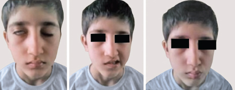
a. Identify the clinical findings in the image.
b. Mention the diagnosis.
c. What history will you elicit?
d. What is the syndrome associated, with the viral exanthem?
Answer
| a. | Right half of the face is paretic, incomplete closure of right upper eyelid, upward and outward rolling of eyes, loss of nasolabial fold on right, and deviation of angle of mouth to the left. |
| b. | Right lower motor neuron (LMN) facial palsy/Bell’s palsy. |
| c. | History of upper respiratory illness (URI) (Epstein–Barr virus, Cytomegalovirus, mumps, and human herpesvirus 6), vaccine exposure (intranasal influenza vaccine), ear pain (otitis media with cholesteatoma) viral exanthem (Ramsay Hunt syndrome), and history of trauma. |
| d. | Ramsay Hunt syndrome. |
THG01-011
This is a 2-month-old baby, mention four primitive reflexes that you can elicit in this baby and how you can elicit them.
Answer
| ■ | The Moro reflex: The Moro reflex is elicited by supporting the infant in a semierect position and then allowing the infant’s head to fall backward onto the examiner’s hand. A normal response consists of symmetric extension and abduction of the fingers and upper extremities, followed by flexion of the upper extremities and an audible cry with opening of the hands. The reflex disappears in 5–6 months. |
| ■ | The palmar grasp reflex: When the baby’s palm is stimulated along the ulnar side, the hand closes. Disappears by 6 months. |
| ■ | Plantar grasp reflex: When the baby’s plantar aspect of foot is stimulated below his toes, there is flexion of his toes. Disappears by 10 months. |
| ■ | Asymmetric tonic neck reflex: When the head of the baby is turned to one side, the arm extends on the same side with flexion of the contralateral knee. The reflex disappears in 6–7 months. |
| ■ | Glabellar tap/blink reflex: Tapping of the child on the forehead between the eyebrows, causes blinking of the eyes, it stops after 4–5 taps. |
| ■ | Rooting reflex: When the corner of the mouth or cheek of the child is stimulated on one side, the child turns toward the same side. It disappears by 4–7 months. |
(Stepping and placing reflex disappears by 6 weeks. Parachute and Landau will not be present in a two-month-old child)
THG01-012
Child presents to the emergency department with uprolling of eyes and tonic posturing of all four limbs. Elicit history from the mother.
Answer
| ■ | Fever preceding seizure/not, if yes, what day of fever, whether within one hour of onset of fever, low grade, or high grade. How many episodes, if more than one, how many within 24 hours? Aborted spontaneously or with medications, duration of the episode. Spontaneous loss of bowel/bladder control. Focal/generalized seizures. Postseizures—sleep/headache/hemiparesis. |
| ■ | Family history of seizures (febrile/unprovoked). |
| ■ | Unprovoked—any history of fall prior to seizures, accidental ingestion of medications or toxins, any history of incessant cry/swaying on feet while walking prior to fall (increased ICP), history of anticoagulants intake/bleeding manifestations (intracranial hemorrhage). |
| ■ | Developmentally normal child/not. History of hypo/hyperpigmented patches over the skin (neurocutaneous syndromes). |
| ■ | Significant birth history? Perinatal asphyxia/neonatal stay/excessive jitteriness. |
| ■ | History of repeated vomiting/loose stools/reduced oral intake (hypoglycemia). |
THG01-013
A 14-year-old child presents with headache, elicit history.
Answer
| ■ | Duration of the headache, pattern (unilateral/bilateral), frequency of attacks, duration of each attack, severity of pain (interference with activities), location of maximal pain (frontal, temporal, occipital, sinus, and periorbital), quality of pain (throbbing, stabbing, squeezing, and pulsating) |
| ■ | Any associated trigger factors: Caffeine, hunger, noise, smell, stress, sleep, change in altitude, travel, etc. |
| • | Warning symptoms |
| • | Symptoms during attacks: Anorexia, nausea, vomiting, light intolerance, noise intolerance, and pallor |
| • | Relieving factors |
| • | Symptoms between attacks |
| ■ | Family history of migraine |
| ■ | History of fever, running nose, tooth pain, dental caries, more of facial pain aggravated by looking down |
| ■ | Watering of eyes/frequent rubbing of eyes, and vision problems |
| ■ | History of weight loss, bleeding symptoms/petechiae [central nervous system (CNS)/hematologic malignancies] |
| ■ | History of fall/double vision, abnormal movement of eyes, sudden loss of consciousness (LOC) [increased intracranial pressure (ICP)] |
THG01-014
A 2-year-old child presents to the emergency room (ER) with history of fall from height. Elicit history from mother and do clinical examination for the child.
Answer
History:
| ■ | Time of fall, height from which the child fell, surface of fall, position of the child post fall (which part of head came in contact with surface). |
| ■ | Complaints of swelling and injuries in the body post fall. |
| ■ | Whether child cried immediately post fall, any LOC, seizures, bleeding from ear, nose, or mouth, any seizures, white fluid from ear/nose, and vomiting. |
| ■ | Complaint of headache, drowsiness, irritability, and swaying while walking. |
Clinical examination:
| ■ | Assess Glasgow Coma Scale (GCS), respiration (irregular respiration), and circulation (bradycardia and hypertension). |
| ■ | Local examination of swelling/injuries. |
| ■ | Eyes—pupils (unilateral/bilateral pupillary dilation) and fundus examination (papilledema). |
| ■ | Focal neurological signs and abnormal posturing. |
| ■ | Examine for signs of basal skull fracture—hemotympanum, rhinorrhea, otorrhea, periorbital bruising (raccoon eyes), and bruising over mastoid (battle sign). |
THG01-015
List out the steps in cranial nerve examination of a child <2 years.
Answer
| ■ | 1st nerve—cannot be tested. |
| ■ | 2nd nerve—pupillary reflex, accommodation reflex, recognizing mother/strangers, recognize primary colors, and finger counting. |
| ■ | 3rd, 4th, and 6th nerve—check eye movements by moving a bright toy vertically, horizontally, to either sides, and diagonally. |
| ■ | 5th nerve—lollipop sucking, drinking water through straw, check for temporalis hollowing, elicit jaw reflex if the child is cooperative (place a finger on the baby’s chin while the mouth is slightly open and tap the finger. A rapid muscle contraction with closure of mouth is the reflex response). |
| ■ | 7th nerve—taste sensation with salt/lemon (squeeze a lemon or place a gauze tipped in salt solution on the anterior two-thirds of the tongue), check facial symmetry while the child smiles/cry, presence of nasolabial fold, and eye closing. |
| ■ | 8th nerve—distract the child and elicit sounds from the side, check whether the child localizes where the sound comes from. |
| ■ | 9th and 10th nerve—check for the position of uvula and tonsillar pillars while the child is crying. Do not test for gag reflex. Check for swallowing on asking the child to drink water. Check for drooling. |
| ■ | 11th nerve—look for prominence of sternocleidomastoid/drooping of shoulder. |
| ■ | 12th nerve—look for atrophy or fasciculation of tongue when the child cries, if the child is cooperative ask the child to protrude tongue and look for deviation of the tongue from midline. |
THG01-016
List out the steps in head to toe clinical examination in a child with cerebral palsy.
Answer
| ■ | Posture of the baby, spontaneous eye-opening present or not. |
| ■ | Head—measure head circumference (microcephaly), look for overriding of sutures (craniosynostosis) or abnormal shape of skull (positional—plagiocephaly), hair (color, texture, and seborrhea), fontanel—anterior open/closed, posterior—open/closed. |
| ■ | Facies—dysmorphism, neurocutaneous markers, and signs of vitamin deficiencies |
| ■ | Eyes—nystagmus, cataract, and squint; ears—discharge; and mouth—teeth dentition, caries, and drooling of saliva |
| ■ | Extremities—cortical thumb, scissoring of legs, flexion contractures (elbow, knee, and ankle), callosities (back), pathologic fractures/dislocation of hips, limb length discrepancies. |
| ■ | Use of diapers, feeding tubes, feeding gastrostomy/jejunostomy, muscle relaxant pumps, calipers, and braces. |
| ■ | Gait—tip–toe walking/circumduction gait, and crawling—commando crawl (the child uses the arms in a normal reciprocal fashion but tends to drag the legs behind more as a rudder rather than using the normal four-limbed crawling movement). |
THG01-017
Elicit history in a child presenting to outpatient department (OPD) at 1 year with complaints of not responding when called by name.
Answer
| ■ | Language/development history: Did child coo/babble, says bisyllabic words or amma, appa. Turns to sound/eye contact with mother (smiles at mother), claps/waves bye bye. Follows one step verbal command with gesture, normal response to nonverbal auditory stimulus. Does he communicate his/her needs. Does he shake his/her head in order to communicate. Is she interested in playing/interacting with mother/father. |
| ■ | Antenatal infections for mother: Fever with rash during the antenatal period, preterm delivery, low birth weight/growth retardation in scans (tetanus/toxoplasma/cytomegalovirus/herpes/torch). |
| ■ | History of neonatal intensive care unit (NICU) stay: Fever/meningitis/ototoxic medications/perinatal asphyxia (Apgar 0–4 at 1 minute and 0–6 at 5 minutes)/hyperbilirubinemia with exchange transfusion/mechanical ventilation for 5 days or longer. |
| ■ | History of trauma and ear discharge. |
| ■ | Family history of hearing disorders/learning disability/speech delay/genetic disorders. |
THG01-018
List out the steps of pediatric gait, arms, legs, and spine (pGALS) examination in a 6-year-old child.
Answer
| ■ | Observe the child standing (front, back, and sides) |
| ■ | Observe the child walking (walk on your heels and walk on your tiptoes) |
| ■ | Hold your hands out straight in front of you |
| ■ | Turn your hands over and make a fist |
| ■ | Pinch your index finger and thumb together |
| ■ | Touch the tips of your fingers |
| ■ | Squeeze the metacarpophalangeal joints for tenderness |
| ■ | Put your hands together palm to palm and put your hands together back to back |
| ■ | Reach up and touch the sky and look at the ceiling |
| ■ | Put your hands behind your neck |
| ■ | Try and touch your shoulder with your ear |
| ■ | Open wide and put three fingers in your own |
| ■ | Feel for effusion at the knee (patella tap or cross fluctuation) |
| ■ | Active movement of knees (flexion and extension) and feel for crepitus |
| ■ | Passive movement of hip (knee flexed to 90° and internal rotation of hip) |
| ■ | Bend forward and touch your toes |
THG01-019
Match the following:
| Hemiparetic gait/spastic gait | Wide-based steppage gait, patient lifts the legs up higher than usual in the swing phase and then slaps the foot down |
| Cerebellar ataxia | Hip girdle weakness with compensatory lordosis, proximal muscle weakness (difficulty in climbing stairs) |
| Sensory ataxia | Spasticity and circumduction of the leg, decreased arm swing on the affected side |
| Myopathic or waddling gait | Wide-based reeling gait like that of a drunk person |
Answer
| Hemiparetic gait | Spasticity and circumduction of the leg, decreased arm swing on the affected side |
| Cerebellar ataxia | Wide-based reeling gait like that of a drunk person |
| Sensory ataxia | Wide-based steppage gait, patient lifts the legs up higher than usual in the swing phase and then slaps the foot down |
| Myopathic or waddling gait | Hip girdle weakness with compensatory lordosis, proximal muscle weakness (difficulty in climbing stairs) |
THG01-020
A 5-year-old male child is brought with complaints of difficulty in breathing following a fall while skating. Assess the child’s respiratory system by inspection and palpation.
Answer
Inspection:
| ■ | Undress the child and look for visible injuries |
| ■ | Assess the shape and symmetry of thorax |
| ■ | Assess the pattern of respiration |
| ■ | Count the rate of respiration |
| ■ | Assess work of breathing |
Palpation:
| ■ | Confirm inspection findings |
| ■ | Assess tracheal deviation: Index finger of the dominant hand placed on the trachea in the suprasternal notch, then the finger is moved laterally left and right in the spaces bordered by the upper edge of clavicle, the inner aspect of the sternocleidomastoid muscle and the trachea. |
| ■ | Measure expansion of chest: Hold the chest and see the distance of thumbs anteriorly. |
| ■ | Look for any tenderness |
| ■ | Tactile fremitus: Ask the child to repeat 1, 2, 3. Use palmar bases of fingers and ulnar aspect of two hands in both sides of chest simultaneously to compare. If one hand is used, move it from one side of chest to the corresponding area on the other side. |
THG01-021
This is a 3-year-old child with complaints of cough for the past 1 month. Elicit history.
Answer
| ■ | Onset of cough—insidious/sudden |
| ■ | Type of cough—dry/wet |
| ■ | Diurnal variation |
| ■ | In case of night cough, what is the timing of cough—immediately after lying down/early night/midnight |
| ■ | Whether associated with fever |
| ■ | History of (H/O) loss of weight or appetite |
| ■ | Symptoms persistent or variable |
| ■ | Any upper respiratory symptom |
| ■ | Any activity limitation |
| ■ | History of allergy or atopy |
| ■ | History of triggers |
| ■ | Past history of cough and interval symptoms |
| ■ | Family history of wheeze |
THG01-022
Regarding chest percussion.
a. Perform chest percussion.
b. Write down the percussion finding in the following conditions:
Answer
| a. | Chest percussion: |
| • | Position the child with the head bent and arms folded over the chest |
| • | On the posterior chest, percuss systematically at about 3 cm intervals from the upper to lower chest, moving left to right, right to left. Avoid scapular and other bony areas. |
| • | Percuss the anterior and lateral chest with the child’s arm positioned over the head. |
| • | Follow rules of percussion: |
| ◆ | Middle finger of the left hand is placed firmly on the part which is to be percussed. No other fingers should touch the chest wall. |
| ◆ | Back of the middle phalanx is then struck with the tip of the middle finger of the right hand. |
| ◆ | Stroke should be delivered from the wrist and finger joints but not from the elbow. |
| ◆ | Percussing finger should be bent so that when the blow is delivered its terminal phalanx is at right angle to the metacarpal bones and strikes the pleximeter finger perpendicularly. |
| ◆ | As soon as the blow has been delivered, the striking finger must be released just as the hammer fall back. |
| ◆ | Blow should be no longer than is necessary. |
| ◆ | Give two blows for each percussion. Pleximeter—middle finger of nondominant hand. Plexor—middle finger of dominant hand. |
| • | Areas of percussion in RS |
| Supraclavicular, Clavicular, Infraclavicular, Mammary, Inframammary, Axillary, infra-axillary, Suprascapular, Interscapular, Infrascapular. |
| b. | Percussion finding in the following conditions: |
| 1. | Right hydropneumothorax—right stony dullness |
| 2. | Left consolidation—left dullness |
| 3. | Right pleural effusion—right stony dullness, right Ellis curve. |
| 4. | Left collapse—left dullness. |
THG01-023
Regarding auscultation of respiratory system.
a. Perform auscultation of respiratory system
b. Write down the auscultatory finding in the following conditions:
Answer
| a. | Auscultation of respiratory system: |
| • | Expose the child |
| • | Auscultate systematically comparing areas in right and left. |
| • | Hear breath sounds |
| • | Look for bronchophony, aegophony and whispering pectoiloquy |
| • | Comment on added sounds. |
b. 1. Right hydropneumothorax: Breath sounds diminished in the right side. Succession splash and coin test+
| 2. | Left consolidation: Tubular breath sounds in the left sides, crackles+, increased vocal resonance. |
| 3. | Right pleural effusion: Diminished or absent breath sounds in the right. |
| 4. | Left collapse: Absent breath sounds in the left. |
THG01-024
A 2-year-old child brought with acute onset stridor. Elicit history. Tabulate the severity classification of croup-based on physical examination.
Answer
History:
| ■ | Onset of stridor—sudden/insidious |
| ■ | H/O associated fever—High or low grade |
| ■ | H/O preceding symptoms of upper respiratory infection |
| ■ | H/O change of voice |
| ■ | H/O irritability or lethargy |
Severity:
| Signs | Mild-to-moderate | Severe | Life threatening |
|---|---|---|---|
| Sensorium | Alert | Lethargic arousable | Agitated, pain responsive or unresponsive |
| Respiratory distress | No | ICR, SCR present | Sternal, SSR, severe or declining in intensity without improvement in conciousness |
| Stridor | No stridor at rest | Stridor at rest | Audible stridor becoming quiet without improvement in consciousness and saturation |
| Heart rate | Normal | Tachycardia | Bradycardia |
| SpO2 | >95% | >95% | <94% |
THG01-025
Name the bronchopulmonary segments. Explain the following:
a. Harrison’s sulcus
b. Traile’s sign
c. S shaped curve of Ellis
d. Kronig’s isthmus
e. Traube’s space
Answer
Bronchopulmonary segments:
■ Right lobe:
| • | Upper lobe: Apical, anterior and posterior. |
| • | Middle lobe: Lateral, medial. |
| • | Lower lobe: Apical, medial, anterior, posterior and lateral. |
■ Left lobe:
| • | Upper lobe: Apical, anterior, posterior, superior lingular, inferior lingular. |
| • | Lower lobe: Apical, anterior, posterior, lateral. |
| a. | Harrison’s sulcus: Depression at the region of the eighth to tenth ribs (site of diaphragm muscle leaving the chest wall) and the bottom of the rib cage will flare. |
| b. | Traile’s sign: Prominence of the tendon of the sternocleidomastoid near the clavicle on one side (due to deviation of trachea on the same side) |
| c. | S shaped curve of Ellis: Seen in pleural effusion. On percussion- uppermost dullness in axilla, lowest in spine posteriorly and near sternum anteriorly- appears to be in the shape of S. |
| d. | Kronig’s isthmus: Band of resonance over supraclavicular fossa. Borders: scalenus muscle medially, acromion process laterally, clavicle anteriorly, and trapezius posteriorly. |
| e. | Traube’s space: Space bordered by lung resonance above, costal margin below, liver in the right and spleen in the left. Content-stomach. |
THG01-026
Regarding breath sounds.
a. What are the types of breath sounds. Explain and give an example.
b. What is the added sound you hear in
Answer
| a. | Types of breath sounds: |
| • | Vesicular: Low pitched, rustling in nature, inspiration > expiration, no pause. |
| Example: No pause, due to filtering effect of lung parenchyma. | |
| • | Bronchial: High-pitched, guttural in nature, inspiration = expiration, pause present. |
Types of bronchial breath sounds:
| • | Tubular: High pitched. Example: Consolidation, above the level of effusion. |
| • | Cavernous: Low pitched, seen in thick-walled cavity with communicating bronchus. |
| • | Amphoric: Low pitched with high tone and metallic overtones. Seen in tension pneumothorax and bronchopleural fistula. |
| • | Bronchovesicular: Expiration more than inspiration, no pause. Heard over root of lungs just below the clavicle. |
| b. | Added sound in: |
| 1. | Asthma: Expiratory polyphonic wheeze |
| 2. | Foreign body: Fixed monophonic wheeze |
| 3. | Pulmonary edema: Late inspiratory fine crackles. |
| 4. | Bronchiectasis: Mid inspiratory coarse crackles. |
THG01-027
Regarding examination of respiratory system.
a. What are the accessory muscles of respiration?
b. Explain characteristic features of pleural rub.
c. Explain D’Espine’s sign.
d. Explain Hamman’s mediastinal crunch.
e. Explain whispering pectoriloquy
Answer
| a. | Accessory muscles of respiration: Sternocleidomastoid, trapezius, alae nasi and abdominal muscles. |
| b. | Pleural rub characters: Rubbing or creaking sound heard close to ear. Audible during both the phase of respiration and disappears on holding the breath. It is confined to localized area, not altered by cough and associated with local pain and tenderness. |
| c. | D’Espine’s sign: Presence of high-pitched tubular breath sounds and whispering pectoriloquy heard below T4. It is due to mass in the middle or posterior mediastinum. |
| d. | Hamman’s mediastinal crunch: It is the clicking rhythmical sound synchronous with cardiac cycle due to mediastinal emphysema. |
| e. | Whispering pectoriloquy: Whispered voice heard loudly and clearly— seen in consolidation. |
THG01-028
Fill in the findings:
| Condition | Movement | Mediastinum | Percussion | Breath sounds | Vooaa resonance |
|---|---|---|---|---|---|
| Consolidation | |||||
| Collapse | |||||
| Pleural effusion | |||||
| Hydropneumothorax |
Answer
| Condition | Movement | Mediastinum | Percussion | Breath sounds | Vvocd resonance |
|---|---|---|---|---|---|
| Consolidation | Diminished | Center | Dull | Tubular, crackles | Increased |
| Collapse | Diminished | Pulled to same side | Dull | Absent | Absent |
| Pleural effusion | Diminished | Pushed to opposite side | Stony dullness, Ellis curve | Absent | Absent |
| Hydropneu-mothorax | Diminished | Pushed to opposite side | Stony dullness. Straight line of dullness | Diminished | Absent |
THG01-029
A 7-year-old child is brought with complaints of recurrent episodes of upper respiratory infection. Elicit history and examine the upper respiratory system.
Answer
History:
| ■ | Onset and progression |
| ■ | Persistent or variable |
| ■ | Nature of nasal discharge—watery or mucopurulent |
| ■ | H/O triggers |
| ■ | H/O sneezing or watering of eyes or redness of eyes |
| ■ | Any diurnal variation. |
| ■ | If associated with cough—nature and timing of cough. |
| ■ | If associated with fever |
| ■ | H/O halitosis/facial puffiness/headache/postnasal dribbling |
| ■ | H/O atopy |
| ■ | Family H/O allergy/atopy/wheeze |
Examination of upper respiratory system:
■ Examination of ear:
| • | Examine digitally |
| • | Examine the inner part of external auditory canal |
■ Examination of nose and paranasal sinuses:
| • | Examine the nose digitally: Document bilateral patency. |
| • | Color and size of the mucosa should be noted (inferior turbinate) |
| • | Any bluish discoloration of the infraorbital region (allergic shiners) |
| • | Bilateral fold of skin below lower eyelid—Dennie-Morgan line can be seen in atopic dermatitis and allergic rhinitis |
| • | Transverse crease running at the junction of the cartilaginous and bony portion of the nose (allergic salute) |
| • | Any mid-facial hypoplasia |
| • | Any nasal polyps |
| • | Any sinus tenderness |
■ Examination of throat:
| • | Examine oropharynx for size and any malformation such as cleft palate or high arched palate |
| • | Any dental caries, halitosis. |
| • | Any large tongue or retrognathia |
| • | Tonsillar size: |
| ◆ | Grading of tonsillar hypertrophy: |
| ◆ | Grade 0: Tonsils within the tonsillar fossa. |
| ◆ | Grade 1: Tonsils just outside of the tonsillar fossa and occupy 25% of the oropharyngeal width |
| ◆ | Grade 2: Tonsils occupy 26–50% |
| ◆ | Grade 3: Tonsils occupy 51–75% |
| ◆ | Grade 4: Tonsils occupy >75% of the oropharyngeal width. |
THG01-030
What is bradypnea, tachypnea, dyspnea, apnea, hyperpnea, periodic breathing, Cheyne–Stokes breathing, Biot’s breathing and paradoxical respiratory movements. Give one example for each.
Answer
| ■ | Bradypnea: Abnormally slow rate of respiration for age. Example: Metabolic alkalosis/CNS depression |
| ■ | Tachypnea: Abnormally rapid rate of respiration for age. Example: Asthma, pneumonia, metabolic acidosis. |
| ■ | Dyspnea: Difficulty in breathing. Example: Pneumonia, asthma |
| ■ | Apnea: Cessation of breathing. Example: Arrest |
| ■ | Hyperpnea: Abnormally deep respiration. Example: Sepsis, exercise |
| ■ | Periodic breathing: Respiratory pauses of less than 6 seconds occur in groups of 3 or more that are separated by less than 20 seconds. Example: Preterm babies. |
| ■ | Cheyne–Stokes breathing: Cycles of increasing and decreasing tidal volumes separated by apnea. Example: CCF, Raised ICT. |
| ■ | Biot’s breathing: Irregular cycles of respiration at variable tidal volumes interrupted by apnea. Example: Severe brain damage. |
| ■ | Paradoxical respiratory movements: Chest wall moves in during inspiration and moves out during expiration. Example: Paralysis of intercostal muscles. |