THG24-001
Fill in the blanks:
a. Visual acuity of newborn is ______.
b. Visual acuity reaches 6/6 or 6/9 by ______ years.
c. Eye coordination is usually achieved by ______ of age.
d. Tears are not present while crying until ______ of age.
e. Baby can fixate near objects by ______ of age.
Answer
| a. | Visual acuity of newborn is (20/400) 6/120. |
| b. | Visual acuity reaches 6/9 or 6/6 by 2–3 years. |
| c. | Eye coordination is usually achieved by 3–6 months of age. |
| d. | Tears are not present while crying until 1–3 months of age. |
| e. | Baby can fixate near objects by 3 months of age. |
THG24-002
Key developmental milestones in vision—match the following:
| Age | | | Mileetoon | | |
| | | | | | |
| 1. | Birth | | a. | Follow rapidly moving objects | |
| | | | | | |
| | | | | | |
| 2. | 4 weeks | | b. | Adjusts position to see objects | |
| | | | | | |
| | | | | | |
| 3. | 12 weeks | | c. | Establishment of binocular vision | |
| | | | | | |
| | | | | | |
| 4. | 16 weeks | | d. | Fixate somewhat and move 45° | |
| | | | | | |
| | | | | | |
| 5. | 5 months | | e. | Fixate and move 90° | |
| | | | | | |
| | | | | | |
| 6. | 6 months | | f. | Fixate intently and move 180° | |
| | | | | | |
| | | | | | |
| 7. | 12 months | | g. | Excites when food is prepared | |
| | | | | | |
Answer
| 1. | d |
| 2. | e |
| 3. | f |
| 4. | c |
| 5. | g |
| 6. | b |
| 7. | a |
THG24-003
Acuity of vision in various age groups and testing tools—various age groups.
a. Acuity of vision—fill in the blanks:
b. What are the usual testing methods?
Answer
a. 1. 3 months—6/60
| 2. | 12 months—6/18 |
| 3. | 3 years—6/12 |
| 4. | 4 years—6/9 |
| 5. | 5–6 years—6/6 |
b. 1. 3–5 years: Tumbling E test, HOTV test, Allen figures, and Lea symbols.
| 2. | Above 6 years: Snellen acuity chart; Snellen letters and Snellen numbers. |
THG24-004
Two children attending the neurology clinic were discovered to have visual field defects.


a. Damage to what part of the visual pathways would produce the visual field defect in 1?
b. Damage to what part of the visual pathways would produce the visual field defect in 2?
c. Damage to what part of the visual pathways would produce the visual field defect in A?
d. Damage to what part of the visual pathways would produce the visual field defect in B?
Answer
| a. | Lesion at right optic nerve produces blindness of right eye with loss of direct light reflex. |
| b. | Lesion at optic chiasma produces bitemporal hemianopia. |
| c. | Lesion at left optic tract produces right homonymous hemianopia—both optic tracts emanating from the lateral geniculate body itself. |
| d. | Lesion in the right temporal lobe damaging the temporal lobe radiation. |
THG24-005
With respect to the extraocular muscles of the eye answer the following:
| Sl. no | Muscle | Nerve supply | Action with eye abducted | Action with eye adducted |
|---|---|---|---|---|
| 1. | Lateral rectus | |||
| 2. | Inferior rectus | |||
| 3. | Inferior oblique | |||
| 4. | Medial rectus | |||
| 5. | Superior rectus | |||
| 6. | Superior oblique |
Answer
| Sl. no | Muscle | Nerve supply | Action with eye abducted | | | Action with eye adducted | | |
| 1. | Lateral rectus | VI nerve = Abducens | Abduction | | | Abduction | | |
| 2. | Inferior rectus | III nerve = Oculomotor | Depression | | | | | |
| • | Depression | |
| • | Extorsion | |
| | | |
| 3. | Inferior oblique | III nerve = Oculomotor | | | | Elevation | | |
| • | Extorsion | |
| • | Elevation | |
| | | |
| 4. | Medial rectus | III nerve = Oculomotor | Adduction | | | Adduction | | |
| 5. | Superior rectus | III nerve = Oculomotor | Elevation | | | | | |
| • | Elevation | |
| • | Intorsion | |
| | | |
| 6. | Superior oblique | IV nerve = Trochlear | | | | Depression | | |
| • | Intorsion | |
| • | Depression | |
| | | |
THG24-006
A 9-month-old female infant brought to family physician with parents having noticed that her eyes do not always seem to be looking in the same direction.
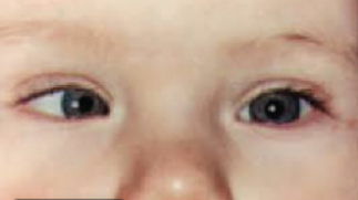
a. What does the child have?
b. Name screening tests of clinical examination.
c. What are its types?
Answer
| a. | Right esotropia (right convergent strabismus/squint) |
| b. | Hirschberg corneal reflex test, cover test, and red reflex test |
| c. | Concomitant and nonconcomitant strabismus |
THG24-007
Q24.7
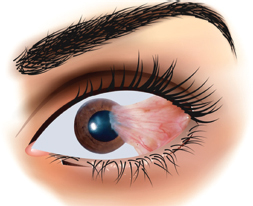
a. What is the diagnosis?
b. What is the typical area where it occurs?
c. What are the pathologic findings?
d. What is the risk factor?
e. What is the treatment?
Answer
| a. | Pterygium. |
| b. | Nasal interpalpebral region. |
| c. | Elastic and hyaline degenerative changes of the conjunctiva. |
| d. | Exposure to ultraviolet light. |
| e. | Usually no treatment; removal if it infringes on cornea or for cosmetic reasons. |
THG24-008
Q24.8
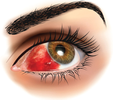
a. What is the diagnosis?
b. What are the causes (any four)?
c. What is the treatment?
Answer
| a. | Subconjunctival hemorrhage. |
| b. | Trauma; contact lens usage; severe sneezing/coughing; inflammation; blood dyscrasias; hypertension, diabetes mellitus. |
| c. | Left alone—self-limiting—no treatment. |
THG24-009
Q24.9
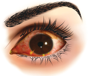
a. What is the diagnosis?
b. What is the pathogenesis behind this?
c. What are the causative factors?
d. What is the treatment?
Answer
| a. | Phlyctenular conjunctivitis. |
| b. | Morphologic expression of delayed hypersensitivity to diverse antigens. |
| c. | Staphylococci and tuberculosis. |
| d. | Topical corticosteroid therapy and treatment of underlying disorder. |
THG24-010
Q24.10
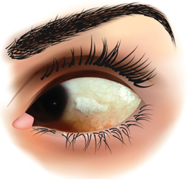
a. What do you see?
b. What is the diagnosis?
c. Name the earliest symptom of this condition.
d. How will you treat?
e. What are the prevention strategies?
Answer
| a. | Bitot spots. |
| b. | Vitamin A deficiency. |
| c. | Night blindness. |
| d. | Three doses of vitamin A are given: 2 lakh units, first one immediately on diagnosis, second dose 24 hours later, and third dose 1–4 weeks later. |
| e. | Prevention strategies: (1) Improving availability and consumption of vitamin A-rich foods, (2) food fortification, and (3) periodic mega-dose vitamin A supplementation to preschool children and during pregnancy. |
THG24-011
Around 50 children in neighborhood have similar symptoms of pain, redness, and tearing of eyes with findings of subconjunctival hemorrhages, follicles, and chemosis.
a. What is the diagnosis?
b. What are the usual causative organisms?
c. What is the treatment?
Answer
| a. | Acute hemorrhagic conjunctivitis. |
| b. | Coxsackie and Enterovirus. |
| c. | Self-limiting illness. |
THG24-012
A 4-year-old girl presented with right red eye, and matting of eyelids on awakening; No history of fever, rhinorrhea; on examination there is right bulbar and tarsal conjunctival erythema with a small amount of mucopurulent discharge at the corner of the right eye. There is purulent fluid visible behind her right tympanic membrane.
a. What is the probable diagnosis?
b. What is the most common organism?
c. What are the other organisms?
d. ______ and ______ are helpful in differentiating specific types.
e. What is the treatment outline?
Answer
| a. | Acute purulent conjunctivitis (conjunctivitis-otitis syndrome) |
| b. | Nontypeable Haemophilus influenzae (80%) |
| c. | Pneumococci (20%) and staphylococci (5–10%) |
| d. | Conjunctival smear and culture are helpful in differentiating specific types. |
| e. | Warm compresses and topical antibiotics. |
THG24-013
A 7-month-old infant brought to emergency room (ER) with inconsolable crying, rubbing of eyes, photophobia, and on examination mild injection of his left conjunctiva and focal haze of cornea of left eye.
a. What is the diagnosis?
b. What are the causative factors?
c. Most serious infection is due to which organism?
d. What are the complications?
e. What is the treatment?
Answer
| a. | Corneal ulcer. |
| b. | Trauma and contact lens wear. |
| c. | Pseudomonas. |
| d. | Perforation, corneal scarring, and blindness. |
| e. | Application of topical ophthalmic antibiotic ointment or drops to the affected eye until the abrasion is healed. |
THG24-014
Child brought to outpatient department with a 3-day history of low-grade fever and a rash around the right eye.
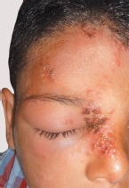
a. What is the most likely diagnosis? What is the causative organism?
b. What is the mechanism?
c. Severe manifestation and recurrence seen in which conditions (any two)?
d. What are the complications in eye (any two)?
e. What is the treatment, drug of choice, and dosage?
Answer
| a. | Herpes zoster ophthalmicus and varicella-zoster virus. |
| b. | Reactivation of varicella-zoster virus in the ophthalmic division of the trigeminal nerve. |
| c. | Immune-compromised children, on immunosuppressive therapy, malignancy, and human immunodeficiency virus (HIV) infection. |
| d. | Corneal hypoesthesia, corneal keratitis, uveitis, sclerokeratitis, central retinal artery occlusion, and optic atrophy. |
| e. | Oral acyclovir 20 mg/kg/dose; maximum: 800 mg/dose; 4 doses/day × 5 days. |
THG24-015
An 8-year-old male presented with 3 days of fever and increased swelling of the eyelid. Physical examination revealed a warm and violaceous discoloration and swelling of the left eyelid which extended to the nose and limited movement of the left eyeball and decreased visual acuity and proptosis of left eye.
a. What is the diagnosis?
b. What is the triad of this condition?
c. What is the most common cause?
d. Which organisms are responsible?
e. What are the complications?
f. What is the imaging of choice?
Answer
| a. | Orbital cellulitis. |
| b. | Proptosis, painful limitation of movement of the eye, and potentially decreased visual acuity. |
| c. | Ethmoid sinusitis. |
| d. | Group A Streptococcus, Staphylococcus aureus, and anaerobes. |
| e. | Visual loss, cavernous sinus thrombosis, meningitis, epidural empyema, subdural empyema, brain abscess, optic atrophy, exposure keratitis, and retinal or choroidal ischemia. |
| f. | CT imaging of the orbit and paranasal sinuses with contrast. |
THG24-016
A 16-month-old male, woke up with a temperature of 37.7°C. Physical examination revealed erythema and swelling of the right eyelid. There was no evidence of ophthalmoplegia, proptosis, or decreased visual acuity.
a. What is the diagnosis?
b. What are the causes (any two)?
c. Which organisms are responsible (any two)?
Answer
| a. | Preorbital cellulitis. |
| b. | Bacteremia, sinusitis, trauma, infected wound in the periorbital region, abscess of the lid or periorbital region-pyoderma, hordeolum, conjunctivitis, dacryocystitis, and insect bite. |
| c. | Haemophilus influenza type B, group A Streptococcus, Pneumococcus, and Staphylococcus aureus. |
THG24-017
A 6-year-old boy presented with proptosis, painful limitation of movement of right eye, and decreased visual acuity.
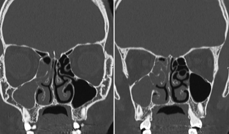
a. What is the CT scan finding?
b. What is the diagnosis?
c. What is the most common cause?
d. What are the causative pathogenic organisms (any three)?
e. What are the complications (any three)?
f. What is the investigation of choice?
g. What are the principles of management?
Answer
| a. | Right maxillary and ethmoid sinusitis. |
| b. | Orbital cellulitis. |
| c. | Paranasal sinusitis. |
| d. | Staphylococcus aureus—methicillin sensitive and methicillin resistant, Streptococcus group A, Streptococcus anginosus, Streptococcus pneumoniae, anaerobes, Bacteroides species, Prevotella species, and Haemophilus species. |
| e. | Optic neuritis, retinal artery occlusion, orbital abscess = visual loss; intracranial extension of infection = cavernous sinus thrombosis or meningitis, epidural or subdural empyema, or brain abscesses. |
| f. | CT imaging of orbit, paranasal sinuses with IV contrast. |
| g. | Hospitalization, intravenous antibiotics, sinus drainage, and drainage of orbit for orbital or subperiosteal abscess. |
THG24-018
Tender swelling
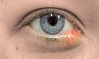
a. What is the diagnosis?
b. Mention the usual agent.
c. What are the types?
d. What is the treatment?
e. What is the complication if left untreated?
Answer
| a. | Hordeolum (stye). |
| b. | Staphylococcus aureus. |
| c. | Internal and external hordeolum. |
| d. | Frequent warm compresses, topical antibiotics, surgical excision, and drainage. |
| e. | Cellulitis of lid or orbit. |
THG24-019
Nontender swelling
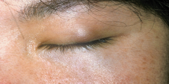
a. What is the diagnosis?
b. This occurs due to?
c. What is the usual outcome?
d. What is the indication for surgery—excision?
e. What is the indication for systemic macrolide?
Answer
| a. | Chalazion. |
| b. | Due to granulomatous inflammation of a meibomian gland. |
| c. | Spontaneous resolution. |
| d. | Indications: Cosmetically unacceptable; they become large enough to distort vision (by inducing astigmatism by exerting pressure on the globe). |
| e. | Significant corneal changes secondary to the underlying blepharitis; frequent chalazion formation. |
THG24-020
Q24.20
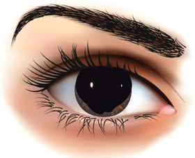
a. What is the defect?
b. What are the two forms?
c. Caused by defect in ______ gene on chromosome ______.
d. What are the associated ocular defects (any four)?
e. What is the tumor associated with this condition and in which form?
Answer
| a. | Absence of iris—aniridia. |
| b. | Familial and sporadic. |
| c. | Caused by defect PAX6 gene on chromosome 11. |
| d. | Macular hypoplasia, optic nerve hypoplasia, decreased vision, nystagmus, small cornea, pannus, cataract, lens dislocation, and glaucoma. |
| e. | Wilms tumor and sporadic aniridia. |
THG24-021
A 2-year-old toddler boy has symptoms of increased tearing, increased sensitivity to light (photophobia), and eyelid squeezing (blepharospasm) of right eye. On examination right cornea is enlarged and edematous.
a. What is the diagnosis?
b. Symptoms are attributed to ______.
c. What are the ocular signs?
d. What are the other associated ocular anomalies?
e. What are the predisposing causes of this condition?
f. What is the treatment modality of choice?
Answer
| a. | Infantile glaucoma. |
| b. | Symptoms are attributed to corneal irritation. |
| c. | Conjunctival injection, ocular and corneal enlargement (buphthalmos), Haab’s striae in cornea (breaks in Descemet membrane), cornea edematous and hazy, cupping of optic nerve. |
| d. | Aniridia, ectopia lentis, spherophakia, and cataract. |
| e. | Trauma, intraocular tumor, ocular inflammatory disease, intraocular hemorrhage, following cataract surgery. |
| f. | Surgical. |
THG24-022
Infant’s eye viewed through ophthalmoscope
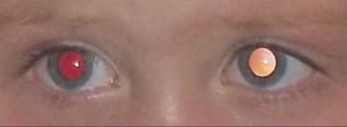
a. What is this test called?
b. How do you perform the test?
c. What do you observe in this infant?
d. What is your interpretation?
e. Mention the likely causes for what you observe in left eye (any four).
Answer
| a. | Red reflex examination. |
| b. | The red reflex test is properly performed by holding a direct ophthalmoscope close to the examiner’s eye with the ophthalmoscope lens power set at “0”. In a darkened room, the ophthalmoscope light should then be projected onto both eyes of the child simultaneously from approximately 18 inches away. |
| c. | Normal red reflex in the right eye and white reflex in the left eye. |
| d. | Leukocoria left eye. |
| e. | Primary causes: Cataract, persistent hyperplastic primary vitreous, retinal detachment, retinoschisis, larval granulomatosis, cicatricial retinopathy of prematurity (ROP), and retinoblastoma. Also to be considered: Endophthalmitis, organized vitreous hemorrhage, leukemic ophthalmopathy, and exudative retinopathy (as in Coats disease). |
THG24-023
Systemic conditions and eye disorders—match the following:
| 1. | Wilms tumor | a. | Lisch nodules | ||
| 2. | Neurofibromatosis | b. | Cataract | ||
| 3. | Marfan syndrome | c. | Aniridia | ||
| 4. | Galactosemia | d. | Ectopia lentis | ||
Answer
| 1. | c |
| 2. | a |
| 3. | d |
| 4. | b |
THG24-024
Hereditary pattern and eye conditions—match the following:
| 1. | Congenital cataract | a. | Mostly sporadic and almost 10% autosomal recessive | ||
| 2. | Retinitis pigmentosa | b. | Most common is autosomal dominant | ||
| 3. | Glaucoma | c. | Usually X-linked | ||
| 4. | Oculocutaneous albinism | d. | Nonheritable forms 60% and heritable forms 40% | ||
| 5. | Ocular albinism | e. | Autosomal recessive | ||
| 6. | Retinoblastoma | f. | Sporadic, autosomal dominant, autosomal recessive, and X-linked | ||
Answer
| 1. | Congenital cataract | b. | Most common is autosomal dominant | ||
| 2. | Retinitis pigmentosa | f. | Sporadic, autosomal dominant, autosomal recessive, and X-linked | ||
| 3. | Glaucoma | a. | Mostly sporadic and nearly 10% autosomal recessive | ||
| 4. | Oculocutaneous albinism | e. | Autosomal recessive | ||
| 5. | Ocular albinism | c. | Usually X-linked | ||
| 6. | Retinoblastoma | d. | Nonheritable forms 60% and heritable forms 40% | ||
THG24-025
A 7-year-old child came with clumsiness in walking and history of recurrent sinopulmonary infections.
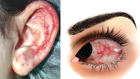
a. What do you see?
b. What is the probable diagnosis?
c. What is the mode of inheritance?
d. Which chromosomal is involved?
e. What is the usual clinical presentation?
f. What are the laboratory abnormalities?
g. What are the complications?
Answer
| a. | Ocular and aural telangiectasia. |
| b. | Ataxia telangiectasia. |
| c. | Autosomal recessive. |
| d. | Long arm of chromosome 11. |
| e. | Progressive cerebellar ataxia: 1–3 years; followed by oculocutaneous telangiectasia and frequent sinopulmonary infections. |
| f. | Immunoglobulin A (IgA) reduced, diminished IgG2, IgG4, and IgE levels increased alpha-fetoprotein. |
| g. | Increased predisposition to malignancies. |
THG24-026
This is the eye of a 7-year-old boy admitted with acute liver cell failure with hemolytic anemia.
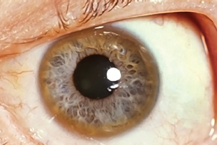
a. What abnormality is seen?
b. What is the most likely clinical diagnosis?
c. Mention other conditions in which this abnormality is seen (any four).
d. What is the mode of inheritance for the most likely clinical diagnosis?
e. What are the relevant investigations you will do to usually confirm diagnosis?
f. What action would you take regarding the rest of family?
Answer
| a. | Yellow-gray ring in cornea surrounding the iris—Kayser–Fleischer (KF) ring. |
| b. | Wilson disease. |
| c. | Cryptogenic cirrhosis, chronic active hepatitis, neonatal hepatitis, primary biliary cirrhosis, cholestatic cirrhosis, hepatocellular disorders (when bilirubin rises acutely above 20 mg/dL), galactosialidosis, Schistosoma infection, alcoholic liver disease, and intraocular foreign body containing copper. |
| d. | Autosomal recessive. |
| e. | Serum ceruloplasmin, serum copper level, and urinary copper excretion per 24 hours. |
| f. | Screening for serum ceruloplasmin. |
THG24-027
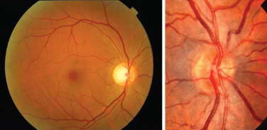
Describe the fundus images. In what condition it is seen?
Courtesy: Radhatri Nethralaya.
Answer
■ Disc:
| • | Margins are sharp |
| • | Color: Yellowish orange to creamy pink |
| • | Shape: round |
■ Vessels:
| • | Normal arteriovenous (AV) ratio |
| • | AV crossing: No indentation |
| • | No arterial light reflex |
■ Fundus background:
| • | No exudates or hemorrhages |
| • | Color: Red to purplish |
■ Macula:
| • | No vessels are noted around macula |
This is a picture of a normal fundus.
THG24-028
Q24.28
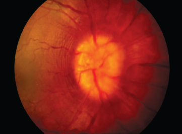
a. Identify the image.
b. What may be the accompanying clinical manifestations?
c. What are common causes (at least four)
d. What is the differential diagnosis?
Answer
| a. | Papilledema. |
| b. | Clinical manifestation: Irritability, headache, vomiting, and altered sensation. |
| c. | Common causes: |
| • | Obstructive hydrocephalus |
| • | Meningoencephalitis |
| • | Intracranial tumors |
| • | Intracranial hemorrhage |
| • | Toxic encephalopathies |
| • | Pseudotumor cerebri |
| d. | Differential diagnosis: |
| • | Pseudopapilledema |
| • | Disc swelling of papillitis in optic neuritis |
| • | Disc changes in hypertension and diabetes mellitus |
THG24-029
Q24.29
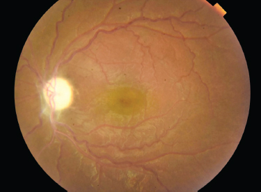
a. Describe the fundus image.
b. In what condition it is seen?
c. Name two principal causes of this condition in children.
d. What are the two main types based on ophthalmoscopic appearance?
Answer
| a. | The disc is pale and whiter than normal, and its edges are unusually sharply demarcated from the retina. The retinal vessels are slightly attenuated. |
| b. | Optic atrophy. |
| c. | Intracranial tumors and hydrocephalus. |
| d. | Primary and secondary. |
THG24-030
Acute loss of vision in a 15-year-old child—fundus image.
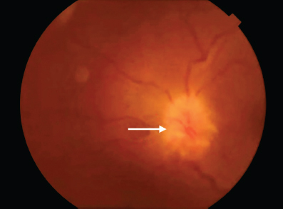
a. What do you see?
b. What is the likely diagnosis?
c. Mention other expected symptoms (any two).
d. Name two infectious diseases/two demyelinating disorders/two toxic or nutritional conditions that may cause this condition.
e. Which drug is used in treatment?
Answer
| a. | Swollen optic disc. |
| b. | Optic neuritis. |
| c. | Pain on movement of the globe, pain on palpation of the globe, decreased visual activity, decreased color vision and contrast sensitivity, and a relative afferent pupillary defect. |
| d. | Infective: Tuberculosis, lyme disease, meningitis, viral encephalitis, HIV |
| Demyelinating diseases: ADEM, multiple sclerosis, neuromyelitis optica | |
| Nutritional: Vitamin B12 deficiency | |
| Toxins: Methanol, ethambutol, lead poisoning, chloramphenicol, vincristine. | |
| e. | Intravenous methylprednisolone. |
THG24-031
Q24.31
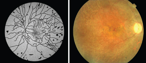
a. What is the likely clinical diagnosis?
b. What are the modes of inheritance?
c. What are the visual defects associated with the same (any three)?
d. Name clinical conditions associated with similar fundus image (any three).
Answer
| a. | Retinitis pigmentosa. |
| b. | Autosomal recessive, dominant, and X-linked recessive. |
| c. | Night blindness, tubular vision, subcapsular cataract, glaucoma, and keratoconus. |
| d. | Laurence–Moon syndrome, Bardet–Biedl syndrome; abetalipoproteinemia, Leber’s congenital retinal amaurosis, Kearns–Sayre syndrome, Usher syndrome, Refsum disease, and mucopolysaccharidoses (particularly Hurler, Hunter, Scheie, and Sanfilippo syndromes). |
THG24-032
Retinitis pigmentosa—regarding prognosis and inheritance. Match the following:
| 1. | X-linked | a. | Favorable prognosis with central vision preserved up to 5th-6th decade | ||
| 2. | Autosomal recessive | b. | Best prognosis with central visual acuity beyond the 6th decade | ||
| 3. | Autosomal dominant | c. | Worst prognosis with severe visual loss beyond 4th decade | ||
Answer
| 1. | X-linked | c. | Worst prognosis with severe visual loss beyond 4th decade | ||
| 2. | Autosomal recessive | a. | Favorable prognosis with central vision preserved up to 5th-6th decade | ||
| 3. | Autosomal dominant | b. | Best prognosis with central vision preserved beyond the 6th decade | ||
THG24-033
Retinitis pigmentosa—systemic associations (mention True or False).
a. Bardet–Biedl syndrome
b. Refsum disease
c. Usher disease
d. Stickler’s syndrome
Answer
Only Stickler’s syndrome is not associated with retinitis pigmentosa = False. All others are associated = True.
THG24-034
Q24.34
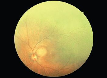
a. What is the likely diagnosis?
b. What are the two patterns of transmission?
c. What is the initial clinical sign? And how it occurs?
d. What is the second most common clinical sign?
e. What investigations you will do to confirm diagnosis?
Answer
| a. | Retinoblastoma. |
| b. | Hereditary and nonhereditary. |
| c. | Leukocoria (white pupillary reflex). Because of the reflection of light off the white tumor. |
| d. | Strabismus. |
| e. | Ultrasonogram, CT scan, and MRI of orbit. |
THG24-035
Q24.35
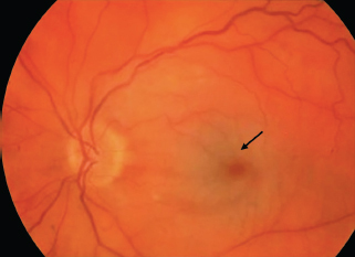
a. What do you see in fundus?
b. What is the explanation for this finding in fundus?
c. Name five conditions in which similar findings are seen in fundus.
d. What are the possible conditions with HSM (any two)?
Answer
| a. | Cherry red spot. |
| b. | Due to loss of transparency of the retinal ganglion cell layer secondary to lipid accumulation/edema. Because ganglion cells are not present in the fovea, the fovea transmits the underlying choroidal color. |
| c. | Mucopolysaccharidosis, Hurler’s disease, Tay-Sachs disease, Farber’s disease, GM1 gangliosidosis, Niemann–Pick disease, Sandoff’s disease, lysosomal storage diseases, metachromatic leukodystrophy, and central retinal artery occlusion. |
| d. | GM1 gangliosidosis, Sandhoff’s disease, and mucopolysaccharidosis. |
THG24-036
Diseases associated with cherry red spot—mention their inheritance pattern.
| 1. | GM1 gangliosidosis | |
| 2. | GM2 gangliosidosis | |
| 3. | Mucolipidosis | |
| 4. | Niemann-Pick disease | |
| 5. | Farber disease | |
Answer
| Autosomal recessive | |||
| 1. | GM1 gangliosidosis | ||
| Autosomal recessive | |||
| 2. | GM2 gangliosidosis | ||
| Autosomal recessive | |||
| 3. | Mucolipidosis | ||
| Autosomal recessive | |||
| 4. | Niemann-Pick disease | ||
| Autosomal recessive | |||
| 5. | Farber disease | ||
THG24-037
Fundus image of preterm baby born at 28 weeks of gestation.
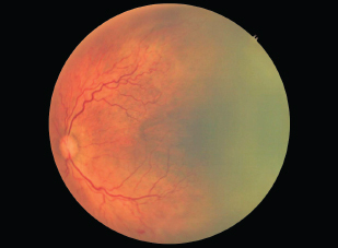
a. What is the diagnosis?
b. What are the risk factors (other than prematurity) associated with this condition (any four)?
c. What is the ideal age at which initial screening ophthalmic examination should be performed in this child?
d. What is the ideal treatment to reduce severe complications of progressive disease?
e. What is screening criteria for screening for this condition according to Rashtriya Bal Swasthya Karyakram (RBSK) (Government of India)?
Answer
| a. | Retinopathy of prematurity. |
| b. | Risk factors: Oxygenation, respiratory distress, heart disease, apnea, bradycardia, hypercarbia, acidosis, anemia and need for transfusion. |
| c. | 4 weeks (chronologic), 32 weeks postmenstrual. |
| d. | Laser photocoagulation of avascular retina. |
| e. | RBSK guideline: Screen <2,000 g weight or <34 weeks; between 34 and 36 weeks but with risk factors and those with unstable clinical course who are at high risk for ROP as determined by neonatologist/pediatrician. |
THG24-038
Q24.38
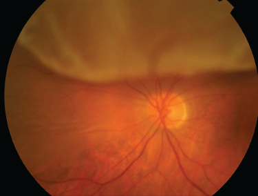
a. What is the diagnosis?
b. What are the types?
c. Mention the causes (any five).
d. What are the usual clinical signs?
e. Mention the investigations to determine cause and appropriate treatment.
Answer
| a. | Retinal detachment. |
| b. | Types: Rhegmatogenous, traditional, and exudative. |
| c. | Rhegmatogenous: Trauma, child abuse, ROP, and after surgery for congenital cataract |
| Traditional: Diabetes, sickle cell disease, and ROP | |
| Exudative: Coats disease, retinoblastoma, and ocular inflammation | |
| d. | Loss of vision, secondary strabismus or nystagmus or leukocoria. |
| e. | Ultrasonogram and CT/MRI. |
Figure Sources
Figures of Q24.6, Q24.14, Q24.18, Q24.19, Q24.22, Q24.25, Q24.26 and Q24.30 are from open source.
Figures of Q24.4, Q24.7, Q24.8, Q24.9, Q24.10, Q24.17 and Q24.20 are from author’s personal collection and drawn by artist.