THG19-001
A 6-month-old child was brought with history of feeding difficulty, chest retractions, and poor weight gain. His weight was 4 kg. SpO2 was 98%. X-ray showed cardiomegaly. ECG is given here.
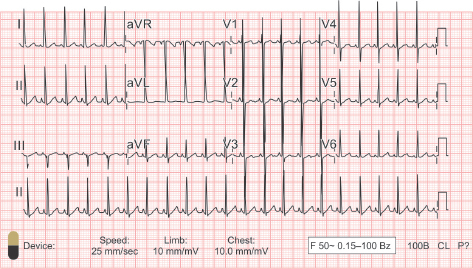
a. Write down the ECG characteristics.
b. What is the likely diagnosis?
c. What is the management you would advise for this child?
Answer
| a. | Sinus tachycardia with HR of approximately 130 beats/min |
| There are large equiphasic QRS complexes in midprecordial leads, suggestive of biventricular hypertrophy—a phenomenon called Katz-Wachtel phenomenon. |
Large equiphasic QRS complexes (R + S >60 mm) in midprecordial leads—V2, V3, and V4. This is usually seen in any large post-tricuspid shunts like ventricular septal defect (VSD), patent ductus arteriosus (PDA), and aortopulmonary (AP) window.
| b. | Clinical signs of heart failure, cardiomegaly, and Katz-Wachtel phenomenon on X-ray, suggestive of large left-to-right shunt |
| c. | Confirmation of the diagnosis on echocardiogram. Early surgery (ideal age for surgery in large post-tricuspid shunts, such as VSD, PDA, AP window—3–6 months of age) |
THG19-002
This is the ECG of a 7-year-old asymptomatic boy.
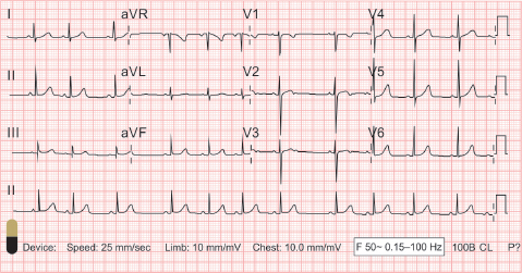
a. What is the ECG diagnosis?
b. Explain the physiologic basis.
c. What is your advice regarding management?
Answer
| a. | ECG shows irregular heart rhythm. There is irregular P-P interval, irregular R-R interval, monomorphic P waves, and regular P-R interval—characteristic of sinus arrhythmia. |
| b. | Sinus arrhythmia is a normal variation seen in children and adolescents. During inspiration, there is decrease in the vagal tone causing an increase in the HR. Conversely, on expiration and increased vagal tone, the HR decreases. |
| c. | This is physiologic in children and adolescents; does not require any treatment. |
THG19-003
A 12-year-old child was diagnosed to have congenital heart disease and planned for cardiac surgery. This is the ECG of the child.
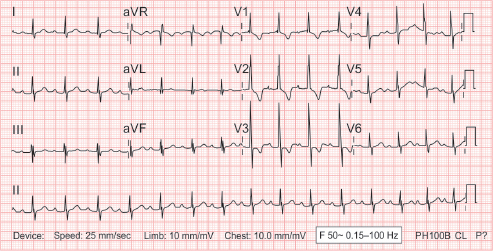
a. What is the ECG finding?
b. What are the congenital heart defects which can be associated with this?
c. What is the treatment?
Answer
| a. | ECG shows HR of around 90 beats/min. Every QRS complex is preceded by a P wave; P-R interval is constant but prolonged (240 ms)—characteristic of first-degree AV block. There is also evidence of incomplete right bundle branch block (RBBB). |
| b. | First-degree AV block can be associated with congenital heart defects, such as ASD, AV canal defect, Ebstein anomaly, and corrected transposition of great arteries. (Acquired heart diseases that can manifest first-degree AV block include acute rheumatic fever, Lyme disease, myocarditis, and cardiomyopathies) |
| c. | Management involves treatment of associated heart defect, if any. No specific intervention is required for first-degree AV block. |
THG19-004
Given below is the ECG of an asymptomatic neonate with bradycardia. Mother gives history of intrauterine death during previous pregnancy.
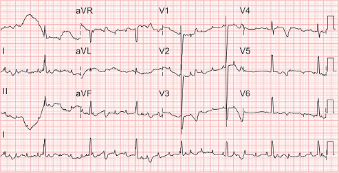
a. What is the diagnosis?
b. What is the most likely etiology?
c. What is the definitive treatment?
Answer
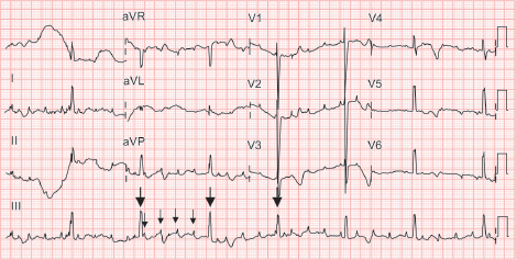
ECG complete heart block.
| a. | ECG shows bradycardia with HR (ventricular rate) of 42 beats/min. Atrial rate is around 170 beats/min. There is no association between P waves (arrow as shown in image) and QRS complexes (bold arrow as shown in image). ECG is diagnostic of complete heart block (third-degree AV block). |
| b. | The most common cause of congenital complete heart block is maternal connective tissue disorder, such as Sjögren syndrome, systemic lupus erythematosus (SLE), rheumatoid arthritis. The maternal autoantibodies cross the placenta and affect the fetal conduction system resulting in complete heart block. |
| c. | Definitive treatment includes permanent pacemaker implantation. |
Indications for pacemaker implantation in congenital complete heart block:
| • | Symptomatic bradycardia |
| • | Asymptomatic with ventricular dysfunction, ventricular ectopy, wide QRS complexes, and prolonged QT interval |
| • | Infant with HR <50 beats/min in a structurally normal heart or <70 beats/min when associated with structural heart defect |
THG19-005
A 3-month-old infant brought to the emergency room (ER) with poor feeding, lethargy, and cold peripheries. On examination, he was drowsy. He had tachycardia, feeble peripheral pulses, and poor perfusion.
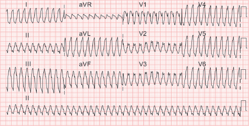
a. Interpret the ECG.
b. What are the possible etiologies?
c. How do you manage this child?
d. What are the investigations you would like to do?
Answer
| a. | ECG shows regular wide complex tachycardia with HR >200 beats/min. There is no discernible P wave. ECG is suggestive of ventricular tachycardia. |
| b. | Ventricular tachycardia in children can be due to underlying structural heart defect, myocarditis, cardiomyopathy, cardiac tumors (like rhabdomyoma), electrolyte derangement, long QT syndrome, drugs, and toxins. |
| c. | This child has ventricular tachycardia (VT) with pulse and has presented with altered sensorium and shock. Treatment includes: |
| • | Maintain airway |
| • | Synchronized cardioversion at 0.5–1 J/kg; can repeat up to 2 J/kg |
| • | Followed by injection amiodarone infusion (5–15 µg/kg/min) or lidocaine infusion (20–50 µg/kg/min) |
| • | Correct dyselectrolytemia |
| • | Work-up for possible etiology |
| d. | Echocardiogram, serum electrolytes, calcium, magnesium, and complete blood count. |
THG19-006
A 10-month-old asymptomatic child referred in view of murmur. ECG is given here.
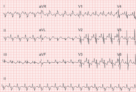
a. Interpret the ECG. What is the likely diagnosis?
b. What are the likely clinical signs in this condition?
c. What are the indications for surgery in this condition?
Answer
| a. | ECG shows HR of 125 beats/min, sinus rhythm, tall P waves (P wave height 5 mm in lead II), and wide low-voltage splintered QRS complexes. P waves are taller than corresponding QRS complexes in lead II—suggestive of Himalayan P waves. |
| Diagnosis: Ebstein anomaly |
(Tall P waves—3 mm or more in amplitude–indicate right atrial enlargement; Himalayan P waves—P wave amplitude taller than corresponding QRS complex; In Ebstein anomaly, tricuspid valve is displaced apically, part of right ventricle (RV) is atrialized, and the effective functional RV is small. Hence, there is intraventricular conduction delay resulting in wide QRS complexes. Splintering means multiple notching on QRS complexes)
| b. | Ebstein anomaly can have a bimodal presentation, with symptoms in neonatal period, then an asymptomatic phase in childhood, and again symptomatic phase in adolescence or adulthood. |
They can present with exertional dyspnea, arrhythmias, heart failure, or cyanosis.
They have multiple heart sounds (wide split S1, wide split S2, and S3), and a pansystolic murmur due to tricuspid regurgitation (TR).
X-ray shows box-shaped cardiac silhouette.
| c. | Surgery is indicated in: |
| • | Symptomatic patients |
| • | Cardiomegaly on X-ray |
| • | Presence of cyanosis |
THG19-007
A 5-year-old asymptomatic child was evaluated for an incidentally detected murmur and wide split-second sound. Her ECG is given below.
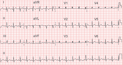
a. Interpret the ECG. What is the likely diagnosis?
b. What are the options available for treatment? What is the ideal age to intervene?
Answer
| a. | ECG shows sinus rhythm with HR of around 120 beats/min. There is rightward QRS axis, rsR’ in V1, and Crochetage sign. (Crochetage sign—notching of R wave in inferior leads—II, III, and aVF. Due to RV dilatation, there is delayed conduction through the right bundle, leading to RBBB pattern in V1—rsR’ pattern) |
| Diagnosis: Atrial septal defect (ASD) | |
| b. | Child is 5 years old. Spontaneous closure is not feasible at this age. Child should be advised to undergo ASD closure, either by device closure or surgical closure. Ideal age for intervention in ostium secundum ASD is 3–4 years. Sinus venosus ASD is repaired at 4–5 years of age. |
THG19-008
A 6-month-old infant was brought with history of irritable cry, tachypnea and bluish discoloration of lips, fingers, and toes. His oxygen saturation was 65%. On auscultation, S2 was single. There was no murmur. There was no history of fever. There was history of similar episodes in the previous week. His X-ray is given here.
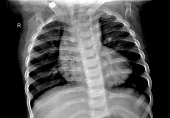
a. Describe the X-ray finding. What is the likely diagnosis?
b. What is the acute management in this child?
c. What is the definitive management after stabilizing the child?
Answer
| a. | X-ray shows boot-shaped heart, RV type of apex, concave pulmonary bay, and decreased pulmonary vascularity. Clinical presentation and X-ray are suggestive of tetralogy of Fallot (TOF) (or other TOF like conditions) presenting with cyanotic spell (irritable cry, tachypnea, cyanosis, decrease in intensity of murmur, or disappearance of murmur are clues for cyanotic spell). |
| b. | Management of cyanotic spell: |
| • | Knee chest position |
| • | Supplemental oxygen (without agitating the child) |
| • | Intravenous fluid bolus 10–20 mL/kg |
| • | Injection morphine IV 0.1–0.2 mg/kg |
| • | Intravenous sodium bicarbonate 1–2 mEq/kg |
| • | If there is no response, |
| ◆ | Injection phenylephrine 0.02 mg/kg |
| ◆ | Esmolol infusion |
| ◆ | Ketamine |
| c. | After stabilizing the child, he is started on oral propranolol 1–4 mg/kg/day q 8–12 hourly, and iron supplementation so as to maintain Hb > 14 g/dL. |
If cyanotic spells recur despite propranolol, he is advised to undergo surgery—complete correction (intracardiac repair) or palliative procedure (BT shunt/right ventricular outflow tract stenting, based on the size of pulmonary arteries on the echocardiogram).
THG19-009
A 6-year-old girl was referred for symptoms of exertional dyspnea. Her oxygen saturation was 87%. On auscultation, she had a wide split second heart sound and ejection systolic murmur. Her X-ray is given here.
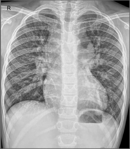
a. Interpret the X-ray.
b. What is the diagnosis?
c. What is the reason for this X-ray finding?
d. What is the management?
Answer
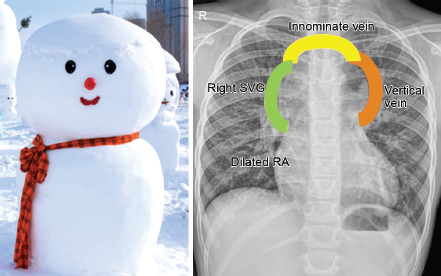
| a. | X-ray chest posteroanterior (PA) view showing classical Snowman appearance or figure of 8 appearance. There is pulmonary plethora. |
| b. | Supracardiac total anomalous pulmonary venous connection (TAPVC) |
| c. | Snowman appearance is usually seen in older children with supracardiac TAPVC (this shape is not seen in young infants with TAPVC). |
The head of the Snowman is formed by the vertical vein on the left, dilated innominate vein superiorly, and dilated superior vena cava (SVC) on the right. The body of the Snowman is formed by the dilated heart chambers.
| d. | Surgical correction of TAPVC |
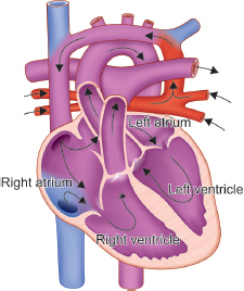
Schematic diagram of supracardiac TAPVC: All Four pulmonary veins join together to form a confluence, which then drains through the vertical vein into the innominate vein. The innominate vein drains into the right SVC and subsequently into right atrium.
THG19-010
An adolescent was referred for cardiac evaluation as the heart sounds were better heard on the right side during a routine auscultation. Her X-ray is given here.
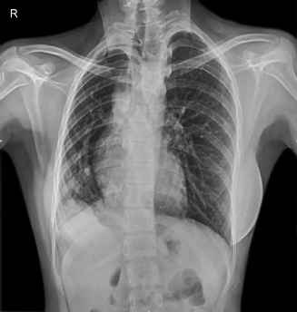
a. Mention the X-ray finding.
b. What is the likely diagnosis?
c. What are the associated lesions in this condition? What is the clinical presentation?
Answer
| a. | X-ray shows mediastinal shift to the right, causing dextroversion of the heart. There is Scimitar sign—Turkish sword appearance noted to the right of heart. This is caused by the anomalous right pulmonary veins which course inferiorly and drain into the inferior vena cava (IVC). (In dextrocardia, the cardiac base-apex axis is toward right. Apex points toward right. In dextroversion, heart is pulled toward the right due to the mediastinal shift. Here, the apex still points toward left. Though the heart is shifted to the right, the base-apex axis is toward left). Right lung volume is reduced. |
| b. | Scimitar syndrome |
| c. | Scimitar syndrome is characterized by partial anomalous pulmonary venous connection of one or both right pulmonary veins to the IVC. It is often associated with hypoplastic right lung, which causes mediastinal shift to the right and dextroversion of the heart. |
It can be associated with intracardiac defects such as ASD and VSD. Clinical presentation is varied. They can present in neonatal period or infancy with severe pulmonary arterial hypertension (PAH) and heart failure symptoms. Some of them are asymptomatic in childhood and present later.
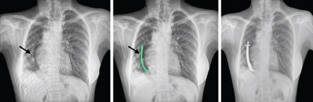
Scimitar sign—Turkish sword appearance.
THG19-011
A 2-month-old neonate was admitted with history of fever, respiratory distress, poor feeding, and lethargy. He was one of twins and had a long neonatal intensive care unit (NICU) stay for 6 weeks. His chest X-ray was normal. A screening echocardiogram was done. The echo image is given here.
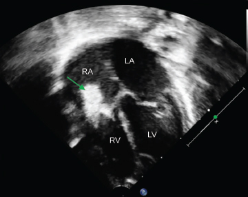
a. What do you see on the echocardiographic image? What is the likely diagnosis?
b. What are the investigations you will do?
c. How will you manage this child?
Answer
| a. | Echocardiogram shows a large vegetation on the tricuspid valve. |
| Diagnosis: Infective endocarditis | |
| b. | Three sets of blood culture at least 30 minutes apart, from three different sites for aerobic, anaerobic, and fungal cultures. Investigations include complete blood count, C-reactive protein (CRP), erythrocyte sedimentation rate (ESR), and urine routine examination. Other investigations like renal and liver function tests to be done. |
| c. | After taking samples for blood culture, start on empirical antibiotics—injection ceftriaxone or injection crystalline penicillin G plus injection gentamycin intravenously. Once the organism is identified on culture, antibiotics to be changed as per the sensitivity report. |
THG19-012
A 14-year-old boy with structurally normal heart had three episodes of supraventricular tachycardia (SVT) in the last 1 year. During every episode, the rhythm reverted to sinus with injection adenosine. This is his baseline ECG during sinus rhythm.
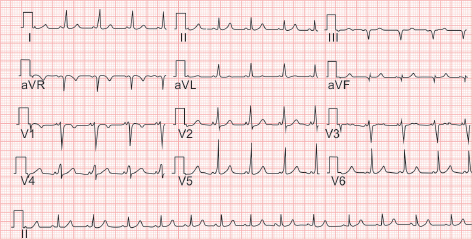
a. Interpret the ECG finding. What is the diagnosis?
b. What treatment would you advise in the given situation?
Answer
| a. | ECG shows sinus rhythm with HR of around 80 beats/min. There is short P-R interval and slurred upstroke of QRS complex—delta wave (arrow as shown in image). This indicates preexcitation through an accessory pathway. |
| Diagnosis: Wolff-Parkinson-White (WPW) syndrome | |
| b. | As he is 14-year-old with WPW syndrome, he may be advised to undergo electrophysiological study and radiofrequency ablation. Medical management with β-blockers is advised till the procedure. In infants and younger children, medical management is advised. |
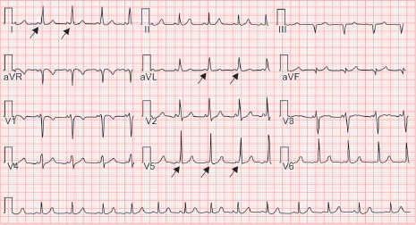
Wolff-Parkinson-White syndrome.
THG19-013
A 9-year-old boy presented with recurrent syncope. He was evaluated and subsequently underwent a cardiac procedure. This is his ECG after the procedure.
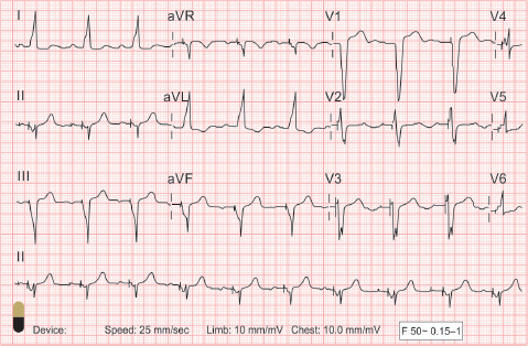
a. What is the ECG finding?
b. What is the procedure he has undergone?
Answer
| a. | ECG shows pacing spikes before all QRS. P waves are normal in most complexes (in few complexes, P waves also have a spike). This ECG shows predominantly atrial-sensed ventricular paced rhythm. |
| b. | Permanent pacemaker implantation |
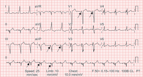
Pacemaker spike.
THG19-014
A 12-year-old girl presented with exertional dyspnea. She was diagnosed to have congenital heart defect in her infancy, but did not undergo treatment and was lost to follow-up. On auscultation, she has a loud P2 and early diastolic murmur at the left upper sternal border. Oxygen saturation in the right upper limb and lower limb was 97% and 78% respectively. Her chest X-ray is given here.
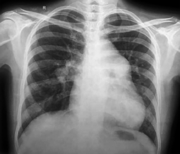
a. Discuss her clinical signs and X-ray features.
b. What is the diagnosis?
Answer
| a. | Lower limb SpO2 78%, upper limb SpO2 97% suggestive of differential cyanosis. Auscultation suggestive of severe PAH. She was diagnosed to have congenital heart defect in her infancy. So, the clinical picture is suggestive of PDA with severe PAH and right-to-left shunt. X-ray shows dilated main pulmonary and central pulmonary arteries with pruning of peripheral vasculature. |
| b. | PDA with Eisenmenger syndrome |
THG19-015
A 1-year-old male child is brought to the pediatric ER with history of lethargy and irritability. On examination, airway was stable; tachycardia, respiratory rate (RR)—50 breaths/min, normal blood pressure (BP), and good peripheral pulses. Electrocardiogram was done.
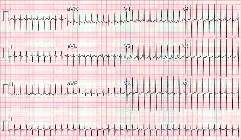
a. Calculate heart rate (HR)?
b. What is the diagnosis?
c. In a resource limited setting with no cardiac drugs what will you do?
d. What is the drug that you will use to restore normal rhythm? What is the mechanism of its action and its half-life? What is the recommended dose of the drug?
Answer
| a. | Heart rate: 250 beats/min |
| b. | Supraventricular tachycardia/narrow QRS complex tachycardia |
| c. | Vagal maneuvers: Ice on face causes block or slowing of conduction in AV node. |
| d. | Adenosine. It acts on adenosine receptor (A1) on sinoatrial (SA) node, AV node, and atrial myocytes. Activation of these receptors hyperpolarizes the cells in SA and AV node and causes transient heart block. Adenosine is rapidly metabolized and its half-life is <10 seconds. Recommended dose is 0.1–0.2 mg/kg/dose. |
THG19-016
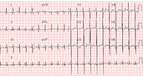
A 3-month-old girl is brought to the pediatric ER with complaints of breathlessness and feeding difficulty. On examination, baby was irritable with HR—170 beats/min, RR—55 breaths/min, SpO2—99%, normal BP, poor perfusion, and hepatomegaly. Chest X-ray was taken. Electrocardiogram was taken few hours after stabilization.
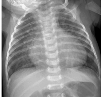
a. What are the findings in chest X-ray?
b. What are the probable congenital cardiac lesions that can present in this manner?
c. What are the findings in ECG?
Answer
| a. | Cardiomegaly and pulmonary plethora |
| b. | Large VSD, large PDA, complete AVSD, large AP window |
| c. | Sinus rhythm, HR—140 beats/min (approximately), left axis deviation, and biventricular hypertrophy |
THG19-017
A 4-month-old infant weighing 3.5 kg was brought to pediatric OPD with history of feeding difficulty, forehead sweating, and poor weight gain. Two months back the baby was diagnosed to have episodes of abdominal colic and advised oral medication. On examination, infant had HR—140 beats/min, RR—60 breaths/min, normal BP, and normal perfusion. Auscultation revealed basal crepitations and S3 at apex. Chest X-ray showed cardiomegaly. Screening ECHO revealed decreased left ventricular function.
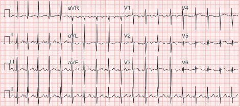
a. What are the salient findings in ECG?
b. What could be the probable diagnosis?
c. What is the definitive treatment?
Answer
| a. | Sinus rhythm, HR—140 beats/min, left axis deviation, and abnormal Q waves in lead I and aVL. |
| b. | Anomalous origin of left coronary artery from pulmonary artery (ALCAPA) |
| c. | Surgical repair: Direct reimplantation of left coronary artery onto the aorta. |
THG19-018
An 8-year-old boy came with history of palpitations of 1 hour duration. He had past history of recurrent palpitations over the last 3 months period. He had one episode of syncope lasting for less than a minute 2 months back. On examination, child was lethargic with HR—140 beats/min, RR—20 breaths/min, weak peripheral pulses, and hypotension.
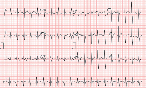
a. What is the ECG finding?
b. What is the preferred treatment?
c. What are the cardiac channelopathies which can present with arrhythmias?
Answer
| a. | Ventricular rate approximately 130 beats/min. Narrow QRS complex tachycardia, and atrial flutter waves seen. |
| b. | Synchronized cardioversion: 0.5 J–2 J/kg |
| c. | Long QT syndrome and Brugada syndrome |
THG19-019
A male infant aged 3 months came with history of breathlessness and feeding difficulty for 2 weeks of age. There was history of recurrent respiratory tract infection. Birth weight was 3 kg. Present weight was 3.7 kg. On examination, baby had HR—150 beats/min, RR—45 breaths/min, normal BP, and SpO2—98%. Chest X-ray showed cardiomegaly with pulmonary plethora. Echocardiogram was done. These features are analyzed in the following questions.
a. Both atrioventricular (AV) valves insert at the same level at the cardiac crux
b. Cleft in the left-sided AV valve component
c. Large atrial septal defect (ASD) (ostium primum type)
d. Large inlet ventricular septal defect (VSD)
a. Which of the above findings in echocardiogram would be suggestive of complete atrioventricular septal defect (AVSD)?
a. a and b only
b. a, b, and d only
c. a, b, and c only
d. a, b, c, and d
b. What among the above findings would be suggestive of partial AV canal defect?
a. a and b only
b. a, b, and d only
c. a, b, and c only
d. a, b, c, and d
c. What is the ideal age of surgical repair for complete AVSD?
d. Familial occurrence of complete AVSD is rare—True/False
Answer
| a. | D |
| b. | C |
| c. | 3–6 months of age |
| d. | True |
THG19-020
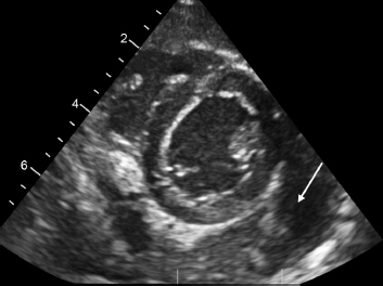
Child aged 5 years was admitted in pediatric ward with history of fever >10 days. On examination, child had tachycardia, tachypnea, and high-grade fever. Total count was raised with neutrophilic predominance. Acute phase reactants were elevated. Chest X-ray showed cardiomegaly. Echocardiogram was done and the fixed frame image is given here.
a. What is the diagnosis from echocardiogram?
b. What are the most common causes of the above mentioned condition? List out the other infectious causes.
c. What are the noninfectious causes for the above mentioned condition?
Answer
| a. | Heterogeneous echogenicity of pericardial space around heart. Probable pyopericardium |
| b. | Bacterial: Staphylococcus, Haemophilus influenzae, Streptococcus pneumoniae, Pseudomonas aeruginosa, Neisseria meningitidis, Escherichia coli, Klebsiella, and Mycobacterium tuberculosis |
| Viral: Enterovirus (coxsackie B), adenovirus, Cytomegalovirus, Epstein–Barr virus, and human immunodeficiency virus (HIV) | |
| Fungal: Candida, Aspergillus, and Blastomyces | |
| Rickettsial: Typhus | |
| Parasitic: Entamoeba histolytica | |
| c. | Hypothyroidism, neoplastic diseases (mostly metastatic), and chronic renal failure |
THG19-021
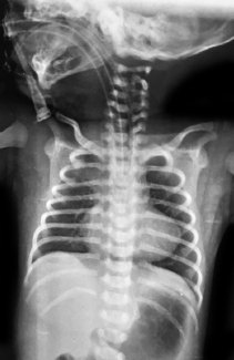
Baby was delivered by emergency lower segment cesarean section (LSCS) for obstetric indication [gestational age (GA): 39 weeks] at a primary health center (PHC) located in a remote village. Birth weight was 3.2 kg. Baby was in postnatal ward with mother. On day 4 of life, baby was found to be lethargic with HR—130 beats/min, RR—40 breaths/min, normal perfusion status, and BP—65/40 mm Hg. Capillary blood glucose was 90 mg/dL. SpO2 (right upper limb) was 70%. System examination was done. Cardiovascular system (CVS): S1—normal and S2—difficult to interpret with no murmur; Respiratory system (RS): normal breath sounds; Abdomen: no organomegaly; Central nervous system (CNS): anterior fontanelle (AF)—normal, pupils equal and reacting to light; and No focal neurological deficit. Within few minutes baby developed apnea and required intubation. Chest X-ray was done.
a. What are the findings in chest X-ray?
b. What could be the probable cardiac condition in this baby?
c. What should the medical officer at PHC do to ensure safe referral of baby to the nearest tertiary neonatal care unit located 100 km away?
d. What should be done by the neonatologist at the tertiary neonatal care unit?
e. What will be the general line of management for such cardiac conditions?
Answer
| a. | Boot-shaped heart and pulmonary oligemia |
| b. | Duct-dependent pulmonary circulation |
| c. | Baby should be started on prostaglandin E1 (PGE1) infusion. |
| d. | Neonatologist should be ready to do a screening ECHO to assess ventricular function, look for PDA flows, and if possible look for any structural cardiac lesion. He should inform the cardiologist for early opinion as a structural cardiac defect is being suspected in the baby. |
| e. | After ECHO confirmation and if the baby is having congenital heart disease with PDA-dependent pulmonary circulation, dose of PGE1 infusion should be adjusted to maintain saturation between 80 to 85%. If baby is dependent on prostaglandin infusion, cardiac intervention will be needed. Stenting of PDA or Blalock–Taussig shunt will be needed in such babies. |
THG19-022
A 3-week-old baby was brought to a PHC with history of refusal of feeds for 2 days and breathlessness for 1 day. Baby was delivered by LSCS in the PHC and was given to mother for breastfeeding soon after birth. Baby and mother were discharged after 5 days. On examination, baby general condition (GC) was poor, mild subcostal retractions, HR—180 beats/min, RR—65 breaths/min, and weak dorsalis pedis and femoral pulses. Soles were dusky in color. Noninvasive blood pressure (NIBP) recorded by staff nurse was 80/60 mm Hg (right upper limb). Capillary blood glucose (CBG) was 75 mg/dL. Medical officer at PHC diagnosed the baby to have shock and started IV saline boluses 5 mL/kg. In spite of two saline boluses, baby did not improve. Dorsalis pedis pulse was still weak and liver span had increased.
a. Can a cardiac condition be suspected in this baby? If so, what could be the probable cardiac diagnosis?
b. What drug should be available in PHC to stabilize the baby before referral to a tertiary neonatal care facility?
c. What are the structural cardiac defects that can present in this manner?
Answer
| a. | Duct-dependent systemic circulation |
| b. | Prostaglandin E1 infusion |
| c. | Critical/severe coarctation of aorta, hypoplastic left heart syndrome, interrupted aortic arch, and critical aortic stenosis |
THG19-023
Male baby aged 5 weeks weighing 3.5 kg was brought to the pediatric casualty with history of breathlessness for past 1 week, difficulty in breastfeeding for past 3 days and failure to gain weight. No history of fever/cough/coryza. Baby was delivered by LSCS and birth weight was 2.9 kg. On examination, baby had HR—170 beats/min, RR— 65 breaths/min, SpO2—99%, normal perfusion, and normal BP. Baby had mild subcostal retractions and an audible systolic murmur at left lower sternal border. Chest X-ray revealed mild cardiomegaly with plethora. Suspecting a cardiac disease, the pediatrician requested for ECHO.
a. What is the pediatrician have in mind when he wanted a cardiologist opinion?
b. What are the common structural cardiac lesions that can present at this age with the above findings?
c. What is the initial line of management for the cardiac lesion diagnosed other than general management? What are the drugs that can be used?
d. Which left-to-right shunt is unlikely to present at this age?
Answer
| a. | Pediatrician is suspecting a large left-to-right shunt lesion or ventricular dysfunction |
| b. | Large left-to-right shunt lesions, such as VSD, PDA, complete AVSD, and AP window |
| c. | Antifailure drugs should be started once these large left-to-right shunt lesions are diagnosed. Furosemide, spironolactone, digoxin, and enalapril |
| d. | Atrial septal defect and partial anomalous pulmonary venous drainage |
THG19-024
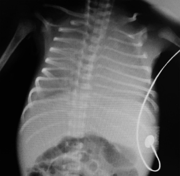
A newborn baby was delivered in a PHC by labour naturale at GA of 36 weeks. Birth weight was 2.4 kg. Baby was noted to have fast breathing with mild retractions at about 6 hours of life. Chest X-ray was done. Baby was given surfactant suspecting hyaline membrane disease. However, baby’s condition did not improve and baby was intubated. SpO2 did not pick up and remained at 80% despite FiO2 >90%.
a. What is the chest X-ray finding?
b. What cardiac lesion should be suspected if X-ray findings remain the same after surfactant therapy?
c. What should the medical officer do next?
d. What will be the definitive line of management for the suspected cardiac lesion?
Answer
| a. | Ground glass like opacification of lung fields |
| b. | Obstructed TAPVC |
| c. | Cardiologist opinion should be obtained and urgent ECHO should be done (if ECHO machine is available) or shift the baby to nearby tertiary neonatal care center where it is possible. |
| d. | If obstructed TAPVC is confirmed, emergency cardiac surgery is needed. |
THG19-025
A 2-year-old male child was brought to pediatric OPD with complaints of two episodes of sudden loss of consciousness in the past 1 week. Each episode lasted for approximately 1 minute. Child regained consciousness in few minutes. On examination, vitals were normal and CVS examination revealed normal S1 and S2 with no murmur. Electrocardiogram was done.
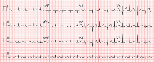
a. What are the ECG findings? What is the diagnosis?
b. What is Bazett formula?
c. What are the syndromes associated with this condition?
Answer
| a. | Sinus rhythm, HR—100 beats/min, normal QRS axis, normal P-R interval, QTc: >500 ms (534 ms) |
| b. | Corrected QT interval = QT interval/√ R-R interval |
| c. | Jervell and Lange-Nielsen syndrome, Romano-Ward syndrome, and Timothy syndrome |
THG19-026
A 6-year-old boy complained of left- and right-sided chest pain which was pinpricking in nature with no radiation. He plays football regularly. There was no history of palpitations/syncope. General and system examination was normal. Vital signs were normal. ECG was taken.
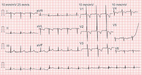
a. Interpret the ECG? What could be the probable diagnosis?
b. What are the supportive findings?
Answer
| a. | Dextrocardia |
b. • Inverted P wave in lead I with upright P in aVR
| • | Negative QRS complex in lead I, upright QRS complex in aVR |
| • | Right axis deviation, normal P-R interval |
| • | Lack of progression in R wave height from V1 to V6 |