THG23-001
An 8-year-old male child presents with episodic headache for the past 6 months. The headache frequency is approximately three episodes per month and each episode lasts for 30 minutes to 2 hours. The headache is associated with nausea, photophobia, phonophobia, and occasional vomiting during the episodes. The headache is prominent usually in the evening and it is relieved with rest. There is family history of intermittent headache in mother.
a. What is the diagnosis?
b. How do you objectively assess the severity of headache in this condition?
c. How will you treat the headache episodes?
d. What investigation would you like to do to confirm the diagnosis?
Answer
| a. | Migraine |
| b. | PedMIDAS score |
| c. | Lifestyle modification and analgesics |
| d. | NIL |
THG23-002
A 10-year-old obese female child presented with intermittent headache for the past 6 months. The headache episode was also associated with often blurring of vision and vomiting. There was no photophobia or phonophobia. The headache was aggravated in the morning once the child woke up. There was no family history of headache. Fundus evaluation revealed papilledema bilaterally. Magnetic resonance imaging (MRI) brain with magnetic resonance venography (MRV) was normal.
a. What is the diagnosis?
b. How would you confirm the diagnosis?
c. How will you treat this condition?
Answer
| a. | Idiopathic intracranial hypertension (pseudotumor cerebri) |
| b. | CSF manometry to check CSF opening pressure |
| c. | Lumbar puncture (therapeutic) and oral acetazolamide |
THG23-003
A 6-year-old female child presented with swaying while walking from 2 years of age. The child was having intention tremors and unsteady gait and it has been gradually progressive. The child was noted to have oculomotor apraxia on examination. The child also has history of frequent respiratory tract infections.
a. What is the likely diagnosis?
b. What specific general examination finding can help you diagnose this condition?
c. What biochemical maker in serum is elevated in this condition?
d. What is the mode of inheritance of this condition?
Answer
| a. | Ataxia telangiectasia |
| b. | Telangiectasia in bulbar conjunctiva (telangiectasia can also be found over bridge of nose, on the ears, and exposed surface of the extremities) |
| c. | Serum α-fetoprotein |
| d. | Autosomal recessive |
THG23-004
A 7-year-old male child presented with failure to thrive, anemia, and progressive swaying, unsteady gait, and intention tremors. The child also has frequent loose stools and bulky sticky stools.
a. What is the likely diagnosis?
b. What hematological abnormality would you expect?
c. What biochemical investigation will give you a clue toward the diagnosis?
d. What ophthalmological abnormality would you expect?
e. Mention the compound used in treatment of this condition?
Answer
| a. | Abetalipoproteinemia |
| b. | Acanthocytes in peripheral smear |
| c. | Lipid profile (decreased levels of serum cholesterol and serum triglycerides) |
| d. | Retinitis pigmentosa |
| e. | Vitamin E |
THG23-005
A 12-year-old boy presented with unsteady gait, frequent falls, and intention tremors and it was gradually progressive over the past 6 months. The child had pyramidal signs but bilateral knee and ankle jerks were sluggish and bilateral plantar was extensor. Echo done in view of exertional dyspnea revealed evidence of cardiomyopathy.
a. What is the likely diagnosis?
b. What is the mode of inheritance?
c. What triplet repeat disease is implicated in this condition?
d. Mention the triplet repeat that occurs in this condition?
e. What endocrine abnormality has to be screened in this condition?
f. What is the most common cause of mortality in this disorder?
Answer
| a. | Friedreich’s ataxia |
| b. | Autosomal recessive |
| c. | GAA |
| d. | Pes cavus and hammer toes |
| e. | Diabetes mellitus |
| f. | Hypertrophic cardiomyopathy leading to congestive cardiac failure |
THG23-006
A 9-year-old female child presented with abnormal involuntary jerky dance like movements of limbs and they are absent during sleep. The child was noted to have emotional lability. Investigations revealed elevated C-reactive protein (CRP), erythrocyte sedimentation rate (ESR), and significant ASO (antistreptolysin O) titer. (Note: A video of the involuntary movements can be displayed in examination instead of description)
a. What is the diagnosis?
b. What is the triad of this condition?
c. What are the signs you would like to elicit?
d. What investigations would you do?
e. How would you treat?
Answer
| a. | Sydenham’s chorea |
| b. | Chorea, hypotonia, and emotional lability |
| c. | Pronator sign, Darting/Jack in the box tongue, milkmaid’s grip |
| d. | ASO titer, ESR, CRP, ECG, and Echo |
| e. | Long-term penicillin prophylaxis, steroids along with drugs used to control chorea like valproate, carbamazepine, and dopamine receptor antagonists |
THG23-007
A 12-year-old female child presented with handwriting change and gradually worsening behavioral change over the past 6 months associated with intermittent abnormal twisting posture of limbs and rapid involuntary jerky dance like movements of limbs.
a. What is the likely diagnosis?
b. What finding in ophthalmological evaluation would help in clinching the diagnosis?
c. What is the mode of inheritance?
d. What is the gene implicated in this condition?
e. What are the expected findings in MRI brain?
f. What investigations would you do?
g. How would you treat the primary illness?
Answer
| a. | Neuro-Wilson disease |
| b. | Kayser–Fleischer (KF) ring |
| c. | Autosomal recessive |
| d. | ATP7B gene |
| e. | Bilateral basal ganglia and brainstem involvement; Face of giant panda sign |
| f. | Serum ceruloplasmin, 24-hour urinary copper, and genetic testing |
| g. | Oral zinc, chelation therapy like d-penicillamine, and copper-restricted diet |
THG23-008
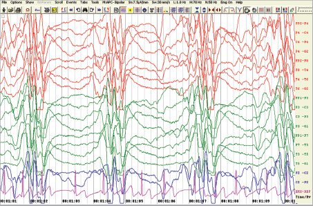
An 8-year-old male child with gradually worsening behavioral problems and cognitive decline for past 5 months. The child has frequent fall due to sudden jerks for the past 3 months. The child is currently admitted with generalized tonic–clonic seizure (GTCS). Electroencephalogram (EEG) was done and it is given in image below.
a. What is your provisional diagnosis?
b. What specific past history can give you a clue for diagnosis?
c. What investigations would you send to diagnose this condition?
d. What does the EEG reveal?
e. How would you treat?
Answer
| a. | SSPE (subacute sclerosing panencephalitis) |
| b. | Past history of measles |
| c. | CSF analysis and CSF for antimeasles antibody, and EEG |
| d. | Periodic complexes |
| e. | No definitive cure, role for isoprinosine (inosiplex), and interferon-α |
THG23-009
A 7-year-old female child presented with gradually progressive oromandibular and limb dystonia from 4 years of age. The child is now nonambulatory. The child does not have any myoclonic jerks or any seizures. Fundus examination revealed evidence of pigmentary retinopathy. MRI brain helped in clinching the diagnosis (Note: The MRI brain image could be given in the examination).
a. What is the most likely provisional diagnosis?
b. What is the gene implicated in the condition?
c. What is the mode of inheritance?
d. What is the expected neuroimaging finding which may have helped in clinching the diagnosis?
e. How would you confirm the diagnosis?
Answer
| a. | Pantothenate kinase-associated neurodegeneration (PKAN) |
| b. | PANK2 (pantothenate kinase 2 gene) |
| c. | Autosomal recessive |
| d. | Eye of the tiger sign |
| e. | Genetic testing (next-generation sequencing) |
THG23-010
A 10-month-old with prior normal development presented with afebrile encephalopathy for 2 days preceded by two episodes of vomiting and running nose for 1 days. The child was noted to have frequent dystonic posturing during the admission. MRI brain revealed that symmetrical basal ganglia and brainstem T2 hyperintensities with diffusion restriction. Magnetic resonance spectroscopy (MRS) which was done along with MRI brain gave a further etiological clue.
a. What is the most likely provisional diagnosis?
b. What is the expected MRS finding which has given the etiological clue?
c. How would you confirm the diagnosis?
d. How would you treat?
Answer
| a. | Leigh’s disease |
| b. | Double inverted lactate peak |
| c. | Genetic testing (whole exome sequencing plus or minus mitochondrial exome sequencing) |
| d. | Mitochondrial cocktail (carnitine, thiamine, riboflavin, biotin, and CoQ), antidystonia medications like trihexyphenidyl, physiotherapy, and stimulation |
THG23-011
A 2-year-old male child with background of developmental delay born to third-degree consanguineous parents presented with seizures and encephalopathy. Perinatal history was uneventful. The child was noted to have alopecia, oral candidiasis, and seborrheic dermatitis. There was history of elder sibling death at 5 months of age with refractory seizures. Retroviral serology was negative.
a. What is the most likely diagnosis?
b. What is the mode of inheritance?
c. What investigations would you do in confirming the diagnosis?
d. How would you treat?
e. What is the possible risk of recurrence in the next pregnancy?
Answer
| a. | Biotinidase deficiency |
| b. | Autosomal recessive |
| c. | Genetic testing, biotinidase enzyme assay in serum |
| d. | Biotin supplementation |
| e. | 25 percentage |
THG23-012
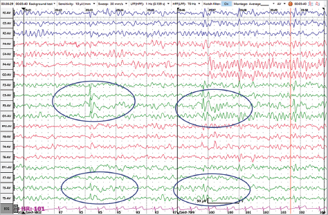
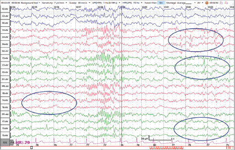
A 5-year-old child was brought with history of getting up in sleep followed by excessive salivation followed by gurgling sounds, then clonic jerking of right side of the body followed by a brief generalized tonic–clonic seizure. After regaining consciousness, he was not able to speak for few minutes.
a. What is the diagnosis?
b. What does EEG1 (as shown in image) show?
c. What does EEG2 (as shown in image) show?
d. What type of epilepsy is it?
e. What is the treatment for this child?
f. What are the drugs used in this condition?
Answer
| a. | Self-limiting childhood epilepsy with centrotemporal spikes (SELECTS)/benign childhood epilepsy with centrotemporal spikes (BECTS)/benign rolandic epilepsy |
| b. | Spike-wave discharges predominantly in left centrotemporal spikes |
| c. | Spike-wave discharge in bilateral centrotemporal region. There is increased frequency and exaggeration of spike-wave in sleep EEG compared to awake EEG |
| d. | Focal epilepsy (mostly benign) |
| e. | Reassurance that this is self-limiting and it usually remits by adolescence. Home management of seizures. |
| f. | Carbamazapine, Oxcarbazepine, Levetiracetam, Sodium valproate. |
THG23-013
A 6-year-old boy brought by parents after teacher complained episodes of staring in the classroom.
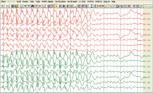
a. What does the EEG (shown in image) show?
b. What is the diagnosis?
c. What are types?
d. If this type of seizure occurs in toddler age group, which disease has to be ruled out?
e. What are the drugs used to treat this condition?
f. What are the drugs avoided in this condition?
g. Which is the bedside provocative test to aid in diagnosis?
Answer
| a. | EEG reveals 3 Hz spike-wave discharge suggestive of typical childhood absence epilepsy |
| b. | Childhood absence epilepsy |
| c. | Typical, atypical, and juvenile absences (typical absence occurs in childhood absence epilepsy, atypical absence occurs in Lennox–Gastaut syndrome, and juvenile absence occurs in juvenile myoclonic epilepsy) |
| d. | Glucose transporter 1 (GLUT1) deficiency |
| e. | Ethosuximide, sodium valproate, and lamotrigine |
| f. | Carbamazepine and phenytoin |
| g. | Hyperventilation |
THG23-014
A 9-month-old infant brought by mother with multiple episodes of sudden forward bending of body, arms, and legs lasting for 1 per 2 seconds, followed by a pause of 5–10 seconds followed by similar episode in clusters. Identify the EEG given here.
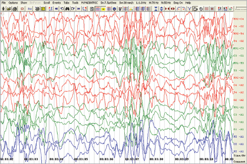
a. Identify the EEG (as shown in image) abnormality.
b. What is the diagnosis? What are its types?
c. What is the treatment?
d. What is the clinical manifestation?
Answer
| a. | EEG reveals hypsarrhythmia (chaotic mixture of asynchronous, very high voltage polymorphic delta slowing, and multifocal spikes) |
| b. | Diagnosis is West syndrome. Types are primary/idiopathic and secondary/symptomatic. |
| c. | Treatment is oral steroids/adrenocorticotropic hormone (ACTH) injection. When it is associated with tuberous sclerosis, treatment is vigabatrin. |
| d. | Infantile spasms (epileptic spasms) with or without developmental delay/regression |
THG23-015
A 3-year-old male child presented with status epilepticus. The child has cognitive issues and behavioral problems. The child had his first seizure which was fever provoked at 14 weeks of life following vaccination. Subsequently the child used to have fever provoked focal seizures every 2 months followed by GTCS after 1.5 years of age. The child has seizures once in every 2 months and started to have unprovoked seizures after 2 years of age along with fever provoked seizures intermittently. The child has intermittent brief blank episodes which last few seconds. Perinatal history is uneventful. MRI brain is normal.
a. What is the most likely diagnosis?
b. What is the most common gene implicated in this condition?
c. What is the most common mode of inheritance?
d. What antiseizure medications are used to control seizures in this condition?
e. What antiseizure medications are to be avoided in this condition?
Answer
| a. | Dravet syndrome |
| b. | SCN1A |
| c. | Autosomal dominant |
| d. | Sodium valproate, clobazam, levetiracetam, and stiripentol |
| e. | Phenytoin, carbamazepine, and lamotrigine |
THG23-016
A 4-year-old child presented with developmental delay, multiple seizure types in the form of jerks, vacant stares, and recurrent head drops and falls. EEG was done.
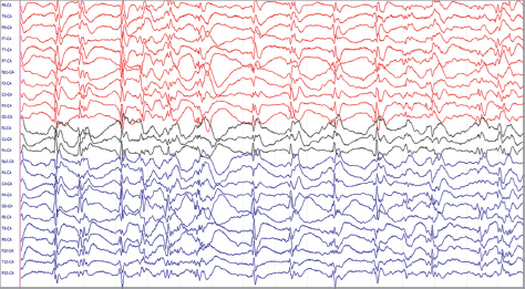
a. What does the EEG show in above image?
b. What is the diagnosis?
c. What is the triad of this condition?
Answer
| a. | EEG revealing diffuse slow spike-wave discharges <2.5 Hz occurring in repetitive sequences. |
| b. | Lennox–Gastaut syndrome |
| c. | Triad |
| i. | Psychomotor delay/regression |
| ii. | Refractory multiple seizure types (tonic seizure, myoclonic astatic, and atypical absences) |
| iii. | EEG findings suggestive of 1–2 Hz slow spike and wave discharge, generalized slowing during wakefulness, and polyspikes/paroxysmal fast rhythms during sleep |
THG23-017
A 9-month-old female child with macrocephaly and motor predominant delay started to have predominant motor neuroregression following a febrile illness along with significant dystonia. The child has refractory dystonia, retrocollis, and opisthotonic posturing. The child is born to second-degree consanguineously married parents.
a. What is the likely diagnosis?
b. What is the classical neuroimaging finding which you would expect in this condition?
c. How will you confirm the diagnosis?
d. How will you treat?
Answer
| a. | Type 1 glutaric aciduria |
| b. | Batwing appearance (bilateral frontotemporal atrophy with open opercula and widened Sylvian fissure), bilateral subdural hygromas may be present and bilateral symmetrical basal ganglia involvement |
| c. | Genetic testing (next-generation sequencing), tandem mass spectrometry (TMS), and urinary gas chromatography mass spectrometry (GCMS) |
| d. | Riboflavin and carnitine supplementation along with lysine-restricted diet |
THG23-018
A 9-year-old girl with decreased scholastic performance presented with seizures. The picture below displays what you have noticed in general examination.
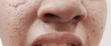
a. What is the likely diagnosis?
b. What is the mode of inheritance?
c. What does the above image show?
d. Which skin finding can be present from birth?
e. Mention two ophthalmological manifestations of this condition.
f. Mention two renal manifestations of this condition.
g. How many major and minor criteria are required for diagnosis?
h. What are the neuroimaging findings in this condition?
i. What drug is used to treat subependymal giant cell astrocytoma (SEGA)?
Answer
| a. | Tuberous sclerosis |
| b. | Autosomal dominant |
| c. | Adenoma sebaceum |
| d. | Ash leaf macule |
| e. | Retinal hamartomas and retinal achromatic patch |
| f. | Renal angiomyolipoma and renal cysts |
| g. | Two major criteria or one major criteria and two minor criteria |
| h. | Subependymal nodules, subependymal calcifications, cortical tubers, and sometimes SEGA |
| i. | Everolimus |
THG23-019
A 7-year-old boy presented with seizures. You notice this during your general examination which is displayed in the image. The father also has similar skin findings.
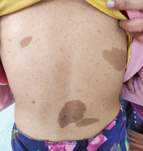
a. What is the most likely diagnosis?
b. What is the mode of inheritance?
c. What do you observe in the above image? When do you consider it significant?
d. What ophthalmological finding would you expect?
e. Name two skeletal manifestations of this condition.
f. Name two endocrine manifestations of this condition.
g. How many criteria need to be present to diagnose this condition?
Answer
| a. | Type 1 neurofibromatosis |
| b. | Autosomal dominant |
| c. | Cafe-au-lait macules (CALMs); six or more CALMs larger than 5 mm in greatest diameter in prepubertal individuals and larger than 15 mm in greatest diameter in postpubertal individuals |
| d. | Lisch nodules |
| e. | Sphenoid dysplasia, cortical thinning of long bones with or without pseudoarthrosis, and scoliosis |
| f. | Precocious puberty and pheochromocytoma |
| g. | If any two out of the seven features in the diagnostic criteria are present |
THG23-020
A 2-year-old female child with background history of developmental delay and cognitive issues presented with status epilepticus. The image below displays what you have noticed in general examination.
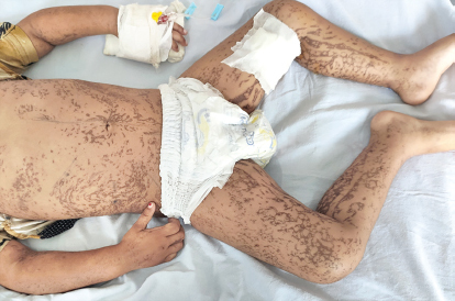
a. What is the most likely diagnosis?
b. What is the mode of inheritance?
c. What are the neurological manifestations?
d. What are the non-neurological manifestations?
Answer
| a. | Incontinentia pigmenti |
| b. | X-linked dominant |
| c. | Seizures, intellectual disability, hemiplegia, hemiparesis, spasticity, microcephaly, and cerebellar ataxia |
| d. | Alopecia, skin pigmentation, ophthalmological manifestation (neovascularization, microphthalmos, and strabismus). |
THG23-021
A 4-year-old child presented with right focal seizure. Child was noted to have port-wine stain on the left side of the face.
a. What is the most likely diagnosis?
b. What is the triad?
c. Where do you usually find the port-wine stain on the face?
d. How do you confirm the diagnosis?
e. What finding do you expect in the computed tomography (CT) brain?
f. Why ophthalmological evaluation is required in this condition?
g. Mention any two neurological manifestations of this condition other than seizure.
Answer
| a. | Sturge–Weber syndrome |
| b. | Facial port wine nevus, ipsilateral leptomeningeal angiomatosis, and glaucoma |
| c. | Region of the distribution of the ophthalmic division of trigeminal nerve |
| d. | MRI brain with contrast |
| e. | Tram-track calcification often with unihemispheric atrophy |
| f. | To detect glaucoma |
| g. | Hemiparesis, transient ischemic attacks, and headache |
THG23-022
A 3-week-old neonate presented with encephalopathy, refractory seizures since day 8 of life and baby is hypotonic. The baby has intractable hiccups and myoclonic jerks. MRI brain done revealed dysgenesis of corpus callosum. EEG done revealed suppression burst pattern.
a. What is the most likely diagnosis?
b. What investigation would help you in identifying this disorder and what is the cut-off?
c. What investigation can confirm the diagnosis?
d. What is the mode of inheritance?
e. What antiseizure medication must be avoided when you suspect this condition?
Answer
| a. | Neonatal glycine encephalopathy (nonketotic hyperglycinemia) |
| b. | CSF and serum glycine levels; CSF glycine/serum glycine ratio >0.08 |
| c. | Genetic testing/next-generation sequencing |
| d. | Autosomal recessive |
| e. | Sodium valproate |
THG23-023
A 2-year-old female child presented with autistic features and microcephaly. The child had normal development till 8 months of age. Gradually after that the child started to develop cognitive regression, autistic features, decline in head circumference, loss of purposeful hand movements, and an abnormal gait.
a. What is the most likely diagnosis?
b. What is the most common gene implicated in this condition?
c. What is the most common mode of inheritance?
d. What is the common cause of death?
Answer
| a. | Rett syndrome |
| b. | MECP2 gene |
| c. | X-linked dominant |
| d. | Cardiac arrhythmia |
THG23-024
A 2-year-old female child presented with cognitive regression and multiple episodes of seizures and repeated myoclonic jerks from the age of 8 months. The child was also noted to have visual disturbance. Fundus done revealed retinal pigmentary change and retinal artery attenuation. MRI brain done revealed diffuse cerebral atrophy and cerebellar atrophy.
a. What is the most likely diagnosis?
b. What is the mode of inheritance?
c. What skin biopsy finding would you expect?
d. What are the two investigations which can help to confirm the diagnosis?
Answer
| a. | Neuronal ceroid lipofuscinosis (NCL) |
| b. | Autosomal recessive |
| c. | Inclusion bodies |
| d. | Enzyme assay and next-generation sequencing/genetic testing |
THG23-025
A-3-year-old male child with normal development till 15 months of age started to have gradual motor neuroregression, stopped walking, standing, and sitting and became nonambulatory by 2.5 years of age. The child was noted to have spasticity and pyramidal signs. However, the ankle jerk was diminished. The child did not have any seizures. MRI brain was done and it is given below.
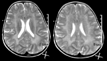
a. What is the most likely diagnosis?
b. What is the mode of inheritance?
c. What is the enzyme deficient?
d. What does the MRI brain image reveal?
e. What could be the reason for the possible diminished ankle jerk?
f. What is the recurrent risk in next pregnancy?
Answer
| a. | Metachromatic leukodystrophy (MLD) |
| b. | Autosomal recessive |
| c. | Arylsulfatase A |
| d. | Symmetrical periventricular white matter T2 hyperintensities sparing the subcortical U-fibers/tigroid appearance |
| e. | Peripheral neuropathy component (MLD is one of the leukodystrophy with peripheral neuropathy) |
| f. | 25% recurrence risk |
THG23-026
A 16-month-old male child presented with developmental stagnation since 7 months of age and cognitive regression since 10 months of age. Child was tremulous and was noted to have pallor and hypopigmented sparse hair. Child is predominantly on breastfeeds.
a. What is the most likely diagnosis?
b. What general examination in limbs would you specifically look for (other than what is mentioned in the question)?
c. What hematological abnormality would you expect?
d. How would you treat?
Answer
| a. | Infantile tremor syndrome due to nutritional B12 deficiency |
| b. | Knuckle hyperpigmentation |
| c. | Macrocytic anemia, hypersegmented neutrophils, and macroovalocytes in peripheral smear |
| d. | Intramuscular B12 therapy, nutritional rehabilitation, and good complementary feeds |
THG23-027
A 7-year-old male child presented with behavioral disturbance for past 8 months, progressive gait abnormality, and increasing stiffness along with vision and hearing disturbance. The child is currently bedbound with significant spasticity, pyramidal signs, unable to speak, on tube feeds, and lost bladder control. MRI brain done is given further.
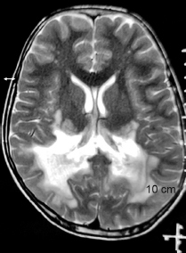
a. What is the likely diagnosis?
b. What is the mode of inheritance?
c. What is the gene implicated in this condition?
d. What general examination finding in skin should one specifically look for?
e. What is the endocrine manifestation of the disease which one should screen for?
Answer
| a. | X-linked adrenoleukodystrophy |
| b. | X-linked recessive |
| c. | ABCD1 gene |
| d. | Hyperpigmentation of skin |
| e. | Adrenal insufficiency |
THG23-028
An 8-month-old male infant presented with delayed head control, epileptic spasms from 5 months of age, and vision impairment. The child was noted to have central hypotonia and macrocephaly. Fundus revealed optic atrophy. MRI brain was done and the image is given below.
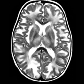
a. What is the most likely diagnosis?
b. What is the mode of inheritance?
c. What is the enzyme deficient?
d. What is the classical pattern of white matter involvement seen in MRI brain?
e. What is the MRI sequence which is displayed in the image?
f. What classical finding is expected in the MRS?
Answer
| a. | Canavan’s disease |
| b. | Autosomal recessive |
| c. | Aspartoacylase |
| d. | Diffuse symmetrical white matter T2 hyperintensities with prominent subcortical U-fiber involvement |
| e. | T2 sequence |
| f. | Giant NAA (N-acetylaspartate) peak |
THG23-029
A 15-month-old male child presented with motor and cognitive neuroregression noted since 6 months of age. The child was also noted to have seizures and vision regression since 8 months of age along with gradually increasing dystonia. The child was noted to have exaggerated startle to noise and was noted to have mild coarseness. There was no organomegaly.
a. What is the most likely diagnosis?
b. What is the mode of inheritance?
c. What fundus examination finding would you expect?
d. How will you confirm the diagnosis?
Answer
| a. | Tay–Sachs disease (GM2 gangliosidosis) |
| b. | Autosomal recessive |
| c. | Cherry-red spot (optic atrophy may also be present) |
| d. | Enzyme assay and genetic testing (next-generation sequencing) |
THG23-030
A 9-month-old male child presented with delayed head control, neuroregression, opisthotonic posturing, strabismus, and massive splenomegaly.
a. What is the most likely diagnosis?
b. What is the mode of inheritance?
c. What enzyme is deficient in this condition?
d. What are the three types this disorder is classified into?
e. How will you confirm the diagnosis?
Answer
| a. | Infantile neuronal Gaucher disease |
| b. | Autosomal recessive |
| c. | Beta-glucocerebrosidase (β-glucosidase) |
| d. | Type 1—Non-neuropathic form, type 2—acute neuropathic form, and type 3—chronic neuropathic form |
| e. | Enzyme assay and genetic testing |
THG23-031
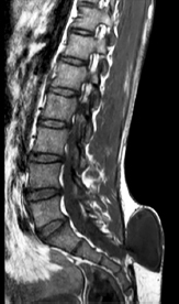
Above image is the sagittal section of MRI of the spine
a. Spot the abnormality (diagnosis).
b. List a few risk factors for development of this condition.
c. What are the indications and timing of surgery?
d. List two long-term complications (neurologic and non-neurologic).
e. What is Chiari crisis? Enlist symptoms of this condition and its management.
Answer
| a. | Lumbosacral meningomyelocele |
| b. | Dietary folic acid deficiency, drugs that antagonize folic acid, such as trimethoprim and the anticonvulsants carbamazepine, phenytoin, phenobarbitone, and valproate |
| c. | All myelomeningocele (MMC) have to be operated; ruptured sac with CSF leak—immediate surgery; otherwise in the first few days of life |
| d. | Neurogenic bladder and bowel, hydrocephalus, club feet, ankle and knee contractures, hip subluxation, and tethered cord |
| e. | Chiari crisis: 15% of infants with hydrocephalus and Chiari II malformation develop symptoms of hindbrain (brainstem) dysfunction, including difficulty feeding, choking, stridor, apnea, vocal cord paralysis, pooling of secretions, and spasticity of the upper extremities, which, if untreated, can lead to death. This Chiari crisis is caused by downward herniation of the medulla and cerebellar tonsils through the foramen magnum. |
THG23-032
A 10-year-old boy comes with acute onset right-sided weakness with aphasia while he was playing on the ground, he also reports two similar such self-improving episodes in the last month.
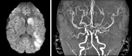
a. Identify the MRI sequences in above image.
b. What is the most likely diagnosis?
c. What is the radiological sign in angiography?
d. What are the conditions associated?
e. What are the treatment options?
Answer
| a. | Diffusion-weighted imaging (DWI) and magnetic resonance angiography (MRA) |
| b. | Moyamoya disease (MMD): Chronic, usually bilateral, vasculopathy of undetermined etiology characterized by progressive narrowing of the terminal intracranial portion of the internal carotid artery. |
| c. | Puff of smoke sign (= Moyamoya in Japanese) |
| d. | Trisomy 21, tuberculous (TB) meningitis, radiation, neurofibromatosis type 1, sickle cell disease, Noonan syndrome, Alagille syndrome, and Williams syndrome |
| e. | Treatment: |
| • | Medical: Aspirin (to prevent recurrent strokes) |
| • | Surgical (definitive) |
| • | Direct revascularization: Superficial temporal artery to middle cerebral artery bypass |
| • | Indirect revascularization: Encephaloduroarteriosynangiosis (EDAS) or encephalomyosynangiosis (EMS) |
THG23-033
Analyze the case vignette, read the MRI (as shown in below image) and answer the questions below:
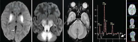
a. Describe the MRI findings.
b. What MRS finding you look for in this case?
c. What characteristic EEG finding is described in this disease?
d. Name the metabolic testing findings in this disorder.
e. What are the four clinical phenotypes of this disorder?
f. What is the treatment (long-term) (do not mention acute crisis treatment)?
Answer
| a. | Classic appearing maple syrup urine disease (MSUD) edema in cerebellum, brain stem, and basal ganglia (diffusion restriction) |
| b. | MRS: Broad peak at chemical shift of 0.9 ppm (TE-26 ms) |
| c. | EEG—Comb-like rhythm |
| d. | Elevated branched-chain amino acids (BCAAs) |
| e. | Clinical phenotypes: Classic, intermediate, intermittent, and thiamine responsive |
| f. | Rx (long-term): Diet restricted in leucine, isoleucine, and valine, while avoiding deficiencies of essential amino acids, fatty acids, and micronutrients; trial of thiamine; liver transplantation. |
THG23-034
A 13-year-old boy presents with a few months of progressively worsening urinary urgency and frequency of insidious onset with few months of low back pain and difficulty in walking. He is found to have asymmetry in feet with calf atrophy and arching of feet and clawing of tows on the right with absent right ankle jerk. Past history is remarkable for spine surgery during the neonatal period.
a. What is the possible diagnosis?
b. Name two spinal congenital malformations that increase the risk of development of this condition.
c. List three cutaneous manifestations/markers associated with this condition.
d. What is management of this condition?
Answer
| a. | Tethered cord syndrome |
| b. | Meningomyelocele, diastematomyelia, syringomyelia, lipoma of the cord, and fatty filum terminale |
| c. | Hemangioma, discoloration of the skin, pit, lump, dermal sinus, or hairy patch |
| d. | Surgery: Detethering of the cord |
THG23-035
An 8-month-old boy is brought for concern of abnormal head shape. He is a preterm neonatal intensive care unit (NICU) graduate and has motor developmental delay. His head circumference and anterior fontanel are normal for age with an unremarkable neurological examination.
a. Mention two other risk factors for development of deformational plagiocephaly in an infant.
b. Mention two techniques for prevention.
c. How do you differentiate between deformational plagiocephaly and craniosynostosis (list three points)?
d. What are the treatment options available for this condition?
Answer
| a. | Male sex, firstborn child, congenital torticollis, prolonged bottle feeding, sleep position—supine, less tummy time, sleeping with head to same side |
| b. | Consistently alternating sleeping position, tummy time for 10–15 minutes at least three times a day |
| c. | Differentiating features: |
| Deformational plagiocephaly | Craniosynostosis |
| Normal head shape at birth | Head shape abnormality at birth |
| Parallelogram shape | Trapezoid shape |
| Ipsilateral ear anteriorly displaced | Ipsilateral ear posteriorly displaced |
| No palpable bony ridges | Palpable bony ridges |
| d. | Repositioning and physiotherapy (RPPT), molding therapy (helmet therapy), and surgery |
THG23-036
An 18-month-old developmentally normal child develops a first episode of generalized tonic–clonic convulsion with fever, lasting for 10 minutes. His neurological examination is normal.
a. What are the indications to perform a lumbar puncture in such a situation?
b. Is an MRI scan of the brain indicated?
c. What are the indications for febrile seizure prophylaxis?
d. What are the major risk factors for recurrence?
e. What are the risk factors for epilepsy (development of unprovoked seizures in the future)?
Answer
| a. | Lumbar puncture should be performed for all infants younger than 6 months of age who present with fever and seizure, or if the child is ill-appearing or at any age if there are clinical signs or symptoms of concern. Optional in infant 6–12 months of age who is unvaccinated with Haemophilus influenzae type b (Hib) or Streptococcus pneumoniae and in those pretreated with antibiotics. |
| b. | Not indicated; can be considered in patients with complex features (prolonged (>15 min), is focal, and/or reoccurs within 24 hours). |
| c. | Complex febrile seizures in children with neurological deficit, strong family history of epilepsy and recurrent simple or complex febrile seizures, febrile status epilepticus, and febrile seizure with a frequency higher than once per quarter |
| d. | Major risk factors for recurrence: Age <1 year; duration of fever <24 hours; low-grade fever (100.4–102.2°F). |
| e. | Presence of neurodevelopmental abnormalities; complex febrile seizures especially focal; family history of epilepsy. |
THG23-037
A 2-year-old boy presents with a 2-day history of high-grade fever, focal seizures, and altered sensorium with signs of meningeal irritation.
a. What are the contraindications to perform a lumbar puncture in this patient?
b. What are the indications for neuroimaging in this scenario?
c. What are the clinical and radiological features to suspect herpes simplex virus (HSV) encephalitis in this patient?
d. List few neurological complications of acute pyogenic meningitis.
e. How does antibiotic pretreatment affect results of cerebrospinal fluid (CSF) analysis?
f. How does a traumatic lumbar puncture influence interpretation of CSF analytes?
Answer
| a. | Contraindications to performing a lumbar puncture include suspected mass lesion of the brain, especially in the posterior fossa or above the tentorium and causing shift of the midline; suspected mass lesion of the spinal cord; symptoms and signs of impending cerebral herniation in a child with probable meningitis; skin infection at the site of the lumbar puncture; and thrombocytopenia with a platelet count <20 × 109/L. |
| b. | If disk edema on fundus examination or focal findings suggest a mass lesion, a head CT should be obtained before proceeding with lumbar puncture to prevent uncal or cerebellar herniation as the CSF is removed. In the absence of these findings, routine head imaging is not warranted. In this child, focal seizures indicate the need for neuroimaging. |
| c. | Focal seizures with high-grade fever and encephalopathy, frontotemporal involvement on neuroimaging |
| d. | Subdural effusion/empyema, brain abscess, ventriculitis, vasculitis and infarction, sinovenous thrombosis, cerebral edema and raised ICP, and acute hydrocephalus |
| e. | CSF obtained from children with bacterial meningitis, after the initiation of antibiotics, may be negative on Gram stain and culture. Pleocytosis with a predominance of neutrophils, elevated protein level, and a reduced concentration of CSF glucose usually persist for several days after the administration of appropriate intravenous antibiotics. |
| f. | Interpretation of CSF leukocytes and protein concentration are affected by lipopolysaccharides (LPs) that are traumatic, although the Gram stain, culture, and glucose level may not be influenced. |
THG23-038
A 6-month-old infant presents with history of rapidly enlarging head size since birth.
a. Name the two most common causes of communicating hydrocephalus in infants.
b. List any two causes of noncommunicating hydrocephalus.
c. What is the mechanism of setting-sun sign in infants with hydrocephalus?
d. In an infant with hydrocephalus and prominent scalp veins, which diagnosis does an audible cranial bruit suggest?
e. Mention two findings on eye examination you will look for while examining an infant with hydrocephalus.
Answer
| a. | Neonatal intraventricular hemorrhage (IVH) and neonatal meningitis |
| b. | Aqueductal stenosis, tumors, hematoma, abscess, vein of Galen malformation, Dandy–Walker malformation, and Chiari malformation |
| c. | The setting-sun phenomenon is an ophthalmologic sign in young children resulting from upward gaze paresis. In this condition, the eyes appear driven downward, the sclera may be seen between the upper eyelid and the iris, and part of the lower pupil may be covered by the lower eyelid. Pathogenesis of this sign is not well understood, but it seems to be related to aqueductal distension with compression of periaqueductal structures secondary to increased ICP. |
| d. | Vein of Galen malformation/arteriovenous malformation |
| e. | Chorioretinitis, papilledema, optic atrophy, cataract, and glaucoma |
THG23-039
A 12-year-old boy is rushed to the emergency room (ER) after sustaining a road traffic accident and injury to the back. His Glasgow Coma Scale (GCS) is 15/15, cranial nerve examination is unremarkable with normal upper extremity power, but is noted to have a power of 0/4 in both lower extremities with ankle and knee areflexia and total loss of all sensation up to the umbilicus. On attempting to rise up from supine, his umbilicus moves upward.
a. To which spinal level do you localize the lesion?
b. What is clinical sign described called?
c. List two important steps of management (treatment) of this patient.
d. In spinal cord lesions above T6 level, what important complication (other than cervical spine/brainstem injury and resulting respiratory failure) is to be anticipated? What are its symptoms and signs?
e. If this child also has weakness in shoulder abduction and flexion, what would you monitor the patient for?
Answer
| a. | Above T10 |
| b. | Beevor sign |
| c. | Immobilization and spine stabilization, IV steroids, and spine decompressive surgery |
| d. | Autonomic dysreflexia: A sudden, exaggerated reflexive increase in blood pressure in response to a stimulus, usually bladder or bowel distension, originating below the level of the neurological injury, severe headache, bradycardia, and facial flushing along with pallor, cold skin, and sweating in the lower part of the body. |
| e. | Diaphragmatic weakness and respiratory failure |
THG23-040
A 5-year-old developmentally normal boy presents with sudden onset of weakness of left side of the face and limbs since the past 4 hours. This occurred suddenly in school while he was seated in class. He reported having such an episode earlier in the morning but it had resolved in few minutes. The parents note a history of a self-limiting viral upper respiratory infection (URI) 1 week back. He does not complain of a headache or vomiting and is in a clear sensorium walking with a limp. He has never had any neurological concerns in the past.
a. Which imaging modality is preferred to identify the pathology in this patient?
b. What are the investigations you would like to perform?
c. Give two other differential diagnoses for acute onset hemiparesis in a child.
d. What is the most likely etiology in this child?
e. What are the treatment options?
Answer
| a. | MRI brain (DWI) |
| b. | MR/CT angiography, Echo, hematological and thrombophilia workup |
| c. | Todd’s paresis (postictal paralysis), demyelination, migraine, encephalitis, and tumor |
| d. | Focal cerebral arteriopathy/transient cerebral arteriopathy |
| e. | Neuroprotection, aspirin, and rehabilitation |
THG23-041
A 6-year-old girl presents with sudden onset weakness of the left side of the face with deviation of the angle of mouth to the right. She recovers with treatment in 3 weeks.
a. What is the most likely diagnosis?
b. How would you differentiate a lower motor neuron (LMN) lesion from an upper motor neuron (UMN) lesion in this case?
c. Treatment of this condition and specific precaution to be observed.
d. Mention the most important complication that may occur occasionally during recovery.
Answer
| a. | Bell palsy (acute idiopathic peripheral facial nerve palsy) |
| b. | Involvement of upper half of the face (raising of eyebrows, closure of eyelids) in addition to the lower half indicates an LMN lesion |
| c. | Oral corticosteroid (prednisone (1 mg/kg/day for 1 week, followed by a 1-week taper) with/without acyclovir for 10 days, protection of the cornea with methylcellulose eye drops or an ocular lubricant to prevent exposure keratitis. |
| d. | Nerve regrowth may be misdirected and result in synkinesis, where activation of one muscle group may produce activation of another inappropriate muscle group; blinking may result in mouth twitching, smiling may cause eye blinking, and lacrimation (crocodile tears) may occur while eating. |
THG23-042
A 6-month-old male infant is admitted with second episode of pneumonia in the last 2 months. He has a history of motor developmental delay with complete lack of head control, her social and language milestones are normal. He is found to be floppy without any elicitable deep tendon reflexes. He is the firstborn child to consanguineous parents with an unremarkable antenatal, perinatal, and family history.
a. Which important physical examination finding would support a diagnosis of spinal muscular atrophy (SMA) in this child?
b. What is the diagnostic test?
c. Mention three new drugs approved for this condition.
d. Name the gene that determines prognosis in SMA.
e. Mention two muscle groups selectively spared in SMA.
f. Mention two long-term complications.
Answer
| a. | Tongue fasciculations |
| b. | PCR-based testing of the SMN1 gene for deletions/duplications [multiplex ligation-dependent probe amplification (MLPA)] |
| c. | Nusinersen (Spinraza), risdiplam (Evrysdi), and onasemnogene abeparvovec (Zolgensma) |
| d. | SMN2 gene (the number of copies determines the severity) |
| e. | Extraocular muscles and sphincters |
| f. | Recurrent aspiration pneumonia/respiratory infections, respiratory failure, feeding difficulty due to bulbar dysfunction, scoliosis, gastroesophageal reflux, contractures, fractures, and hip dislocation |
THG23-043
An 8-year-old boy presents with 1 day of lethargy and seizures following 3 days of an acute self-limited viral URI.
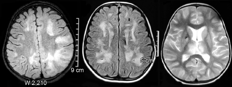
a. What is the diagnosis?
b. Name the antibody associated with this entity.
c. Mention two common neurological structures affected other than brain.
d. What are the treatment options?
Answer
| a. | Acute disseminated encephalomyelitis (ADEM) |
| b. | Anti-MOG (myelin oligodendrocyte glycoprotein) antibody |
| c. | Optic nerve and spinal cord |
| d. | IV steroids, intravenous immunoglobulin (IVIg), and plasmapheresis |
THG23-044
A 1-year-old boy presents with fever, seizures, and altered sensorium.
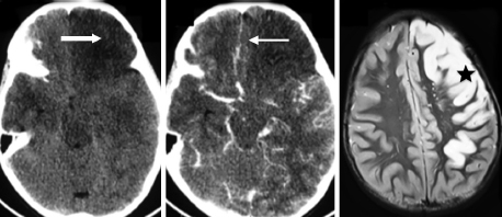
a. What is the diagnosis?
b. What is the confirmatory test?
c. What is the EEG finding?
d. What is the treatment?
Answer
| a. | Probable HSV encephalitis |
| b. | CSF HSV DNA PCR |
| c. | Periodic lateralized epileptiform discharges (PLEDs) |
| d. | IV acyclovir × 21 days |
THG23-045
A 10-year-boy with chronic kidney disease (CKD) presents with seizures and altered sensorium.
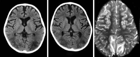
a. What is the diagnosis?
b. What are the radiological differential diagnoses?
c. Discuss the pathophysiology.
d. What are the risk factors?
e. What is the treatment?
Answer
| a. | Posterior reversible encephalopathy syndrome (PRES) |
| b. | Differential diagnoses: Hypoglycemic brain injury; cerebral sinus venous thrombosis; posterior circulation stroke. |
| c. | Pathophysiology: Hypertension—hyperperfusion hyopethesis |
| d. | Risk factors: Hypertension, renal disease, rheumatologic/collagen vascular disorder, post-transplantation, immunosuppressive, and chemotherapeutic drugs. |
| e. | Treatment: Gradual reduction of blood pressure; treating the underlying etiology/withdrawing the offending agent and antiseizure medications. |
THG23-046
Read the case vignette and MRI and answer the questions below:
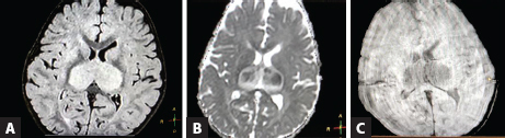
a. Identify the sequences given in image A to C
b. What is the diagnosis?
c. Name the serum biochemical markers of this disorder.
d. Mention two important viruses causing similar clinical and radiological picture
e. What is the treatment?
Answer
| a. | A: Fluid-attenuated inversion recovery (FLAIR); B: Apparent diffusion coefficient (ADC); and C: Gradient-recalled echo (GRE) |
| b. | Acute necrotizing encephalopathy of childhood (ANEC) |
| c. | Elevated liver enzymes, low platelet counts, elevated serum and CSF interleukin 6 (IL-6); severe cases can have systemic inflammatory response syndrome-acute kidney injury (SIRS-AKI), acute liver failure (ALF), disseminated intravascular coagulation (DIC), and shock |
| d. | Dengue, Japanese encephalitis, herpes group of viruses, and coronavirus disease 2019 (COVID-19) |
| e. | IV pulse steroids, IVIg, and tocilizumab |
THG23-047
A 14-year-old girl with fever and headache of 1 week duration.
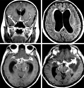
a. What are the imaging findings (as shown in above image)?
b. What is the diagnosis?
c. Explain the complete treatment plan.
Answer
| a. | Hydrocephalus, tuberculomas, basal exudates, left basal ganglia infarction, and vasculopathy/vasculitis |
| b. | TB meningitis with complications |
| c. | Antitubercular therapy (ATT) (12 months) + Steroids + Ventriculoperitoneal (VP) shunt |
THG23-048
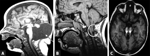
Spot the MRI findings and diagnosis given in image A to C
Answer
| A: | Large posterior fossa cyst with cerebellar agenesis: Dandy–Walker malformation |
| B: | Tonsillar herniation: Arnold–Chiari malformation |
| C: | Molar tooth sign: Joubert syndrome |
THG23-049
Read the following MRI of a 2-year-old boy with seizures and developmental delay and answer the questions below:
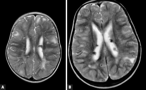
a. Identify the findings in image A and B.
b. What is the diagnosis?
c. What are the cutaneous manifestations?
d. Name two non-neurological manifestations.
e. What is the targeted pharmacologic treatment available?
Answer
| a. | A: Cortical tubers; B: Subependymal nodules |
| b. | Tuberous sclerosis |
| c. | Hypomelanotic macules (ash-leaf); shagreen patch, angiofibromas, and subungual fibromas |
| d. | Cardiac rhabdomyoma, renal angiomyolipomas/cysts, retinal achromatic patches/hamartomas, and lymphangioleiomyomatosis |
| e. | Mammalian target of rapamycin (mTOR) inhibitors: Everolimus and sirolimus |
THG23-050
Below is the noncontrast CT (image A) and contrast MRI brain of a 14-year-old boy who presented with progressive headache for 2 months:
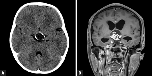
a. Describe the imaging features in above image.
b. What is the diagnosis?
c. Explain the common clinical symptoms at presentation.
d. What is the treatment option?
Answer
| a. | Suprasellar lesion showing solid and cystic components; solid component enhances with contrast; CT shows calcifications |
| b. | Craniopharyngioma |
| c. | Craniopharyngioma often presents with endocrinologic abnormalities such as growth failure and delayed sexual maturation. Visual changes can occur and may include decreased acuity or visual field abnormalities. |
| d. | Surgery is the primary treatment modality with gross total resection curative in small lesions. |