VVK20-001
a. What major problems can occur in using a cast?
b. What are the signs that the cast is too tight?
c. How do you prevent a tight cast?
d. How long does it take for a cast to harden?
Answer
a. A cast that is too tight can cause serious neurovascular compromise.
b.
- • Pain when moving the figures/toes
- • Pallor, or pale/white distal part due to lack of blood supply
- • Paresthesias and possible numbness due to nerve compression and lack of circulation
- • Paralysis due to loss of nerve function.
c.
- • Use sufficient padding
- • Split the cast on both sides and spread the cast to allow for swelling (bivalving)
- • Split the padding to the skin
- • Elevate the limb to prevent dependent edema and inflammatory swelling
d. Plaster cast 2–8 minutes Fiber glass 4–5 minutes
VVK20-002
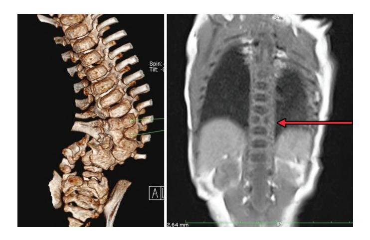
a. What is the radiological anomaly?
b. What is the incidence?
i. 1:300
c. What can be the clinical abnormality associated with above abnormality?
Answer
- a. Hemi vertebrae-fusion defect of marked vertebrae
- b. B ( 1:3000)
- c. Kyphosis- DORSAL
Scoliosis
Lordosis
VVK20-003
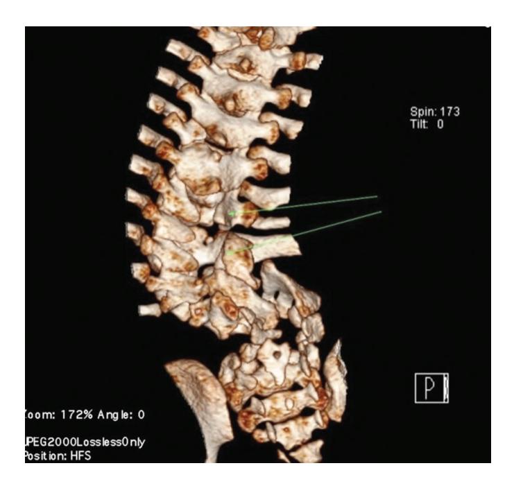
a. What is the radiological finding?
b. What is the diagnosis?
c. What are common physical signs of above diagnosis?
d. What is the single most important prenatal/antenatal factor leading into above abnormality?
Answer
- a. Unfused posterior element of marked vertebrae
- b. Spina bifida
c.
- i. Leg weakness and paralysis
- ii. Orthopedic abnormalities, i.e. club foot, hip dislocation, scoliosis
- iii. Bladder and bowel control problems, including incontinence, urinary tract infections, and poor kidney function
- iv. Pressure sores and skin irritations
- v. Abnormal eye movement
- c. Not having enough folic acid during pregnancy.
VVK20-004
A 10-year-old presents with history of weakness in the legs; low back pain; scoliosis; and incontinence.
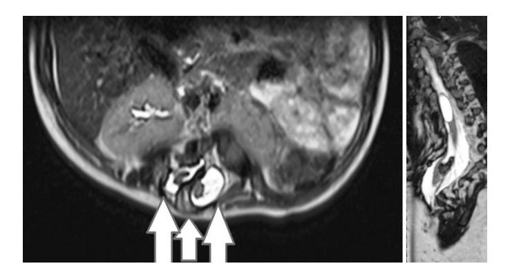
a. What is the radiological finding?
b. What is the diagnosis?
c. What is the treatment?
Answer
- a. Bony spur noted between 2 divided cords
- b. Diastematomyelia
- c. Surgery-decompression (surgery) of neural elements and removal of bony spur.
VVK20-005
Attached are pre and 2 weeks postoperative X-rays of a baby with development dysplasia of hip.
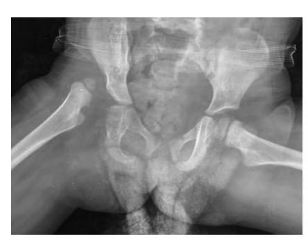
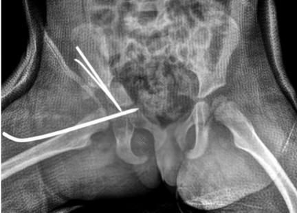
a. Which of the following is a risk factor for DDH? True/False
i. First born
b. Enlist 2 orthopedic conditions associated with DDH
c. Name 2 screening tests on examinations used to confirm the diagnosis.
Answer
- a.
- i. True
- ii. False
- iii. True
- iv. True.
- b. Torticollis, congenital hyperextension of knee, club foot, skull and facial deformities.
- c. Ortolani
Barlows
Note: Please practise ortolani and Barlows manuver as these can be asked for demonstration in observed OSCE stations.
VVK20-006
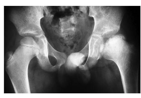
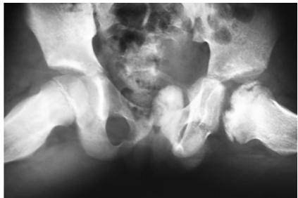
a. What are the radiological findings?
b. What is the diagnosis?
c. What is the pathological process?
d. True false
i. Most commonly occur in children of 5–9 years age.
Answer
- a. Uniformly increased density of left femoral head.
- b. Legg-calve-perthes disease.
- c. Idiopathic avascular necrosis of femoral head.
- d.
- i. True
- ii. False
- iii. False
- iv. False.
VVK20-007
Attached are MRI images of an 8-year-old female.
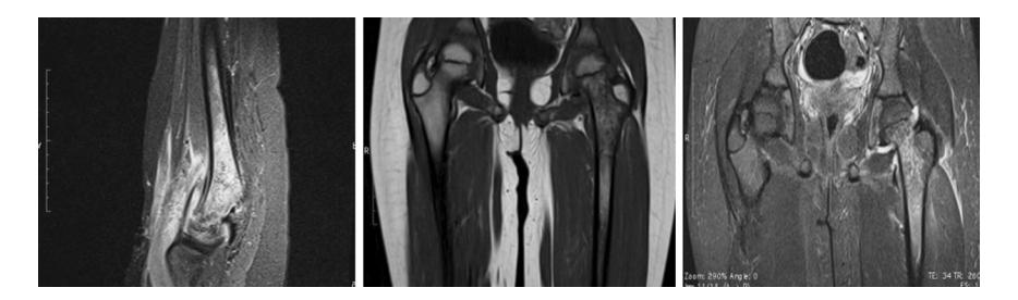
a. What are the radiological findings?
b. What is the diagnosis?
c. What is the treatment?
d. Enlist 3 complications associated with above diagnosis.
Answer
- a. Heterogenous hyperintense signal of neck and upper end of femur.
- b. Osteomyelitis.
- c. Antimicrobial therapy.
- d.
- • Septic arthritis
- • Distant seeding leading into pneumonia, septic pericarditis, etc.
- • Pathological fracture
- • Physeal bars
- • Recurrent infections.
VVK20-008
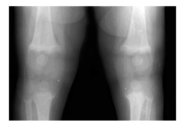
a. What is the diagnosis?
b. This infant is 10 months old, what is the most likely sub type?
c. What is the earliest sign of this disorder?
d. What is the first radiological change that occurs in response to specific therapy?
e. How could this have been prevented?
f. What are the non-specific urinary findings in this disorder? (at least 2).
Answer
- a. Rickets
- b. Vitamin D deficiency
- c. Craniotabes
- d. Appearance of provisional zone of classification
- e. Supplement of 400IU of vitamin D from birth
- f. Generalized aminoaciduria
- • Glycosuria
- • Phosphaturia
- • Elevated urinary citrate
- • Impaired renal acidification.