THG28-001
A 10-month-old child presents with history of bleeding per rectum, incessant cry, and refusal of feeds for one day duration.
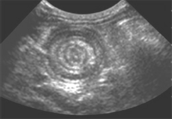
a. What is the probable diagnosis?
b. What is classical triad of symptoms with the most common cause?
c. Name the signs seen on ultrasound.
d. What are the different lead points of the condition?
e. Mention the treatment modalities.
Answer
| a. | Intussusception (ileocolic) |
| b. | Intermittent colicky pain abdomen (incessant cry), vomiting, and bleeding per rectum. Peyer’s patch hypertrophy is the most common cause. |
| c. | Doughnut sign/pseudokidney sign |
| d. | Meckel’s diverticulum/polyp/duplication cyst/lymphoma/Henoch–Schönlein purpura (HSP) |
| e. | Pneumatic reduction/hydrostatic reduction/surgical reduction |
THG28-002
A 3-year-old playful child presents with history of bilious vomiting, pain abdomen which is continuous dull aching, refusal of feeds of 1 day duration. The CT abdomen is shown in given image.
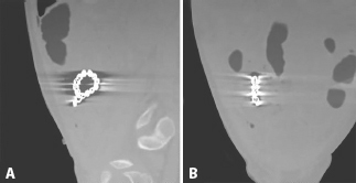
a. What are the clinical signs to be elicited?
b. What are the findings seen in the CT abdomen supine?
c. After identifying the nature of findings, how would you advice the parents?
d. What is the treatment offered?
Answer
| a. | Abdominal distension/visible intestinal loops/tenderness in the right iliac fossa |
| b. | Dilated small bowel (ileal-characterless loops) loops/foreign body (chain of magnetic beads in right iliac fossa/distal collapsed bowel/no signs of pneumoperitoneum |
| c. | Chain of magnetic beads is the foreign body |
| d. | Single magnetic bead is harmless as it will expel with normal bowel transit. Multiple magnets as they travel down the bowel will tend to attract each other with changing directions and attract adjacent bowel loops causing pressure necrosis. If the child is attended late, it would have caused gangrene bowel with perforation. Surgical removal of the foreign body with or without resection of the bowel depending on the presentation. |
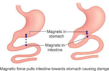
Single and multiple magnets in the intestine.
THG28-003
An 8-year-old male child presents with pain and redness in the left scrotum for 1 day duration. History of cycling and trivial trauma present. He had similar episodes in the past treated with symptomatic relief.
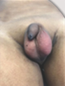
a. Name the surgical emergency expected.
b. Name the key clinical sign to look for.
c. What is the differential diagnosis for this condition?
d. What are the systemic medical conditions which can have this?
Answer
| a. | Acute scrotum—torsion left testis to be ruled out |
| b. | Absent cremasteric reflex is the sign suggestive of torsion testis. |
| c. | Torsion testis, torsion-appendage testis, epididymo-orchitis, and traumatic hematocele are the differential diagnoses. |
| d. | HSP/mumps orchitis can have acute scrotum as part of systemic symptoms. |
THG28-004
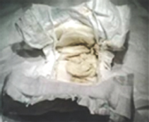
A 6-month-old infant presents with painless abdominal distension, excessive sleepiness, and yellowish discoloration of urine and body for 4 months duration. History of passing clay-colored stools (seen as in given image).
a. List the clinical findings seen in the infant.
b. Give any three differential medical diagnoses.
c. Name the surgical diagnosis primarily to be ruled out.
d. List the supportive investigations needed.
e. Name the surgical procedure which can be offered.
Answer
| a. | Icteric child with acholic stool/abdominal distension with flank fullness (probably ascites)/sleepy child (hepatic encephalopathy)—clinically all are pointers to features of liver failure. |
| b. | Neonatal hepatitis/toxoplasmosis, others (syphilis, hepatitis B), rubella, cytomegalovirus, herpes simplex (TORCH) infections/sepsis are the three important differential diagnoses to be ruled out in workup. |
| c. | Extrahepatic biliary atresia (EHBA) should be the primary diagnosis. |
| d. | USG abdomen—to see for absent gallbladder and triangular cord sign/hepatobiliary iminodiacetic acid (HIDA) scan—for drainage of nuclear material into duodenum in 24 hours suggesting presence of patent biliary tree/peroperative cholangiogram is the definitive investigation to rule out EHBA. |
| e. | Kasai portoenterostomy is the suggested treatment for EHBA to ensure bile drainage at initial 2 months of life before liver cirrhosis sets in. Liver transplant is the only surgical option for children with hepatic failure. |
THG28-005
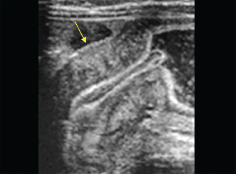
A 2-month-old male infant presented with history of nonbilious projectile vomiting of 5 days duration. Mother finds the child always hungry to drink milk but vomits every single feed. Mother also noticed some fullness in the upper abdomen. Child has reduced urine output. Ultrasound picture is given below:
a. Name the condition suspected in a child with “nonbilious projectile vomiting”.
b. What are the clinical findings you would expect?
c. Name the electrolyte abnormality exclusive for this condition.
d. What is the ultrasonography (USG) finding with the diagnostic criteria?
e. What is the differential diagnosis of the condition?
Answer
| a. | Infantile hypertrophic pyloric stenosis (IHPS). It is classical to present in first born male child at 6 weeks of life. |
| b. | The visible gastric peristalsis/pyloric mass or olive can be palpable. |
| c. | Hyponatremic hypokalemic hypochloremic metabolic alkalosis with paradoxical aciduria in severe dehydration when all the solutes are being lost entirely, instead of preserving H+ ions, kidney loses it, causing paradoxical aciduria to conserve sodium. |
| d. | Pyloric mass is detected/thickened pyloric canal—17 mm length/4 mm thickness is diagnostic of IHPS to be operated upon. |
| e. | Severe gastroesophageal reflux disease (GERD)/pyloric atresia (only in newborn)/pyloric web are differential diagnoses to IHPS. |
THG28-006
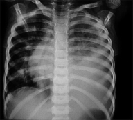
A 7-year-old child was brought with history of cough, cold, and fever of 7 days duration. Child had received oral antibiotics and cough syrup for 4 days on an outpatient basis. On examination, child has tachypnea, nasal flaring, and moist cough. On auscultation, bronchial breath sounds are heard in the left hemithorax.
a. What is the diagnosis?
b. Define the stages of evolution.
c. Name the most common organisms involved in the condition.
d. List the standard criteria to differentiate the uncomplicated from complicated one.
e. What are the modalities of treatment in each stage?
Answer
| a. | Empyema thoracis left |
| b. | Stage 1—exudative stage/stage 2—fibrinopurulent/stage 3—organized stage |
| c. | Staphylococcus aureus/Streptococcus pneumoniae are the most common organisms involved in pneumonia. |
| d. | Pleural fluid aspiration: Biochemical analysis criteria—PH <7.2, protein—2.5 mg/dL, lactate dehydrogenase (LDH) >1,000 units/L, sugar—<60 mg/dL—suggestive of exudative effusion in complicated pneumonia. |
| e. | Modalities of treatment in each stage: |
| • | Antibiotics and supportive care in stage 1 |
| • | Intercostal drainage with or without fibrinolytics in stage 2 |
| • | Thoracoscopic [Video-assisted thoracoscopic surgery (VATS)] or open decortication in stage 3. |
THG28-007
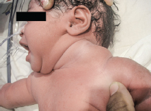
A newborn presented with clinically visible swelling in the left side of the neck. Crying well and no evidence of respiratory distress. Swelling is transilluminant on examination.
a. Identify the anomaly in the newborn.
b. Name the defect in embryology.
c. What are the modalities of treatment?
d. Name the agent commonly used in intralesional treatment.
e. Where is it derived from?
Answer
| a. | Cystic hygroma/lymphangioma of the neck—most common sites being neck, axilla, groin, and retroperitoneum. |
| b. | Abnormality of jugular lymph sac development leads to cystic hygroma in neck. |
| c. | Intralesional sclerosant injection/surgical excision of the lesion/laser diathermy ablation of the cyst wall. |
| d. | OK-432 or Picibanil is the sclerosant commonly used to inject for lymphangioma. Other agents are hypertonic saline, bleomycin, absolute alcohol, etc. |
| e. | Picibanil is derived from low virulent strain of Streptococcus pyogenes. It is not available in India. Bleomycin is commonly used in India. |
THG28-008
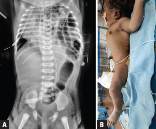
A 12-hour-old male newborn presented with clinically visible fullness of the chest and scaphoid abdomen. He was depressed at birth and hence ventilated immediately. He had passed meconium. Mother had no antenatal ultrasounds done. X-ray chest and abdomen is shown below.
a. Identify the anomaly in the X-ray newborn.
b. Name the defect associated and its name.
c. Name the poor prognostic factor in clinical outcome (antenatal as well as postnatal).
Answer
| a. | Congenital diaphragmatic hernia (CDH): Bowels ascending into thorax cause mediastinal shift with chest fullness/scaphoid abdomen clinically. X-ray shows bowel loops in the left hemithorax with mediastinal shift to right/minimum lung expansion seen in the right hemithorax/only single loop seen in the abdomen. Nasogastric tube seen in abdomen (stomach in abdomen—not thorax). |
| b. | Classical posterolateral defect in the diaphragm—Bochdalek hernia (CDH). |
| c. | Antenatally CDH is detected by 20 weeks anomaly scan and lung hypoplasia is poor prognostic factor to be monitored in the USG. The lung/head ratio <1 has poor prognosis denoting lung hypoplasia. LHR ratio (observed/expected lung-head ratio) is the newer criteria to prognosticate antenatally—<45% should be given guarded prognosis. Postnatally persistent pulmonary hypertension (PPHN) requiring higher supportive management has poor prognosis. |
THG28-009
A 3-hour-old neonate noticed to have swelling and redness in the left forearm at birth. On examination, has got pitting edema in some areas. Distal vascularity of the left limb is good. Minimal movement restriction in the arm involved.
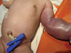
a. Identify the lesion and name the associated syndrome.
b. What is the clinical presentation in the newborn period?
c. List the various modalities of treatment offered.
d. Is there a role for topical application?
Answer
| a. | Hemangioma of the left forearm/Kasabach–Merritt syndrome (KMS) |
| b. | Hemangioma with thrombocytopenia will be the clinical presentation. Consumptive coagulopathy/hemolysis/disseminated intravascular coagulopathy are the severe forms of presentation. |
| c. | Supportive care—limb elevation/minimal handling |
| Treatment—systemic medications to reduce the vascularity to the lesion and prevent episodes of bleeding. Natural course of hemangioma is to involute during infancy with simple residual lesion obliterated later. Oral proponolol (2 mg/kg/day) for 6 month is the preferred modality of treatment. High-dose steroids/alpha interferon/selective embolization of the feeding vessel if any by magnetic resonance angiogram imaging are the other modalities that can used to treat the newborn. | |
| d. | Topical triamcinolone or timolol can be tried in smaller lesions for local absorption. |
THG28-010
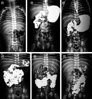
A 6-year-old child presented with intermittent pain abdomen and bilious vomiting with progressive weight loss. Pain intensifies after heavy meal and gets relieved with vomiting. No admissions earlier. Bowel habits normal. The following procedure was performed as part of evaluation.
a. What is the clinical diagnosis suspected?
b. What is the imaging and its findings?
c. What is the screening tool asked for?
d. Name the surgical emergency expected with this?
e. What is the treatment offered?
Answer
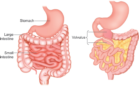
| a. | Midgut malrotation is the working diagnosis. |
| b. | Upper gastrointestinal (GI) contrast study is the barium study until duodenojejunal (DJ) flexure—barium meal follow through is done in the follow-up (last two images) |
| Findings: Scout film is normal/stomach normal/no evidence of GERD/contrast flow in the duodenum is seen entirely to the right of the spine suggesting the “C” loop of the duodenum is not properly formed/contrast is filling the small bowel entirely to the right of the spine. The later images of barium meal follow through suggestive of large bowel with appendix seen on the spine and running contrast to the left of spine. This is a classical contrast study of malrotation showing the small bowel on the right and large bowel in the left side of the abdomen. | |
| c. | USG abdomen is the best screening tool available to identify the alteration of superior mesenteric artery/superior mesenteric vein (SMA/SMV) axis suggestive of malrotation. |
| d. | Midgut volvulus is the surgical emergency anticipated. |
| e. | Elective laparotomy and Ladd’s procedure—derotation of the volvulus if any/division Ladd’s bands which hinders the “C” loop of duodenum from widening/widening of small bowel mesentery/elective appendicectomy because appendix lies in the left side abdomen)/reposition of bowel as earlier. |
THG28-011
Antenatal mother comes for counseling at 32 weeks of gestation with ultrasound of fetus showing bilateral pelvicalyceal dilatation of 5 mm diameter. Bladder appears normal. Amniotic index is also normal. Answer the following questions on counseling of the fetal ailment.
a. How frequent is the antenatal USG required?
b. Advise on nature of delivery.
c. What are the factors to be monitored in antenatal USG?
d. What is the Society of Fetal Urology (SFU) grading?
e. When will be the postnatal evaluation with USG?
f. What is the differential diagnosis?
Answer
| a. | Mild grades 1/2 in society of fetal urology (SFU) grading will need only USG on 5th/7th/9th month of gestation. Higher grades 3 and 4 in SFU grading will need monthly follow-up with USG. |
| b. | Normal vaginal delivery can be advised as per maternal indication. |
| c. | Anteroposterior diameter of pelvis/unilateral or bilateral/ureteric dilatation/bladder emptying and amniotic index |
| d. | SFU guidelines: SFU grading table as follows: |
| Grade 0: No dilatation | |
| Grade 1: Dilatation of renal pelvis without dilatation of calyces, no parenchymal atrophy | |
| Grade 2: Mild dilatation of renal pelvis and calyces, no parenchymal atrophy | |
| Grade 3: Moderate dilatation of renal pelvis, blunting of fornices and flattening of papillae, mild cortical thinning | |
| Grade 4: Gross dilatation of renal pelvis and calyces, loss of borders between the renal pelvis and calyces, renal atrophy seen as cortical thinning | |
| e. | Anytime in the first week of life after the physiological dehydration (48 hours of life) is over. This baseline ultrasound for the newborn will guide on the interval needed for further follow-up. |
| f. | Transient hydronephrosis/bilateral or unilateral hydronephrosis/vesicoureteric reflux (VUR). |
THG28-012
Male baby, primi mother comes for antenatal counseling at 20 weeks of gestation with ultrasound of fetus showing bilateral hydroureteronephrosis, distended urinary bladder, and oligohydramnios.
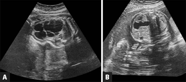
a. What is the working diagnosis?
b. How frequent is the antenatal USG required?
c. What is the classical sign found in the USG, in above diagnosis?
d. How would you investigate further?
e. How do you prognosticate the disease in fetus?
Answer
| a. | Bladder outlet obstruction (BOO): It is never labeled as posterior urethral valve (PUV) in ultrasound as it will expose the gender of the fetus (it is not legally allowed to declare the sex of fetus antenatally in our country). |
| b. | Close follow-up with monthly follow-up in second trimester/bimonthly follow-up in third trimester is needed for amniotic index assessment. |
| c. | Key hole sign is classical evidence of PUV. Distended bladder with dilated posterior urethra with resembles a keyhole in USG imaging. |
| d. | Amniocentesis is suggested to get the urinary indices of the fetus. Amniotic fluid is entirely contributed by the fetal urine. The poor urinary indices as given in below Box are indirect evidence of the renal dysplasia attributing the renal insufficiency in PUV children. |
| Unfavorable urinary indices |
|---|
| Na+ >100 mEq/L |
| Cl- >90 mEq/L |
| Osmolarity >210 mOsm/L |
| Elevated urinary calcium (>1.2 mmol/L) |
| Elevated levels of urinary beta-2 microglobulin |
| e. | PUV identified with oligohydramnios is always given a guarded prognosis. Oligohydramnios in third trimester if not managed well, will lead onto preterm delivery and its complication due to lack of lung maturity. The renal insufficiency documented in antenatal period will definitely lead the child to chronic kidney disease and renal replacement therapy postnatally. |
THG28-013
Antenatal mother comes for counseling at 28 weeks of gestation with ultrasound of fetus showing unilateral pelvicalyceal dilatation of 15 mm diameter. Ipsilateral ureter appears dilated. Bladder appears normal. Amniotic index is also normal.
a. How frequent is the antenatal USG required?
b. What are the factors to be monitored in USG?
c. What is the working diagnosis?
d. Postnatal evaluation and follow-up?
Answer
| a. | SFU grade 4 warrants monthly follow-up with ultrasound imaging. |
| b. | Bladder cycling, anteroposterior diameter of pelvis and amniotic index to be followed up. |
| c. | Unilateral hydroureteronephrosis with or without BOO. The differential diagnosis can be VUR/obstructive megaureter/unilateral vesicoureteral reflux and renal dysplasia (VURD) in posterior urethral valve (PUV). |
| d. | Postnatal ultrasound in the first week of life. If poor urine stream is present clinically, will need micturating cystourethrogram (MCU) to rule out PUV in a male child. |
THG28-014
A newborn presented with antenatally detected bilateral hydroureteronephrosis. Amniotic index was 10 until 37 weeks. Near term delivery at 37 weeks with birth weight of 2.8 kg. Observed to have poor urine stream at birth. Postnatal USG had bilateral hydroureteronephrosis. The above investigation was asked for.
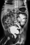
a. Read the findings in the study.
b. Name the syndrome involved.
c. What is the immediate investigation and management required?
d. Name any three pop-off mechanisms of the condition.
Answer
| a. | MCU (oblique view)—narrow anterior urine stream/dilated posterior urethra with valve impression/trabeculated bladder/diverticulum seen in the left side/left VUR/intrarenal reflux (sunray appearance of contrast into renal parenchyma). |
| b. | Duckett’s syndrome or VURD (vesicoureteric reflux-renal dysplasia syndrome) |
| c. | Renal parameters: Blood urea nitrogen (BUN)/creatinine to be checked if kidney injury is seen at birth. Child needs immediate catheterization by pediatric surgeon and decompression of the renal system. If the creatinine is declining after catheterization, child will undergo primary valve fulguration. If the renal parameters are increasing, child will need a urinary diversion for a better wide drainage that will help in reduction of creatinine in majority of children. |
| d. | Pop off mechanisms—VUR/diverticulum/patent urachus/perirenal urinoma/urinary ascites. |
THG28-015
An 8-year-old girl presented with history of swelling in the left neck of 2 weeks duration, increasing in size. No history of fever, loss of weight, or appetite. No family history of hypothyroidism.
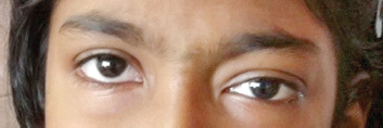
a. Identify the clinical signs and the working diagnosis.
b. Name the syndrome associated and its components.
c. What is the urinary index asked for clinical corroboration of the tumor suspected?
d. What is the genetic prognostic factor in the condition?
Answer
| a. | Left eye ptosis. In the background history of left sided neck mass, probable diagnosis is thoracic neuroblastoma with pressure symptoms. |
| b. | Horner’s syndrome—anhidrosis, miosis, ptosis, and enophthalmos are the four symptoms caused by the sympathetic nerve involvement. |
| c. | 24 hours urinary vanillylmandelic acid (VMA) level—elevated urinary catecholamine level is a definitive tumor marker for neuroblastoma. |
| d. | N-MYC oncogene amplification in the tumor has a poor prognosis with <50% survival prediction. |
THG28-016
A 1-year-old male child with antenatally detected unilateral pelvicalyceal dilatation was missed for follow-up. Child developed simple febrile seizures and admission evaluation with ultrasound showed right hydronephrosis with anteroposterior (AP) diameter of 2.5 cm incidentally. The following investigation was asked for.
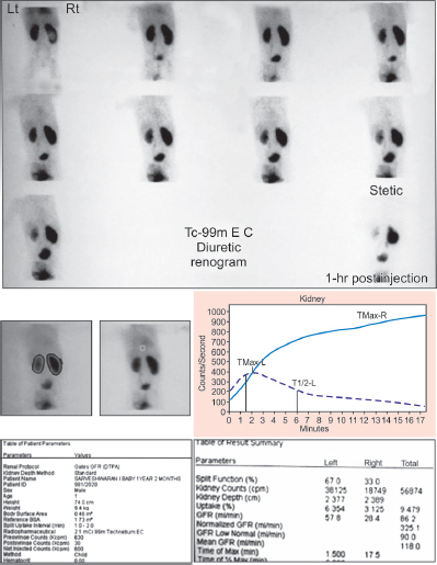
a. What is the investigation named and what is the tracer used?
b. Name the indication for which the above investigation is asked.
c. What are the important points to look for in the study?
d. What are the parameters to look for in individual function of the kidney?
Answer
| a. | Diuretic renogram is the investigation. It is a dynamic study to identify an obstructed system as in pelviureteric junction (PUJ) obstruction or obstructive megaureter. Ethylene cysteine (EC) is the radionuclear tracer used for the study. |
| b. | Any suspected obstruction to urinary drainage warrants EC scan. |
| c. | Delay in drainage of the tracer/holdup of tracer in later image/obstructive drainage pattern curve (a rising curve denotes obstruction to drainage). |
| d. | Differential renal function and individual glomerular filtration rate (GFR) is marked in the EC study. |
THG28-017
A newborn with absent anus is seen at resuscitation. Child was observed to pass black colored urine as seen in the given image.
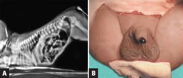
a. What is the clinical finding and probable diagnosis?
b. Name the only investigation to diagnose the type of condition.
c. When should the investigation be done?
d. What is the modification of the investigation followed now?
e. What are the modalities of treatment in each type?
Answer
| a. | Meconium passage via urethra is the finding. High anorectal malformation (ARM) is the working diagnosis. |
| b. | Wangenstein–Rice invertogram is the plain X-ray done to delineate between high and low ARM. The distance between the distal most air shadow and the anal pit site will give an idea about the high or low ARM (via pubococcygeal (PC)/ischial (IC) lines). |
| c. | The invertogram should be done at 24 hours of life allowing the filling of air shadow until the distal most loop of bowel. |
| d. | Prone translateral view X-ray is the modification to invertogram as hanging the baby upside down to conduct invertogram is considered a cruel practice. |
| e. | Diversion colostomy for high ARM and primary anoplasty for low ARM. |
THG28-018
A 3-year-old child with urinary dribbling in daytime and bedwetting history brought for evaluation. Child had one episode culture positive urinary tract infections 2 months back. He is not so regular in bowel habits. Child had bilateral hydroureteronephrosis by ultrasound examination. Child had 40 mL postvoid residue also.
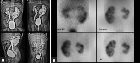
a. What are the clinical signs in the child to look for and why?
b. Read the investigations of the given images.
c. What is the other investigation to be asked for?
d. What is the aim of the treatment required?
Answer
| a. | Clinical signs: Palpable bladder for overflow incontinence/any scar in the back for meningomyelocele surgery/abnormal hairy patch or swelling for occult spinal dysraphisms/spinal deformities. This will point toward neurogenic bladder in the child. |
| b. | MCU: Anterior stream is good/bladder is over distended with multiple diverticulitis/bilateral grade 4–5 VUR/significant postvoid residual contrast |
| Dimercaptosuccinic acid (DMSA) scan: Global scarring with shrunken right kidney/left kidney with polar scars. | |
| Both the investigations pointing toward neurogenic bladder with massive VUR and its impact in reduced renal function with scars. | |
| c. | MRI spine to identify the spinal anomalies. |
| d. | Better urinary drainage/avoid increased pressure in bladder/reduce the VUR and its damage to kidney/reduce the postvoid retention urine. This in turn will reduce the incidence of urinary tract infection. This child needs clean intermittent catheterization (CIC) for better drainage. |
THG28-019
An 8-year-old child presents with sudden onset swelling in the neck. There are complaints of occasional pain and discharge from the swelling. No history of loss of weight and appetite. History of similar episodes in the past twice treated symptomatically.

a. What is the clinical finding noticed?
b. What is the working diagnosis?
c. What is the embryological defect?
d. What are the investigations?
e. What is the differential diagnosis?
f. What is the treatment of this condition?
Answer
| a. | Well-circumscribed swelling in the neck with no signs of erythema or edema. A punctum seen at the summit of the swelling. |
| b. | Branchial sinus. |
c. Second branchial cleft. Defect in the six pairs of branchial arches from the foregut—most common is second branchial cleft anomaly.
| A classical history of sinus opening seen at birth on either side of the neck/salivary discharge from sinus opening/infective episodes clinch the diagnosis. | |
| d. | CT sinogram/MRI neck will confirm the diagnosis. |
| e. | Dermal sinus/tuberculous sinus |
| f. | Surgery: Branchial sinus excision |
THG28-020
A 5-year-old girl child presented with recurrent urinary tract infection four episodes (culture positive) in last 1 year. There is history of daytime urinary enuresis. No bedwetting. Ultrasound imaging has anatomically normal kidneys and bladder.
a. What are the three important points in history to be elicited?
b. What is the working diagnosis?
c. How are the holding maneuvers labeled?
d. What are the supportive imaging asked for?
e. What is the treatment offered?
Answer
a. 1. History on bowel habits to rule out constipation and encopresis
| 2. | History on other lower urinary tract symptoms (LUTS)—dysuria, urge incontinence, postponement, holding maneuvers/vaginal voiding |
| 3. | History on food habits to ensure adequate intake of fiber diet and water consumption. |
| b. | Bladder bowel dysfunction (BBD)—spectrum of LUTS along with constipation/encopresis constitute—“dysfunctional elimination syndrome” or BBD. |
| c. | Vincent curtsy—classical holding maneuvers for LUTS |
| d. | USG abdomen will identify bladder wall thickening with postvoid residue. X-ray abdomen will identify the loaded colon with fecal material. |
| e. | Urotherapy consist of nonpharmacological treatment of the bladder and bowel regulation by parental education and dietary modification. |
THG28-021
Antenatally detected cystic lesion in fetus at 36 weeks of gestation in a mother. She gave birth to a term baby girl. Postnatal clinical evaluation suggested a right iliac fossa mass as shown in given image. USG abdomen showed a clear cystic lesion of 6.2 cm occupying the right lumbar and iliac fossa. No clear site of origin could be made out.
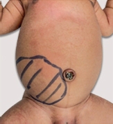
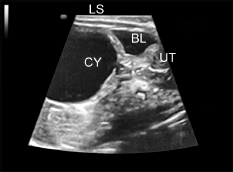
a. What is the working diagnosis?
b. What should be the minimum size of the lesion to be considered as pathological?
c. List the symptoms caused.
d. What is the differential diagnosis?
e. What is the other endocrine evaluation needed?
f. What treatment is offered?
Answer
| a. | Ovarian cyst is the first diagnosis always in a female newborn—neonatal ovarian cyst (NOC) as commonly referred. |
| b. | 2 cm diameter lesion is pathological and needs follow-up. |
| c. | Pressure symptoms like fever, infection, vomiting, intestinal obstruction with abdominal distension, urinary retention, bleeding, torsion, etc. |
| d. | Mesenteric cyst, omental cyst, urachal cyst, duplication cyst, and cystic teratoma |
| e. | Hypothyroidism should be evaluated. Elevated thyroid-stimulating hormone (TSH) behaves like luteinizing hormone/follicle-stimulating hormone (LH/FSH) and known to be associated with ovarian stimulation and cyst formation. Van Wyk–Grumbach syndrome is a classical example of this. |
| f. | Surgery: Ovarian cystectomy saving the ovary involved. |
THG28-022
A 13-year-old girl child presents with history of delayed puberty/flat chest/not attained menarche. She is 123 cm in height. Her weight is 22 kg. Also has webbing of neck and short stature.
a. When do you call the puberty is delayed?
b. Which is the central axis involved in puberty?
c. Are the height and weight appropriate?
d. What is the working diagnosis?
e. What is the workup required?
f. What treatment is offered?
Answer
| a. | Delay in breast development by 12 years (thelarche)/failure of mensuration by 15 years (amenorrhea) is defined as delayed puberty. |
| b. | Hypothalamic-pituitary-gonadal axis (HPG axis) |
| c. | It is below two standard deviation by growth chart. |
| d. | Turner syndrome should be evaluated. |
| e. | Hormonal evaluation—estrogen, LH, FSH/karyotyping—for 46XO—monosomy/cardiac/spinal anomalies/learning disabilities/USG for renal abnormalities. Bone age evaluation for the short stature. |
| f. | Supportive care for the primary ovarian failure, hormonal replacement for the general physical wellbeing, and growth hormone as per the case. |
THG28-023
Newborn presents with abnormal umbilicus. History of sticky green discharge noticed from the umbilicus. Child passed meconium and urine within 6 hours of birth. Antenatal ultrasound and antenatal checkups reported normal.
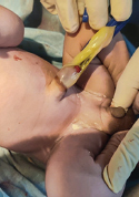
a. What is the clinical finding noticed?
b. What is the precaution while resuscitation in labor room?
c. What is the working diagnosis?
d. What is the differential diagnosis?
e. What treatment is offered?
Answer
| a. | Newborn umbilical cord with protrusion of abnormal tissue at the base/cord clamp seen. |
| b. | Umbilical cord clamp should be placed well away from the protruding tissue to prevent iatrogenic injury. |
| c. | Patent vitellointestinal duct discharging meconium through umbilicus. |
| d. | Exomphalos minor/hernia of the umbilical cord is differential diagnosis. |
| e. | Surgery—laparotomy/patent vitellointestinal duct excision and anastomosis of the ileum. |
THG28-024
A 3-year-old child presents with sudden onset swelling in the neck. No history of fever cough or cold. No history of feeding or breathing difficulty.
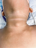
a. What is the clinical finding noticed?
b. What is the working diagnosis?
c. What are the clinical signs to be elicited?
d. What are the investigations required?
e. What is the differential diagnosis?
f. What treatment is offered?
Answer
| a. | Well-circumscribed midline swelling in the neck with no signs of erythema or edema. |
| b. | Thyroglossal cyst |
| c. | Movement of the swelling with deglutition/movement of the swelling with protrusion of tongue |
| d. | Three investigations are mandatory: (1) Thyroid function test to ensure the euthyroid hormonal status, (2) ultrasound of neck to ensure the presence of normal thyroid gland on either side of midline, and (3) thyroid nuclear scan to ensure the uptake in normal thyroid gland and absence of uptake in the midline swelling. An ectopic thyroid can present as midline swelling mimicking thyroglossal cyst, which will be confirmed in nuclear scan with uptake. This investigation will avoid an iatrogenic removal of ectopic functioning thyroid tissue. |
| e. | Ectopic thyroid/dermoid cyst/thyroid mass or nodule/branchial cyst |
| f. | Surgery: Thyroglossal cyst excision |
Figure Sources
All the figures are from author’s personal collection.