THG26-001
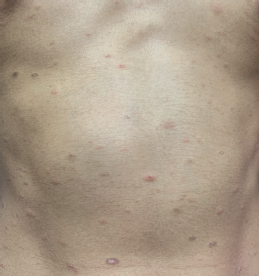
A 13-year-old boy presented with mildly itchy skin lesions all over the body for 2 weeks. Ten days prior to the onset of these lesions, he had a single lesion over the back.
a. What is the diagnosis?
b. What are the points for diagnosis?
c. What is the proposed etiology?
d. Describe the characteristic morphology of the lesions in this condition.
e. What are the various clinical types?
f. What are the drugs that can cause pityriasis rosea-like eruptions?
g. What is the differential diagnosis?
h. How do you treat this condition?
Answer
| a. | Pityriasis rosea, a self-limiting, and papulosquamous disorder |
| b. | An adolescent boy presenting with a single lesion earlier followed by crops of lesions over the trunk and extremities is the typical evolution in pityriasis rosea. |
| c. | The exact etiology of this condition is unknown. Viral etiology with human herpesvirus 6 (HHV-6) or human herpesvirus 7 (HHV-7) has been considered. |
d. • The initial lesion, known as the herald patch, mother’s patch, or medallion patch, is seen as well-demarcated erythematous patch with a collarette of scales in the periphery typically over the trunk, neck, or upper extremities.
| • | Within a few days to 2 weeks, this is followed by the emergence of crops of oval, erythematous plaques with a peripheral collarette of scales and central clearing over the trunk, neck, and extremities. |
| • | Lesions on the trunk align with the skin cleavage lines, producing a “Christmas tree” pattern |
| e. | Classical, inverse, papular, vesicular, purpuric, urticarial, erythema multiforme-like, localized, gigantic pityriasis rosea, and pityriasis circinata et marginata of Vidal are the various clinical types. |
| f. | Drugs, such as metronidazole, omeprazole, isotretinoin, barbiturates, ketotifen, and vaccinations such as BCG and hepatitis B cause pityriasis rosea-like eruptions. |
| g. | Differential diagnosis for the herald patch is tinea corporis. Differential diagnosis for generalized involvement is viral exanthems, guttate psoriasis, morbilliform drug eruption, and secondary syphilis |
| h. | Reassurance about spontaneous resolution and explained that the course lasts for 4–8 weeks. Drug treatment includes calamine lotion, topical corticosteroid creams, erythromycin, oral acyclovir, and oral antihistamines. |
THG26-002
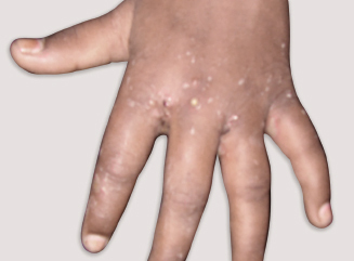
A 3-year-old boy was brought by his parents with history of itchy skin lesions in the finger web spaces, wrists, and genital for 2 weeks. His elder sibling and mother also have similar complaints.
a. In scabies, what does “circle of Hebra” denote?
b. What are the various clinical types of scabies?
c. How does animal scabies differ from classical scabies in distribution of lesions?
d. What are the consensus criteria for the diagnosis of scabies, issued by the International Alliance for the Control of Scabies?
e. What is the differential diagnosis?
f. What are the complications?
g. What are the newer techniques used to demonstrate the mites causing scabies?
h. How do you manage children with scabies?
Answer
| a. | Involvement of an imaginary area around the web space, hands, wrists, elbows, axillae, periumbilical region, and genitalia |
| b. | Classical, neonatal, infantile, nodular, crusted or Norwegian, bullous, animal scabies, scabies in clean, and scabies incognito |
| c. | In animal scabies, lesions are seen in the sites of contact with pets like forearms, abdomen, trunk, thighs, and legs. |
| d. | One among the following should be present: Burrows, typical lesions over the male genitalia, and typical lesions in a typical distribution along with the history of itching and contact history. |
e. • Infantile scabies: Papular urticaria, eczema herpeticum, viral exanthems, and atopic dermatitis
| • | Crusted scabies: Psoriasis, contact dermatitis, eczema, and Darier’s disease |
| f. | Secondary pyoderma, eczema, and poststreptococcal glomerulonephritis |
| g. | Dermoscopy, epiluminescence microscopy, and reflectance confocal video dermatoscopy |
| h. | General measures: |
| • | Treat all family members and close contacts simultaneously |
| • | Patients and contacts should be advised to cut nails and take bath in hot water at night, wipe dry and then apply the medication from behind the ears and below the chin over the entire body. |
| • | In children aged less than two years, face and scalp to be included. |
| • | It should be left overnight (8–12 hours) and the next day morning, hot water bath should be taken. |
| • | Clothing and bed linen should be washed in hot water and dried in sunlight. |
| • | This procedure has to be repeated after one week. |
| • | Antiscabicidal agents: |
| ◆ | 5% permethrin cream (safe from 2 months of age) |
| ◆ | 1% gamma benzene hexachloride (not used in children below 2 years) |
| ◆ | 10% crotamiton cream |
| ◆ | 3–6% precipitated sulfur can be applied for three consecutive nights in infants who are <2 months old, bath should be given 24 hours after the last application. |
| ◆ | Topical ivermectin has also been prescribed. Systemic ivermectin is used in children aged >5 years at 200 µg/kg body weight. The second dose to be given after 1 week. Antihistamines to be continued for 2–4 weeks, as the itching persists in scabies posttreatment due to the delayed hypersensitivity reaction. |
THG26-003
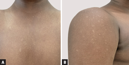
A 13-year-old boy presented with asymptomatic hypopigmented skin lesions over the chest and arms for 3 months. Dermatological examination revealed well-defined hypopigmented macules and patches with borders like pencil-drawn and fine bran-like scales.
a. What is the diagnosis?
b. What are the common causative organisms?
c. What are the reasons for hypopigmentation?
d. What are the most common sites and why?
e. What are the other clinical presentations of this infection?
f. What are the signs that are elicited in this infection?
g. What is the differential diagnosis?
h. Though it is a clinical diagnosis, if there is a clinical suspicion, what is the test done to confirm the diagnosis? What will be the findings?
i. What will be the appearance of the skin lesions in this infection, when visualized under Wood’s lamp?
j. What are the treatment options for localized and extensive infection?
k. Describe any role for general measures?
Answer
| a. | Achromic pityriasis versicolor |
| b. | Malassezia, lipophilic yeast-like fungi and commensals in the skin, under appropriate conditions, cause infection. Malassezia globosa, Malassezia sympodialis, Malassezia furfur, and Malassezia restricta are the common etiological agents. |
| c. | Malassezia produces azelaic acid, a dicarboxylic acid, which competitively inhibits tyrosinase resulting in hypopigmentation. Melanosomes are smaller in size. Scales in the lesion act like sunscreen. |
| d. | Malassezia, being lipophilic, has an affinity for the sebum-rich areas like the trunk, face, and neck. |
| e. | Chromic pityriasis versicolor seen as hyperpigmented macules and patches, follicular form, and red pityriasis versicolor |
| f. | Coup d’ongle sign or the scratch sign and Zireli’s sign |
| g. | Pityriasis alba, polymorphic light eruption (PMLE), and seborrheic dermatitis |
| h. | 10% potassium hydroxide wet mount examination of the scales will show the characteristic appearance of Malassezia—hyaline, short rod-like, or angulated hyphae with groups of spores giving the “spaghetti and meatball” or “banana and grapes” appearance. |
| i. | Pale-yellow fluorescence |
| j. | Localized pityriasis versicolor—topical azole creams for 3–4 weeks. Alternatively, topical ciclopirox olamine, amorolfine, and Whitfield ointment can be used. |
| Extensive or recurrent involvement—2% ketoconazole lotion or 2.5% selenium sulfide lotion has to be applied over the neck and trunk, 10 minutes before bath for 2 weeks. A single dose of oral fluconazole at 8 mg/kg body weight can be given. | |
| In patients with recurrent episodes, monthly once dose of fluconazole 8 mg/kg can be given. | |
| k. | Patients should be advised to maintain good personal hygiene, as sweating is one of the predisposing factors for pityriasis versicolor. |
THG26-004
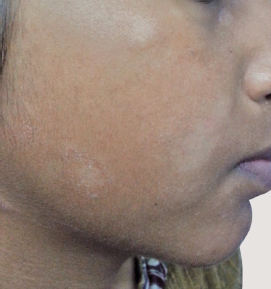
An 8-year-old girl presented with asymptomatic hypopigmented skin lesions over the right cheek for 3 weeks.
a. What is the diagnosis?
b. How will you describe the lesion?
c. Which is the age group most commonly affected?
d. What is the differential diagnosis?
e. What is the treatment?
Answer
| a. | Pityriasis alba, an endogenous eczema, often associated with atopic dermatitis, can present in nonatopic children also. |
| b. | Single or multiple ill-defined hypopigmented patches with fine scales, mostly seen on the cheeks and rarely over the trunk. |
| c. | Pityriasis alba is seen in children in the age group between 3 and 16 years. |
| d. | Differential diagnosis: |
| • | Achromic pityriasis versicolor—well-defined hypopigmented macules and patches. One can stretch the skin and see if the border is well made out. The use of a magnification lens facilitates the differentiation. |
| • | Polymorphic light eruption—scaly condition, commonly seen on the face, associated with itching after exposure to sunlight, seen as a rim of hypopigmentation around the hyperpigmented patch |
| • | Early vitiligo |
| • | Indeterminate Hansen |
| • | Postinflammatory hypopigmentation |
| e. | Emollients and low-potent corticosteroids such as hydrocortisone or desonide creams twice daily for 2–4 weeks. |
THG26-005
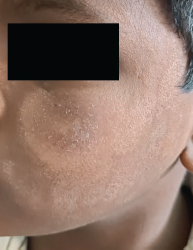
A 7-year-old boy presented with skin lesions over the cheeks with history of itching after exposure to sunlight for 4 weeks. He had recently participated in sports training in school.
a. What is the diagnosis?
b. What are the points that favor the diagnosis?
c. What are the various clinical presentations of this condition?
d. Why does this condition occur?
e. How should it be managed?
Answer
| a. | Polymorphic light eruption |
| b. | A school-going child, who recently had increased exposure to sunlight during sports training, developed skin lesions on the cheeks and itching after exposure to sunlight. |
| c. | Papules, plaques, lichenoid, or urticarial lesions |
| d. | It occurs due to delayed hypersensitivity reaction of endogenous cutaneous antigens, on exposure to UV light causing a reduction in the neutrophil infiltration into the skin. It results in reduced expression of interleukin-4 (IL-4), IL-10, and tumor necrosis factor-α (TNFα), suppressed infiltration of macrophages, and resistance to Langerhans cells, eventually resulting in a nonsuppressive microenvironment in the skin. |
e. • General measures of photoprotection between 9 AM and 4 PM such as the use of covered clothing (loose, long, sleeve shirts, and pants), broad-brimmed hat, and umbrella.
| • | They should be advised to apply broad-spectrum sunscreens 30 minutes before exposure to the sun. |
| • | If the lesion is on the face, desonide cream could be applied twice a day for 2 weeks. If the lesion is on the forearm, fluticasone or mometasone cream could be applied at night for 2 weeks. |
| • | Antihistamines may be given if itching is troublesome. |
THG26-006
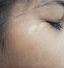
A 6-year-old girl was brought with an asymptomatic hypopigmented skin lesion over the right cheek since birth. Dermatological examination revealed a nonscaly hypopigmented patch.
a. What is the diagnosis?
b. What are the points to substantiate the diagnosis?
c. What is the treatment?
d. What are the differential diagnoses for nonscaly hypopigmented skin lesions?
Answer
| a. | Nevus achromicus or nevus anemicus. Diascopy has to be done to differentiate between the two conditions. |
| b. | Lesion was present since birth. On examination, it is a hypopigmented skin lesion with a serrated border. When diascopy is done, the borders of the lesion merge with the adjacent area in the case of nevus anemicus, whereas in the case of nevus achromicus, the border is well made out. |
| c. | Parents have to be counseled and reassured. Cosmetic camouflage can be tried. An excimer laser may be an option. |
| d. | Differential diagnoses for nonscaly hypopigmented skin lesions are early vitiligo, indeterminate Hansen, and postinflammatory hypopigmentation. |
THG26-007
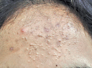
A 13-year-old girl presented with skin lesions on the cheeks and forehead for 2 months.
a. What is the diagnosis?
b. What are the points for diagnosis?
c. How do you grade?
d. What is the pathogenesis of this condition?
e. What is the differential diagnosis?
f. How do you differentiate from acneiform eruption?
g. What is the management?
Answer
| a. | Acne vulgaris |
| b. | An adolescent girl with comedones, papules, and pustules over the cheeks and forehead. |
c. • Grade 1 (mild): Open and closed comedones with few inflammatory papules and pustules
| • | Grade 2 (moderate): Papules and pustules, mainly of the face |
| • | Grade 3 (moderately severe): Numerous papules and pustules and occasional inflamed nodules, also on the chest and back |
| • | Grade 4 (severe): Many large painful nodules and pustules |
| d. | Pathogenesis of acne is multifactorial. Under the influence of androgen, there is increased sebum production, abnormal keratinization, colonization of Cutibacterium acnes and release of inflammatory mediators. |
| e. | Acneiform eruption, acne rosacea, milia, pityrosporum folliculitis, and adenoma sebaceum |
| f. | Acneiform eruption is characterized by the presence of monomorphic papules. There will be no comedones. Drugs that cause acneiform eruption include anticonvulsants, corticosteroids, INH, rifampicin, etc. |
| g. | These children should be first counseled about face hygiene and a low glycemic diet. |
| • | Mild acne: |
| ◆ | Comedonal: Topical adapalene |
| ◆ | Mixed/few papular/pustular: Topical adapalene + topical antimicrobial (Clindamycin) |
| • | Moderate: |
| ◆ | Mixed/papular/pustular: Topical retinoid + Benzoyl peroxide + Oral doxycycline |
| ◆ | Nodular: Topical retinoid + Benzoyl peroxide + Oral azithromycin pulse—3 days a week |
| • | Severe acne: |
◆ Nodular: Oral isotretinoin
THG26-008
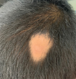
A 7-year-old boy was brought with three asymptomatic patches of hair loss for 2 months.
a. What is the diagnosis?
b. How will you describe the morphology of the lesion?
c. What are the points in favor of the diagnosis?
d. What are the various clinical patterns?
e. What is the differential diagnosis?
f. What are the various associations of this condition?
g. What are the factors for poor prognosis?
h. What is the treatment?
Answer
| a. | Alopecia areata is a chronic, organ-specific autoimmune disorder characterized by the presence of well-circumscribed patchy hair loss with sudden onset. |
| b. | Well-circumscribed/oval smooth patch with loss of hair and normal skin. There is no scarring. |
| c. | Sudden onset of asymptomatic, patchy loss of hair. Dermatological examination reveals the presence of a well-circumscribed, smooth, and nonscarring patch with loss of hair. |
| d. | Ophiasis, Sisaipho, reticulate, diffuse, alopecia totalis, and alopecia universalis |
| e. | Smooth patch of baldness type of tinea capitis, trichotillomania, and frictional/traction alopecia; Diffuse alopecia areata—telogen effluvium |
| f. | Other autoimmune conditions such as vitiligo and thyroid disorders, atopic dermatitis, Down syndrome, collagen vascular diseases, anxiety, and stress |
| g. | Poor prognostic factors: |
| • | Onset at an early age |
| • | Multiple patches with rapid progression |
| • | Ophiasis, sisaipho, alopecia totalis, and alopecia universalis |
| • | Family history |
| • | Associations—autoimmune conditions, atopic dermatitis, Down syndrome, and nail dystrophy |
| • | Poor response to treatment in the past |
h. • First and foremost, children with alopecia areata should be counseled and reassured, as it is associated with low self-esteem and peer bullying.
| • | Topical corticosteroid: In this child, mometasone ointment could be applied once a day for 4 weeks with monitoring. |
| • | Other topical treatment options include contact sensitizers, minoxidil, tacrolimus, and anthralin |
| • | Multiple patches/ophiasis/sisaipho—systemic therapy—oral corticosteroids, cyclosporine, methotrexate, and tofacitinib |
THG26-009
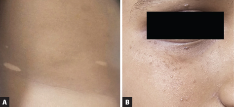
A 6-year-old boy presented with asymptomatic hypopigmented skin lesions over the back since birth and lesions over the face for the past 3 months which are increasing in number. History of seizures from the age of 3 years and does not have any other skin lesions in the body. Dermatological examination revealed hypopigmented lesions over the back and multiple skin-colored papules over the face.
a. What is the diagnosis?
b. What is the mode of inheritance?
c. What is the pathophysiology of this condition?
d. What are the characteristic skin and mucous membrane changes?
e. What are the systemic associations?
Answer
| a. | Tuberous sclerosis complex |
| b. | Autosomal dominant inheritance |
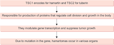
| c. | Arises from inherited or spontaneous mutation of gene TSC1 (9q34) and TSC2 (16p13.3). |
| d. | Skin lesions: Ash leaf macules, angiofibroma, shagreen patches, and fibromas |
| • | Ash leaf macules—seen at the time of birth, single or multiple, oval or may have broad end on one side and tapering on other side. |
| • | Angiofibromas—seen after birth or later in age, firm red brown discrete telangiectatic papules commonly seen over the nasolabial furrow, cheeks, and chin area. |
| • | Shagreen patch—seen over the lumbosacral region as soft elevated, skin-colored plaque with elephant hide or orange-peel appearance. |
| • | Molluscum pendulum—large, soft, and pedunculated papules resembling large skin tags are seen over the axillae, neck, and inguinal folds. |
| • | Periungual fibromas (Koenen’s tumor): Fibromatous tumors of gum, palate, tongue, larynx, and pharynx that appear at or after puberty. |
| • | Enamel pits over the teeth and poliosis are seen. |
e. • CNS: Mental retardation, epilepsy, behavioral disorders, and psychotic symptoms. Subependymal nodules and giant cell astrocytomas occur.
| • | OCULAR: Hypopigmented spots in iris, pigmentary abnormalities of retina, and retinal phacomas occur. |
| • | CVS: Rhabdomyomas and Wolff–Parkinson–White syndrome in cardiovascular system. |
| • | Renal: Angiomyolipomas and cystic renal disease may be associated. |
| • | Pulmonary: Recurrent pneumothorax, chylothorax, and lymphangiomyomas. |
| • | Gastrointestinal tract (GIT): Colonic polyps |
THG26-010
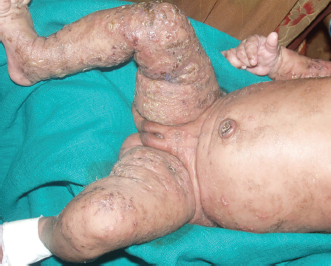
A female neonate presented with vesicles and bullae since birth all over the body and a few warty lesions over the body.
a. What is the diagnosis?
b. What are the other synonyms?
c. What is the basic defect and mode of inheritance?
d. Which gender is commonly involved?
e. Mention the skin manifestations.
f. List the central nervous system (CNS) manifestations.
Answer
| a. | Incontinentia pigmenti |
| b. | Bloch-Sulzberger syndrome, Bloch-Siemens syndrome, melanoblastosis cutis, Pigmented dermatosis Siemens-Bloch type, and nevus pigmentosus systematicus |
| c. | X-linked dominant inheritance |
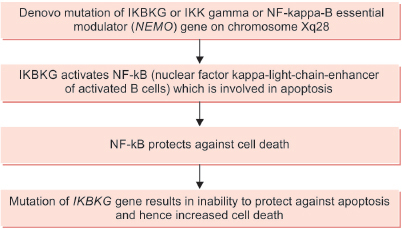
| d. | Common in females and lethal in males. Male cases are reported in literature probably due to somatic mosaicism, hypomorphic mutations, or presence of extra X chromosome like Klinefelter syndrome. |
| e. | Skin lesions are seen in four stages. |
| • | Stage 1: Inflammatory stage starts after birth or within 8 weeks of life and may last for 4–18 months and disappear. Vesicular lesions with erythematous base are seen commonly over limbs and trunk. |
| • | Stage 2: Verrucous stage appears within first few months of life as first stage disappears. Warty papules and plaques are seen along the lines of Blaschko’s lines. |
| • | Stage 3: Hyperpigmented stage is seen from 6 months to 1 year as second stage disappears. It is located over trunk, axilla, and groin in the form of whorled slate gray hyperpigmented plaques and most of the lesions disappear by adolescence. |
| • | Stage 4: It is seen over the posterior calves and arms as pale, hairless linear patches, and plaques. |
In the hair, vertex alopecia in a milder form and rarely agenesis of eyebrows and eyelashes may occur. Nail changes—nail dystrophy, ridging, and pitting. Subungual and periungual keratotic tumors present between puberty and third decade of life. Microdontia, anodontia, hypodontia, peg-shaped teeth, and delayed eruption of permanent teeth may occur.
| f. | CNS involvement in 30% of patients. It is due to ischemic or vaso-occlusive causes. Infantile spasms, seizures, and spastic paralysis can occur. Rarely, congenital hearing loss, cerebellar ataxia, muscular paralysis, and aseptic encephalomyelitis can be seen. |
THG26-011
A 3-year-old boy presented with skin lesions over the sun-exposed areas and photophobia after one and half year of age. He developed hyperpigmented skin lesions over the sun-exposed areas.
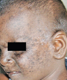
a. What is the diagnosis?
b. What is the mode of inheritance?
c. What are the different types?
d. What are the clinical features?
e. What are the tumors associated with this condition?
f. What are the eye changes expected?
g. What are the neurological features associated?
h. Mention the steps of management.
Answer
| a. | Xeroderma pigmentosum |
| b. | Autosomal recessive genodermatosis. Nucleotide excision repair system is capable of removing the ultraviolet light damaged DNA but due to the mutation this function is lost, leading to xeroderma pigmentosum. |
| c. | Subtypes of A-G and XP variants each with varying mutations in nucleotide excision repair are described: |
| • | XPA: It codes for DNA damage-binding protein 1 (DDBP1) on chromosome 9q22, presents with skin and neurological disease. |
| • | XPB: It codes for excision repair cross-complementing 3 (ERCC3) on chromosome 2q21. It correlates with Cockayne syndrome and trichothiodystrophy. |
| • | XPC: It codes for endonuclease on chromosome 3p25. Mutated endonuclease is unable to sense the damage to DNA which results in sun sensitivity and malignant tumor formation. No neurological involvement. |
| • | XPD: It codes for ERCC2 and is on chromosome 19q13. It correlates with Cockayne syndrome and trichothiodystrophy. |
| • | XPE: It codes for DDB2 |
| • | XPF: It codes for ERCC4 and is on chromosome 16p13. |
| • | XPG: It codes for ERCC5 and is on chromosome 13q33. |
| • | XPV: It is a variant in mutation of XP, not involved in nucleotide excision repair but is involved in postreplication repair, and codes for polymerase in chromosome 6p21. |
d. • Cutaneous features include freckles and dryness occurring over the face, hands, neck, and other sun-exposed areas.
| • | Irregular patches of pigmentation due to fusion of freckles occur |
| • | Angiomas and telangiectasias are common along with atrophic patches. |
| • | Vesiculobullous lesions over the sun-exposed areas are common and when it heals it can lead to scars, contractures, and disfigurement. |
| e. | Tumors could be benign or malignant. Benign tumors such as keratoacanthoma, seborrheic keratosis, and actinic keratosis and malignant tumors such as basal cell carcinoma, squamous cell carcinoma, melanoma, fibrosarcoma, and angiosarcoma are common. |
| f. | Eye changes: Photophobia, conjunctivitis, vascular pterygium, corneal opacities, ectropion, symblepharon, epithelioma of lids, conjunctiva, cornea, and pigmented macules on the conjunctiva. |
| g. | Neurological changes: Patients in groups A, D, and a few in C develop neurological changes. Central nervous system features include mental retardation, areflexia, hyporeflexia, ataxia, spasticity, sensorineural deafness, dysphagia, and abnormal EEG findings. |
| h. | Genetic counseling, protective clothing, sunscreens, and protective sunglasses. |
THG26-012
A 4-year-old girl child presented with multiple asymptomatic skin lesions over the face for the past 1 month.
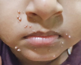
a. What is the diagnosis?
b. What is the description of the lesion?
c. Mention the causative organism.
d. Mention the mode of spread.
e. List the clinical features.
f. How will you treat this infection?
Answer
| a. | Molluscum contagiosum or water warts |
| b. | Dome-shaped, round and sometimes associated with dimple in the center. The molluscum contagiosum lesions in the skin are called Mollusca. |
| c. | Molluscum contagiosum is caused by double-stranded DNA poxvirus called Molluscum contagiosum virus (MCV). There are four types: MCV1 is the most common cause in children, while MCV2 is common in children with HIV. MCV3 and 4 are seen in Asia and Australia. |
| d. | Transmission occurs through skin-to-skin contact or through fomites, towels, etc. It may disseminate by autoinoculation. |
| e. | Incubation period is 2 weeks to 6 months. It presents as dome-shaped lesions associated with a dimple. It infects only keratinocytes in the epidermis and do not have systemic dissemination. It usually asymptomatic but occasionally itchy and painful. |
| f. | It is self-limiting but may take months to disappear. Removal can be done by needling, liquid nitrogen freeze, and electrocautery. |
THG26-013
A 13-year-old girl presented with asymptomatic lesion on the forearm for 3 months.
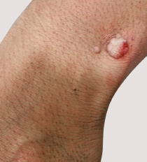
a. What is the diagnosis?
b. Mention the causative organism.
c. What is the mode of spread?
d. What are the different types?
e. How will you treat?
Answer
| a. | Viral wart |
| b. | Small DNA viruses which have got adapted to infect skin and mucous membrane. The most common subtypes causing the warts are HPV 2, 27, 57, and 63 but HPV 1, 4, 10, 45, 65, 88, and 95 were also detected. |
| c. | Transmission is through skin-to-skin contact and fomites. |
| d. | Plane warts, verrucous wart, filiform wart, mosaic wart, plantar wart, palmar wart, mucous membrane wart, etc. |
| e. | Most lesions resolve spontaneously but take months. Topical application of salicylic acid/tretinoin, cryotherapy, electrocautery, intralesional BCG/PPD, and lasers. |
THG26-014
A 12-year-old boy presented with hypopigmented skin lesions with decreased sensation over the right arm and forearm for 3 months. There was no history of similar complaints among the family members. Dermatological examination revealed the presence of four well- to ill-defined hypopigmented and hypoanesthetic patches over the right arm and forearm. Right ulnar nerve was uniformly thickened and was not tender.
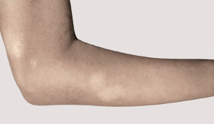
a. What is the diagnosis?
b. What are the points for diagnosis?
c. What are the investigations?
d. What are the treatment options for this infection in children?
e. What should be the responsibility of the pediatrician/dermatologist who sees this child?
Answer
| a. | Borderline tuberculoid Hansen not in reaction |
| b. | Well to ill-defined hypopigmented, hypoanesthetic patches, trophic changes with unilateral, uniform right ulnar nerve thickening, and no signs of inflammation |
| c. | Slit-skin smear. Biopsy is done only when there is clinical dilemma or for academic purpose. |
d. • Paucibacillary leprosy: Monthly supervised dose of capsule rifampicin 450 mg and tablet dapsone 50 mg on day 1, followed by daily dose of dapsone 50 mg from day 2 to 28, for 6 months.
| • | Multibacillary leprosy: Duration—1 year; Monthly supervised dose of capsule rifampicin 450 mg, tablet dapsone 50 mg and capsule clofazimine 150 mg on day 1, followed by daily dose of dapsone 50 mg and alternate day dosage of clofazimine 50 mg from day 2 to 28. |
In children younger than 10 years, weight-based dosage has to be given:
| Capsule Rifampicin | 10 mg/kg body weight |
| Tablet Dapsone | 2 mg/kg body weight |
| Capsule clofazimine | 2-3 mg/kg body weight once a month followed by 1 mg/kg body weight on alternate days |
| e. | Childhood leprosy is an indicator of the active disease in the community, and hence, it is the responsibility of the concerned physician to inform the health authorities, who will in turn, conduct a contact surveillance, and detect patients with Hansen’s disease. |
THG26-015
A 2-year-old child had convulsions at the age of 23 months and was started on phenytoin sodium. He developed peeling of skin over the trunk and extremities with oral erosions. Erosions were seen over 50% of body surface in the next 24 hours.
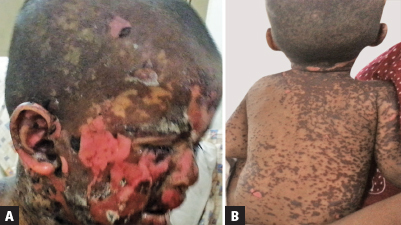
a. What is the diagnosis?
b. What are the other drugs which can lead to this condition?
c. What is the pathology?
d. Mention any scoring system associated with this condition.
e. What are the factors for poor outcome?
f. How to treat?
Answer
| a. | Toxic epidermal necrolysis |
| b. | Any drug can cause this condition. The common drugs implicated are: |
| • | Antiepileptics (phenytoin, carbamazepine, valproic acid, phenobarbital, and lamotrigine) |
| • | Nonsteroidal anti-inflammatory drugs |
| • | Antibiotics (commonly trimethoprim with sulfamethoxazole, aminopenicillins, tetracyclines, and cephalosporins) |
| • | Immune check point inhibitors (nivolumab and pembrolizumab) |
| c. | Hypothesis for drugs generating immunological response leading to TEN: |
| • | Hapten/prohapten concept suggest that drugs will covalently bind to proteins in serum, forming complexes. These complexes are recognized by certain human leukocyte antigen (HLA) molecules and presented to T cells triggering an immune response. |
| • | Pharmacological interaction concept—chemically inert drugs directly bond to HLA molecules, leading to T-cell activation. |
| • | The altered peptide concept says that drug binds inside HLA-binding pockets altering self-protein presentation to T cells. Hence, they are not recognized as self, prompting an immune response. Keratinocyte death was mediated by Fas ligand interacting with Fas receptor on the keratinocyte surface. |
| • | Recent studies have identified granulysin as an important factor in apoptosis. |
| d. | SCORTEN score |
| e. | Increasing age, extent of epidermal detachment, number of medications, elevation of urea, creatinine and glucose levels, neutropenia, lymphopenia and thrombocytopenia, and late withdrawal of the causative drug. |
f. • Cessation of the causative drug
| • | Fluid and electrolyte management |
| • | Infection control and wound care |
| • | Role of corticosteroid as monotherapy is controversial. |
| • | Intravenous immunoglobulin (IVIg) alone in the treatment is controversial. Combination of IVIg and corticosteroids are beneficial. |
| • | Use of cyclosporine with or without steroids |
| • | IVIg with plasmapheresis and etanercept has better outcome. |
THG26-016
A 2-year-old child developed an impetiginous lesion over the right side of the face since 2 days followed by small blisters over the neck and axillae which led to peeling of skin over perioral area, axillae, groin, and trunk. There was no history of oral or genital ulceration and no history of recent intake of new drug prior to the onset of the condition.
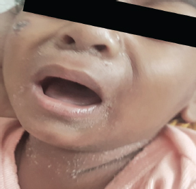
a. What is the diagnosis?
b. What is the causative organism?
c. Mention the pathophysiology.
d. What are the clinical features?
e. Mention the steps in the management.
Answer
| a. | Staphylococcal scalded skin syndrome or Ritter disease |
| b. | Staphylococcus aureus of phage group II types 3A, 3B, 3C, 55, and 71 are the strains that produce exfoliatin A and B. |
| c. | Exfoliatin A and B are serine proteases released at the site of infection, which spreads hematogenously to the stratum granulosum, accumulate and digest the desmoglein-1, thus damaging the adhesion between keratinocytes causing denudation and formation of superficial bullae. |
Desmoglein-3 is seen in the lower epidermis and mucous membrane which maintain the cellular adhesion. The exfoliatin A and B do not lyse the desomogelin-3 and hence mucous membrane is not involved.
d. • Prodrome of fever, fatigue, and irritability as the Staphylococcus localizes in the conjunctiva, nose, throat, etc.
| • | Within 1–2 days, tender erythematous patches occur over face, axilla, groin, and neck. |
| • | A few hours later, fragile blisters develop and the fluid inside the blister may be sterile or pus. |
| • | These blisters enlarge and form bullae which rupture and cause desquamation |
| • | The skin appears wrinkled due to the formation of flaccid bulla which is called sad man facies. |
| • | A positive Nikolsky sign may be present. |
| • | Skin heals without scarring within 2 weeks. Sometimes biopsy is performed to prove the diagnosis. |
| e. | Treatment: |
| • | Penicillinase-resistant penicillins—nafcillin or oxacillin, 100–150 mg/kg/day is given intravenous in four divided doses with a maximum dose of 12 g/day. |
| • | Cefazolin 50–100 mg/kg/day in three divided doses can be given. 2 g of cefazolin intravenous given every 8 hours. |
| • | Vancomycin 45 mg/kg/day in three divided doses with a maximum dose of 2 g/day |
| • | When clinical improvement occurs, systemic therapy is stopped and oral antibiotics such as cephalexin or dicloxacillin are given. The total duration of antibiotic therapy is for 10 days. |
| • | Intravenous fluids to prevent dehydration |
| • | Rarely IVIg in a dose of 0.4 g/kg/day can be given for 5 days. Impetigo is treated with topical mupirocin/fusidic acid. |
THG26-017
A 15-year-old boy presented with few light-brown-colored skin lesions since birth, which have increased in size and number through the years, and a few swellings varying in size from 0.5 × 0.5 cm to 3 × 3 cm.
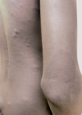
a. What is the diagnosis?
b. What is the mode of inheritance?
c. Mention the systems involved.
d. Mention the criteria for diagnosis of type 1.
e. What treatment can be offered?
Answer
| a. | Neurofibromatosis (NF) |
| b. | Autosomal dominant with complete penetrance |
| • | Neurofibromatosis type 1 (NF1) is due to de novo or inherited loss-of-function mutation of the NF1 gene located on 17q11.2. |
| • | NF type 2 (NF2) is due to loss-of-function mutation of the NF2 gene located on 22q12 and codes for merlin. |
c. • Skin: Dermal and plexiform neurofibroma, café au lait macules, and axillary and inguinal freckling; rarely juvenile xanthogranuloma and nevus anemicus. Learning disabilities, both verbal and nonverbal disabilities may occur.
| • | Ocular: Plexiform neurofibroma, Lisch nodules, optic gliomas, and choroidal nodules occur. |
| Brain tumor in the form of pilocytic astrocytomas is one of the common intracranial neoplasms. | |
| • | Skeletal: Bone dysplasias, pectus deformity, scoliosis or kyphosis, and anteromedial bowing of legs can occur. Sphenoid wing dysplasia, non-ossifying fibromas, short stature, headaches, macrocephaly, and rarely hypertension can occur. |
| d. | Diagnostic criteria for neurofibromatosis type 1: Must have two or more of the following: |
| • | Six café au lait macules that measure >0.5 cm in prepuberty and >1.5 cm in adults |
| • | Freckling of the axillary and/or inguinal areas |
| • | A plexiform neurofibroma or two or more neurofibromas |
| • | Two or more Lisch nodules |
| • | Pathognomonic skeletal dysplasia—tibial or sphenoid wing dysplasia |
| • | An affected first-degree relative |
Criteria for neurofibromatosis type 2:
| • | Bilateral vestibular schwannomas seen by magnetic resonance imaging (MRI) scan |
| • | A first-degree relative with NF2 or any two of the following: schwannoma, meningioma, glioma, neurofibroma, and posterior subcapsular lenticular opacities. |
| • | Unilateral vestibular schwannoma and any of the following: schwannoma, meningioma, glioma, neurofibroma, and posterior subcapsular cataract. |
| • | Multiple meningiomas with unilateral vestibular schwannoma or any two of the following: glioma, schwannoma, and posterior subcapsular cataract. |
e. • Complete physical examination twice a year which includes height, weight, head circumference, blood pressure, and observation for bony abnormalities, and ophthalmological examination.
| • | Monitoring for learning disabilities and early intervention |
| • | Methylphenidate is useful in attention deficit problems in children with NF1. |
| • | Dermal neurofibromas may be excised particularly when disfiguring |
| • | Imatinib has been tried to decrease the progression of plexiform neurofibromas. |
| • | In case of pain associated with plexiform neurofibromas, sirolimus has helped. |
| • | Genetic counseling |
THG26-018
An 11-year-old girl presented with itchy skin lesions over the trunk, groin, and face for 2 months. There was history of similar lesions in the mother.
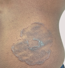
a. What is your diagnosis?
b. Describe the characteristic morphology.
c. What is the difference between steroid modified tinea and tinea incognito?
d. What is the differential diagnosis?
e. In patients with clinical suspicion of this infection, how will you confirm the diagnosis?
f. How will you counsel the patient?
g. How should the topical creams be applied?
h. What are the various systemic drugs available for the treatment of this infection?
i. What are the systemic drugs to be avoided in children with systemic lupus erythematosus (SLE) and this infection?
Answer
| a. | Tinea corporis |
| b. | Classical morphology of tinea corporis is a well-defined scaly plaque with papules in the periphery (active margin) and central clearing. |
| c. | Application of topical corticosteroid creams alters the morphology of tinea. In the case of steroid-modified tinea, it is recognizable as tinea, whereas in tinea incognito, morphology is altered to that extent that it is not recognizable as tinea. |
| d. | Herald patch of pityriasis rosea, nummular eczema, psoriasis, granuloma annulare, etc., are the differential diagnosis considered. |
| e. | Scraping of the skin scales from the active border or where scales are present should be collected in a glass slide and 10–20% potassium hydroxide added. Microscopic examination of this wet mount will reveal the presence of dermatophytes seen as hyaline, long, branching, and septate hyphae. |
| f. | First and foremost, it is important to counsel the parents and children, regarding the general measures such as taking bath twice daily, washing clothes in hot water and drying in good sunlight, avoidance of sharing of fomites, avoidance of wearing synthetic tight clothing/waist and wrist bands, and adherence to drug intake until complete clinical resolution. |
| g. | Topical antifungal creams should be applied twice daily (except luliconazole, oxiconazole, ketoconazole, and naftifine), from 2 cm beyond the outer margin, inward, until 2 weeks beyond clinical resolution. |
| h. | Drug | Dosage | Minimum duration |
|---|---|---|---|
| Griseofulvin | 10 mg/kg | 6 weeks | |
| Fluconazole | 3 mg/kg daily | 4 weeks | |
| Itraconazole | 5 mg/kg | 4 weeks | |
| Terbinafine | <20 kg-62.5 mg 20-40 kg-125 mg >40 kg-250 mg | 4 weeks |
| i. | Terbinafine and griseofulvin are to be avoided in children with SLE and dermatophytosis. |
THG26-019
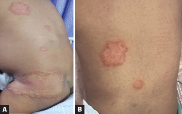
An 18-month-old male baby was brought with 10 itchy skin lesions over the trunk for 2 months. History of application of creams procured from the pharmacy was present. History of similar skin lesions was present in the grandmother.
a. What is the diagnosis?
b. Describe the clinical morphology.
c. What are the combination creams available over-the-counter?
d. What is the impact of these creams on this infection and host skin?
e. What are the common causative organisms of this infection?
f. Mention steps in the management. What is the influence of the diagnosis on the treatment?
Answer
| a. | Steroid-modified tinea |
| b. | Well-defined erythematous plaques with minimal scales and indistinct central clearing. In the larger plaque, there are smaller erythematous plaques within. |
| c. | Topical corticosteroid—antifungal-antibacterial creams containing clobetasol or mometasone are being misused in the treatment of dermatophytosis. |
| d. | These corticosteroid combination creams, by virtue of the anti-inflammatory response, give a symptomatic relief, following which patients tend to discontinue the application of these creams. As a result of corticosteroid effect, there is localized immunosuppression, increased multiplication of dermatophytes, and incomplete clearing, and with onset of symptoms again, the patients start applying the same creams. This results in the ring within ring appearance of steroid-modified tinea lesions. Further, topical corticosteroids increase the transepidermal water loss and reduce the ceramide content of the skin, making the skin dry and itchy. This worsens the initial infection. Parents or caretakers with dermatophytosis who misuse the topical corticosteroid creams, apply these on the children too, when they get infected. |
| e. | Dermatophytes namely Trichophyton mentagrophytes spp. complex, Trichophyton rubrum, and Microsporum canis. |
f. • Topical application of miconazole cream twice daily over the patch and 2 cm beyond the lesion
| • | Oral griseofulvin (10 mg/kg) divided in two doses after a fatty meal for 6 weeks for better absorption. Steroid modified tinea needs to be treated for a longer duration. |
| • | Good personal hygiene and following the general measures in the management of tinea |
| • | Treatment of infected family members |
THG26-020
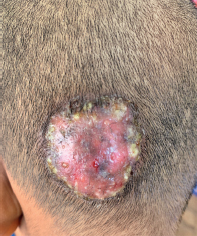
A 7-year-old boy presented with swelling with pustules over the occipital region of the scalp for 4 weeks. Parents gave history of tonsure 3 weeks prior to the onset of lesion. There was no history of pet animals.
a. What is the diagnosis?
b. What are the points for diagnosis?
c. What are the various clinical types of this infection?
d. What is the differential diagnosis?
e. How do you confirm the diagnosis?
Answer
| a. | Kerion—inflammatory type of tinea capitis |
| b. | A 7-year-old boy presented with swelling and pustules over the scalp, 3 weeks after tonsure. Clinical image is of a swelling on the scalp with pustules and broken bits of hair. Characteristic feature of kerion is that these broken hairs are easily pluckable, unlike in bacterial abscess. |
| c. | Noninflammatory types of tinea capitis are gray patch, black dot, alopecia areata-like, seborrheic, and glabrous type of tinea capitis. Inflammatory types are kerion, abscess, pustules, and agminate folliculitis. |
d. • Gray patch—psoriasis and seborrheic dermatitis. There is no hair loss in these two conditions.
| • | Kerion—bacterial abscess |
| • | Alopecia areata-like—alopecia areata |
| • | Patchy hair loss—trichotillomania (loss of hair in accessible areas with hair being of varying lengths) and traction alopecia |
| e. | Microscopic examination of the scraping of the scalp scales and hair root in potassium hydroxide will reveal the presence of endothrix or ectothrix invasion of hair. |
THG26-021
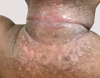
A 4-year-old boy presented with intensely itchy skin lesions over the face, neck, flexures, and trunk for 2 years with history of remissions and exacerbations. Mother is a known case of bronchial asthma.
a. What is the diagnosis?
b. What are the characteristic features?
c. What are the different phases of this condition in children?
d. What are the various minor manifestations?
e. What is the differential diagnosis?
f. What is the gold standard investigation for the diagnosis?
g. What are the general measures to be followed?
h. What is the treatment?
Answer
| a. | Atopic dermatitis |
| b. | Presence of pruritus, peculiar morphology in the different age groups of children, presence of exacerbations and remissions, and family history of xerosis |
| c. | Infantile phase refers to the period between 3 months and 2 years. It can present as early as 3 months, with the onset of erythematous papules on the cheeks and dry skin. Trunk may be involved, but the groin is spared, which helps to differentiate from infantile seborrheic dermatitis. There is a predilection for extensor aspects of the extremities. These babies are irritable due to the itching component. During the childhood phase that ranges from 2 to 12 years of age, flexural involvement is seen as in adults. Erythematous plaques with oozing at times with secondary pyoderma may be the presenting lesion. Lichenification may be seen. |
| d. | Pityriasis alba, juvenile plantar dermatosis, cheilitis, Dennie–Morgan fold, anterior neck folds, hyperlinearity of the palms, keratosis pilaris, recurrent conjunctivitis, pallor of face, white dermatographism, and ichthyosis are the various minor features of atopic dermatitis. |
| e. | Infantile phase of atopic dermatitis has to be differentiated from infantile seborrheic dermatitis. Depending on the clinical presentation, the other conditions for differential diagnosis include scabies, allergic, and irritant contact dermatitis. |
| f. | None |
| g. | Parents, caretakers, and children, if they can understand should be counseled about the general measures, such as avoidance of bathing in hot water, use of mild skin cleansers, short duration of bath, application of emollients postbath within 3 minutes after patting dry, liberal application of emollients, and avoidance of predisposing factors, such as pet animals, pollens, and woollen clothing. |
| h. | Treatment: Saline compresses |
| • | Frequent application of emollients |
| • | Topical corticosteroids to be prescribed for a period of 2 weeks—desonide cream for the face and flexures; fluticasone cream for the few lesions on the trunk. |
| • | Antibiotics: Cephalexin for 5 days and antihistamines |
| • | Child to be reviewed after 2 weeks. Subsequently depending on the response, topical corticosteroids may be slowly tapered and topical tacrolimus 0.03% may be introduced. |