THG34-001
A 2-year-old boy fell from height and presented to emergency room (ER) with complaints of vomiting thrice with altered sensorium. CT brain done is displayed here.
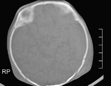
a. Identify the CT findings.
b. What is the primary survey in trauma resuscitation? List the parameters you assess in primary survey.
c. What is PECARN rule used for?
d. What is the PECARN recommendation for child <2 years of age?
Answer
| a. | CT brain plain: Bone window |
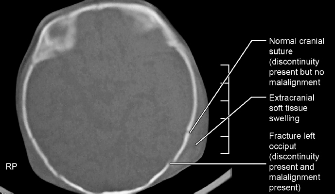
| b. | Primary survey in trauma resuscitation is the order of priority in the initial assessment and its management. It includes: |
| • | A—airway maintenance with cervical spine stabilization/protection |
| • | B—breathing and ventilation |
| • | C—circulation with bleeding/hemorrhage control |
| • | D—disability—neurologic status evaluation |
| • | E—exposure (complete visualization of the patient for rapid identification of injuries in other parts) and environmental control (hypothermia prevention) |
| c. | Pediatric Emergency Care Applied Research Network (PECARN) rule is used to clinically predict/identify the children with clinically low risk factors with traumatic brain injury (TBI) for CT scan recommendation. |
| d. | PECARN rule for age <2 years is as follows: |
| Age <2 years | | | | | |
| CT recommendation | | | Clinical condition | | |
| CT recommended | | | IF Glasgow Coma Scale (GCS) <14 Or Altered mental status Or Palpable skull fracture | | |
| | | | If GCS >14: | | |
| • | Observe or do CT | | • | No altered mental status | |
| • | Do CT if multiple findings, worsening of signs/symptoms during observa-tion, age <3 months, parental preference | | • | Occipital/parietal/temporal scalp hematoma | |
| | | | • | Loss of consciousness (LOC) >5 seconds | |
| • | Severe mechanism of injury | |
| | | |
PECARN rule for age ≥2 years age is as follows:
| Age >2 years | | | | | |
| CT recommendation | | | Clinical condition | | |
| CT recommended | | | If GCS <14 Or Altered mental status Or Basilar skull fracture (hemotympanum, CSF otorrhea or rhinorrhea, raccoon eyes—periorbital bruising, and battle sign—bruising over the mastoid area | | |
| | | | If GCS >14: | | |
| • | Observe or do CT | | • | No altered mental status, no basilar skull fracture signs | |
| • | Do CT if multiple findings, worsening of signs/symptoms during observation, age <3 months, parental preference | | • | History of LOC/severe headache/vomiting/severe mechanism of injury | |
| | | | | | |
THG34-002
A 2-year-old girl met with a road traffic accident (RTA), had loss of consciousness for 5 minutes following which she regained consciousness but became drowsy after 2 hours. She sustained a lacerated wound in left temporal region. This is the CT brain image of this child.
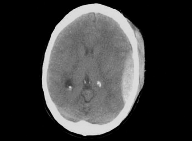
a. Mention the CT findings and diagnosis.
b. Which vessel injury occurs in this condition?
c. What is the complication you expect here?
d. What clinical sign described in the above scenario gives a clue for this diagnosis?
e. What will be your management steps?
Answer
| a. | CT brain plain-brain window showing soft tissue swelling in left temporal region, biconvex hyperdense lesion in left temporal-parietal region—suggestive of extradural hemorrhage. |
Remember mnemonic:
BCCT for hyperdense lesion in CT brain:
Blood,
Calcification,
Contrast-enhanced ring lesion,
Tuber
Easy to remember (mnemonic):
Epidural hematoma (EDH)— side view of iddly (South Indian dish)
| b. | It is an extra-axial blood collection between dura mater and inner table of skull occurs mostly due to arterial bleed from branch of middle meningeal artery. |
| c. | Mass effect if significant bleeding occurs, herniation, and seizures. |
| d. | Lucid interval following a head trauma child had transient loss of consciousness and regains normal consciousness temporarily and lapses back to deteriorating level of consciousness. This is significant in EDH. |
| e. | Airway, breathing, and circulation management, neuroprotective measures—head-end elevation, maintaining normothermia, normoglycemia and normal electrolyte level and maintaining normal hydration, here the CT does not show midline shift so need to monitor for raised intracranial pressure (ICP) features, if ICT raises/midline shift/EDH volume progressively increases—plan for craniotomy and hematoma evacuation. |
THG34-003
A 10-year-old boy fell down from a height and following which he complained headache and had nonprojectile vomiting. He was brought to ER 10 hours after injury with active seizure for 20 minutes.
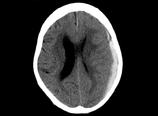
a. What is the CT finding here and what is the diagnosis?
b. Which vessel injury will cause this type of CT image?
c. How do you differentiate this injury between acute and chronic?
d. How will you manage this case?
Answer
| a. | CT brain plain: There is an extra-axial hyperdense concavo-convex (crescent shaped) in left temporal region with compression of left lateral ventricle and midline shift. It is subdural hemorrhage in left temporal region with midline shift. |
Easy to remember:
Subdural hematoma (SDH)—crescent shaped (concavo-convex)
| b. | Bleeding in subdural space occurs when tearing of bridging veins and accumulation of blood occurs between the dura and arachnoid space. |
| c. | Acute SDH will be hyperdense in CT whereas chronic SDH will be hypodense. |
| d. | Airway, breathing, and circulation management, management of seizures and raised ICP, neuroprotective measures, and surgical management—craniotomy and hematoma evacuation. |
THG34-004
This 2-month-old boy previously well infant presented with sudden onset of seizures and lethargy with irritability and refusal of feeds. He was delivered at home and birth vaccination was given at 2 weeks of birth. On arrival to hospital, he had active seizures, bulging anterior fontanelle, and pallor. The child was intubated, seizure controlled with medication, and CT brain was taken.
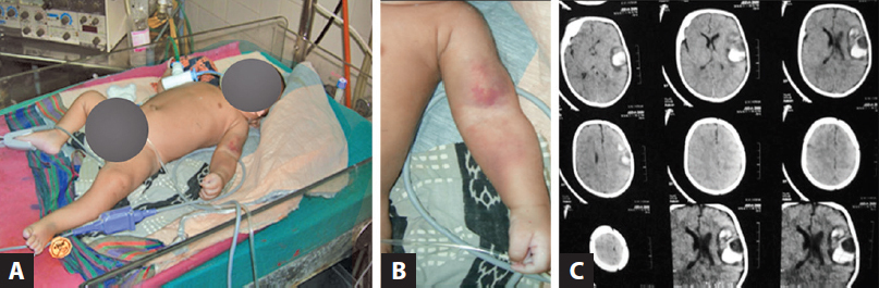
a. Any clue from the images A and B? What is the clinical finding and probable diagnosis?
b. What is the CT finding?
c. What are the possible causes you consider for this condition?
d. Can you correlate the clinical image with radiology and give a diagnosis?
e. What measures could have prevented this?
f. What is the biomarker used in this condition?
Answer
| a. | Bruises in the left arm and forearm, raising a possibility of intracranial bleed. |
| b. | CT brain plain-brain window: Hyperdensity showing intraparenchymal bleed in left temporal region (B in B C C T) with a central hypodensity showing edema. |
| c. | Delayed hemorrhagic disease of newborn, clotting factor deficiency like hemophilia, allo/autoimmune thrombocytopenia, nonaccidental injury, and uncommonly, an arteriovenous malformation. |
| d. | This child is home delivered and with the history of delayed vaccination, there is a possibility of vitamin K was not administered. There is cutaneous bruising and intracranial bleed suggesting delayed hemorrhagic disease of newborn. |
| e. | Vitamin K injection 1 mg intramuscular immediately after birth. |
| f. | Proteins induced by vitamin K absence (PIVKA). |
THG34-005
A 16-year-old boy met with a road traffic accident (RTA). He was riding a bike and it collided with a lorry. This CT brain was taken after 12 hours (image A). He remained unconscious for 3 days, hence MRI brain was done on day 4 (images B and C).
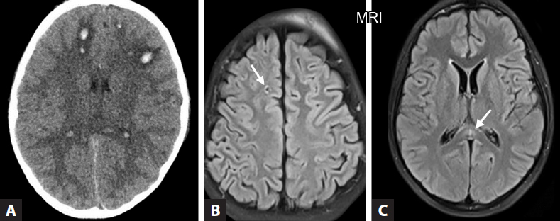
a. What are the radiological findings?
b. What is the diagnosis?
c. What is the mechanism of injury and extent of the lesion?
Answer
| a. | CT brain plain: Scalp hematoma both sides, multiple petechial hemorrhages (dot like hemorrhages) seen at the junction of gray and white matter. No skull fractures. No EDH/SDH/intracerebral hemorrhage (ICH). No midline shift. |
| MRI brain: Punctate hyperintense foci (white arrow) seen in white matter of right frontal lobe and splenium. | |
| b. | Diffuse axonal injury (DAI) |
| c. | Shearing of the axons of the brain that happens when the brain is injured as a result acceleration/deceleration or rotational injury inside the bony skull. Scattered lesion occurs over a widespread area. |
THG34-006
A 10-month-old infant presented with developmental delay and microcephaly presented with recurrent seizures. CT brain image is displayed here.
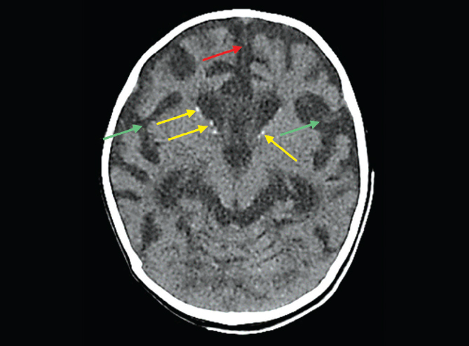
a. What are the CT findings?
b. What is the diagnosis?
c. What finding will you expect in ophthalmological evaluation?
Answer
| a. | Widening of interhemispheric fissure (red arrow), widening of Sylvian fissure (green arrow) suggestive of brain atrophy, dot like calcification (yellow arrow) seen in the periventricular region surrounding anterior horn of lateral ventricle |
| b. | Congenital cytomegalovirus infection. |
| c. | Chorioretinitis |
Remember mnemonic:
BCCT for hyperdense lesion in CT brain:
Blood,
Calcification,
Contrast-enhanced ring lesion,
Tuber
THG34-007
An 8-year-old, developmentally normal girl with no previous history of seizures, presented with complaints of left focal seizures in school for 20 minutes following which she regained consciousness. CT brain plain (A) and contrast (B) images are displayed here.
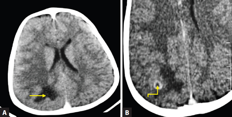
a. Identify the CT findings.
b. Give differential diagnoses based on the image findings and points to differentiate among the differentials.
c. What is your treatment plan here?
Answer
| a. | CT brain plain—hypodense lesion (yellow) seen in right parietal region |
| CECT brain—thick-walled ring enhancing lesion (red arrow) with surrounding edema (yellow arrow) in right parietal region. |
Remember mnemonic:
A E I for hypodense lesion in CT—Abscess, Edema, Infarct
| b. | Neurocysticercosis (NCC) and tuberculoma. Diagnosis here is tuberculoma (>2 cm, thick-walled cyst with no scolex, and surrounded by edema) |
| Neurocysticercosis | | | Tuberculoma | | |
| | | | | | |
| • | Round in shape and thick walled | | • | Irregular in shape and thin walled | |
| • | <20 mm with visible scolex | | • | >20 mm | |
| • | Cystic | | • | Solid | |
| • | Located mostly supratentorially | | • | Supra- or infratentorial | |
| • | Cerebral edema less hence not enough to produce midline shift or focal neuro deficit | | • | Associated with severe perifocal edema and hence prone for midline shift and focal neuro deficit | |
| | | | | | |
| c. | Antituberculosis treatment (ATT) (2 HRZE + 4 HRE) with steroids like prednisolone at 1–2 mg/kg/day or dexamethasone at 0.6 mg/kg/day or its equivalent are used for 2–4 weeks and then are tapered over the next 4 weeks or any steroid in equipotent doses can be used. |
THG34-008
These are the neuroimaging of three different children with same diagnosis.
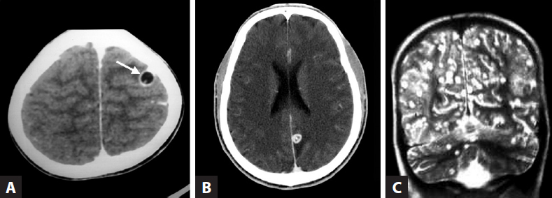
a. What is the most likely diagnosis and can you mention the difference among them?
b. What are the findings in each image?
c. What is the causative organism?
d. A person consumes only vegetarian diet cannot get this disease—True or False?
Answer
| a. | Diagnosis: Neurocysticercosis (NCC), these are three CT images shows different stages of NCC. |
| b. | Image A: CECT brain—small thick-walled cyst without surrounding edema and a scolex seen inside it: Vesicular stage (asymptomatic stage). |
| Image B: MRI brain—thick-walled cyst with surrounding edema: Colloidal vesicular stage (most symptomatic stage). |
Over the year the parasite dies with or without treatment and the fluid in the cyst becomes turbid and membrane becomes leaky causing surrounding edema. This is the most symptomatic stage of NCC.
| Image C: MRI brain—multiple calcified cysts as remnants with no edema: Nodular calcified stage. | |
| c. | Taenia solium (pork tapeworm). |
| d. | True. Eating undercooked pork, eating food/drinking water contaminated with eggs of Taenia solium either through poor hygiene or soil with contaminated human feces are all the source. |
THG34-009
A 2-year-old girl presented with refractory left focal seizures not responding to antiepileptic drugs. She has a red birth mark in the right side of face involving forehead and eyelids.
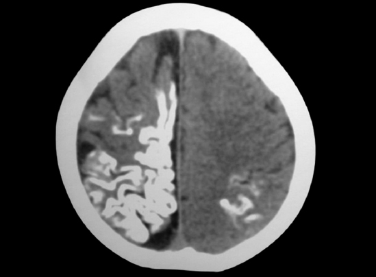
a. What is the CT finding?
b. What is the reason behind the CT finding?
c. What is the diagnosis?
d. What is the ophthalmological complication expected here?
e. Does this condition get inherited to the next generation?
Answer
| a. | Tram-track sign—gyriform calcification in right fronto-temporo-parietal region and left parieto-occipital region. |
| b. | Leptomeningeal angioma cause impaired blood flow in the brain and causes brain atrophy and calcification below the angioma. |
| c. | Sturge weber syndrome |
| d. | Choroidal hemangioma, buphthalmos, and glaucoma |
| e. | No. It is due to a somatic mutation and so it will not be inherited. |
THG34-010
A 5-year-old boy presented with intellectual disability and seizure. He has multiple small red papular lesions over the cheeks and nose. CT brain was done during evaluation.
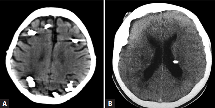
a. What is the CT finding?
b. What is the diagnosis?
c. What are the cutaneous manifestation in this condition?
d. What are the other systemic manifestations in this condition?
Answer
| a. | Image A: Multiple cortical calcifications (calcified cortical tubers) |
| Image B: Calcification seen along the ependymal surface of left lateral ventricle suggestive of calcified subependymal nodules | |
| b. | Tuberous sclerosis |
| c. | Facial angioma, shagreen patch, ash leaf macules, and subungual fibroma |
| d. | Renal: Angiomyolipoma, renal cyst, polycystic kidney, proteinuria, and renal cell carcinoma |
| Cardiac: Rhabdomyoma | |
| Lung: Pulmonary cyst, lymphangioleiomyomatosis causing recurrent pneumothorax, and chylous pleural effusion | |
| Spleen: Splenic hamartoma | |
| Liver: Liver angiomyolipoma | |
| Eye: Retinal hamartoma and palpebral conjunctival angiofibroma | |
| Others: Cortical tubers, subependymal nodules, subependymal giant cell astrocytoma (SEGA), epilepsy, autism spectrum disorder, and cognitive impairment |
THG34-011
A 1.5-year-old boy child presented with 1 week of recurrent vomiting and irritability and a history of progressively increasing head size noted since early infancy.
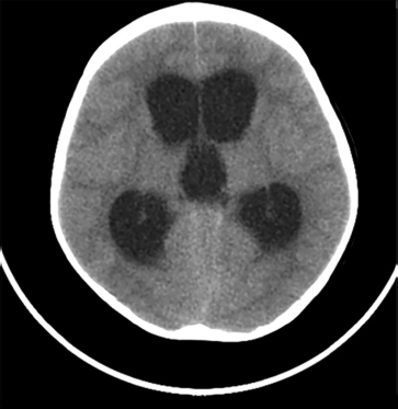
a. What are the CT findings?
b. What is the diagnosis?
c. Name one common cause.
d. Write the normal cerebrospinal fluid (CSF) pathway.
Answer
| a. | CT findings: CT brain plain—enlarged anterior and posterior horn of bilateral lateral ventricles and 3rd ventricle. Fourth ventricle not seen. |
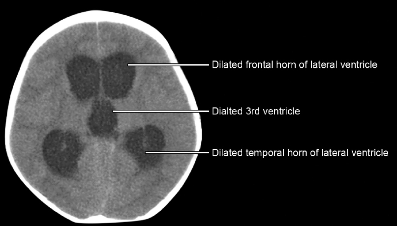
| b. | Obstructive or noncommunicating hydrocephalus (since only the lateral and 3rd ventricle seen dilated) |
| c. | Congenital aqueductal stenosis |
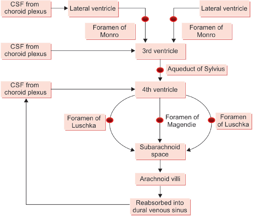
| d. | CSF pathway is as follow: |
THG34-012
One-year-old child with increasing head size, irritability and vomiting. Hence CT brain was done.
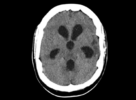
a. Identify the CT findings and diagnosis.
b. What is the pathophysiology here?
c. What is the management for the complication developed here?
Answer
| a. | Communicating hydrocephalus: Lateral ventricle, 3rd and also 4th ventricle is dilated. |
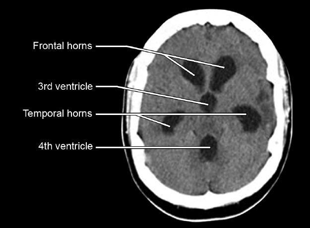
| b. | Block in CSF flow/drainage beyond the level of fourth ventricle, at the level of arachnoid granulations because of impaired absorption. |
| c. | Ventriculoperitoneal shunt and endoscopic third ventriculostomy |
THG34-013
This 4-year-old girl, known case of congenital cyanotic heart disease, was admitted for 10 days of fever, left focal seizures, and left focal deficit.
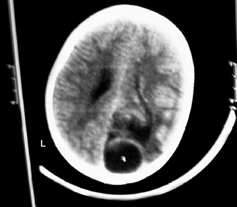
a. Identify the CT findings.
b. What is the probable diagnosis?
c. What are the risk factors/causes for this complication to occur?
d. What are the most common organisms to cause this complication?
e. What is the indication for surgery in these type of cases?
Answer
| a. | CT brain plain: Round hypodense lesion in left occipital region—abscess in right occipital lobe (remember A in A E I). |
| b. | Brain abscess in left occipital lobe. |
| c. | Chronic suppurative otitis media (CSOM), mastoiditis, cyanotic congenital heart disease with right to left shunt, and meningitis. |
| Frontal lobe abscess usually occurs as a complication of sinusitis and orbital cellulitis. | |
| Temporal lobe or cerebellar abscess usually occurs as a complication of mastoiditis. | |
| d. | Group A and B Streptococcus, Enterococcus faecalis, Escherichia coli, Staphylococcus, and Haemophilus species. |
| Citrobacter is common in neonates. | |
| Fungus (Aspergillus and Candida), Nocardia, and Listeria are common in immune compromised children. | |
| e. | Brain abscess is treated with intravenous (IV) antibiotics for 4–6 weeks. Surgical excision is indicated when the abscess size is >2.5 cm in diameter, gas is seen in the abscess, multiloculated lesion, causing mass effect or raised ICP, lesion seen in the posterior fossa or if the organism is fungus. |
THG34-014
A 14-year-old boy presented with complaint of headache and progressive visual defect. On examination, he was short for his age and had bitemporal hemianopia. Plain CT showed hyperdense calcified lesion found anterior to pons.
a. Can you give a diagnosis based on the clinical features and CT findings mentioned?
b. What are the endocrinological issues you expect in this child?
Answer
| a. | Craniopharyngioma (calcified suprasellar mass). Suprasellar mass presents with visual field defect, seizures, and focal deficit; and infrasellar mass presents with multiple cranial nerve defect, raised ICP, and cerebellar signs. |
| b. | Short stature due to growth hormone deficiency, delayed puberty due to gonadotropin deficiency, hypothyroidism due to thyroid-stimulating hormone (TSH) deficiency, weight loss/hypoglycemia due to adrenocorticotropic hormone (ACTH) deficiency, and diabetes insipidus due to vasopressin deficiency. |
THG34-015
An 8-year-old girl child was brought with seizure. There is a history of skin lesion and reddish discoloration of urine 2 days prior to seizure. Her blood pressure (BP) was 160/100 mm Hg.
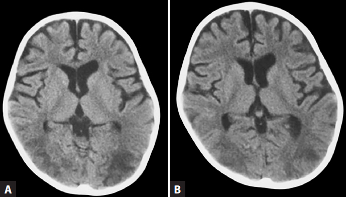
a. Identify the neuroimaging with its findings.
b. What is the diagnosis?
c. What is the predisposing condition for this complication to arise?
Answer
| a. | CT brain plain: Hypodense lesion (edema) in subcortical region of bilateral occipital lobe (remember mnemonic A E I). |
| b. | Posterior reversible encephalopathy syndrome (PRES). |
| c. | Hypertension due to acute glomerulonephritis. |
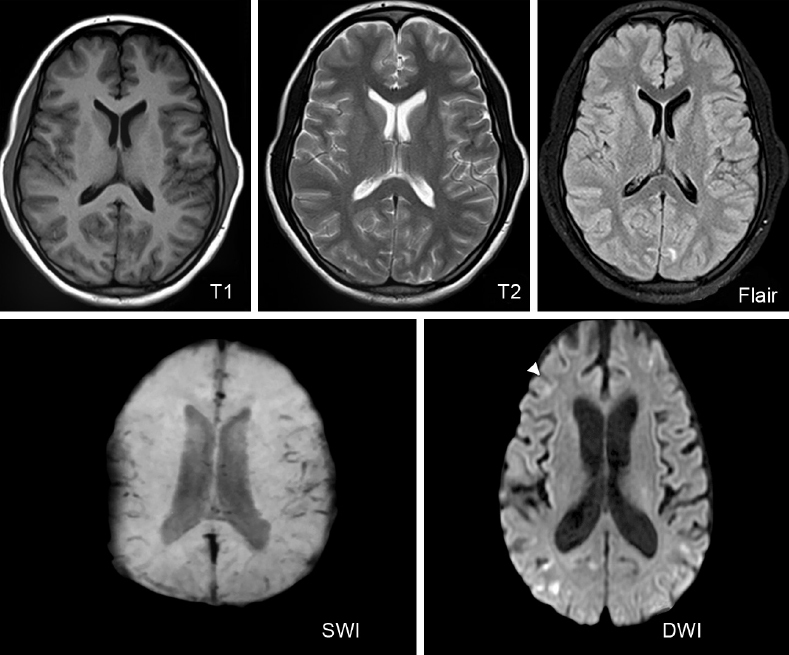
■ SOME BASICS TO UNDERSTAND MRI BRAIN
| ■ | Done either with or without contrast |
| ■ | Three planes: Axial, sagittal, and coronal |
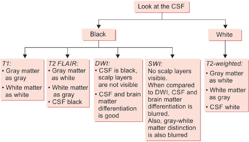
| ■ | Many sequences: T1W, T2W, FLAIR, DWI, SWI, MRS, MRA, and MRV |
| • | T1-weighted (T1W) image (plain) for identifying anatomical defects |
| • | T1-weighted image for postcontrast enhancement, as in infections |
| • | T2-weighted (T2W) for identifying pathology like demyelination |
| • | T2 fluid-attenuated inversion recovery (FLAIR) to define periventricular lesions |
| • | Diffusion-weighted imaging (DWI) for infarct (white) |
| • | Susceptibility-weighted imaging (SWI) for microbleed (black) |
| • | Magnetic resonance spectroscopy (MRS): Spectroscopy identifies the presence particular metabolites and compares the chemical composition of abnormal brain tissue with normal. Horizontal axis represents the parts per million (ppm) and Y axis reflects the quantitative peak of the chemicals such as lactate, lipid, N-acetylaspartate (NAA) creatinine, and choline |
Hunter’s angle in MRS:
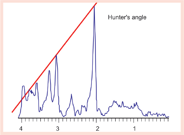
| A term coined from Hunter Sheldon, a neurosurgeon. He used his pocket comb. He placed his comb on the MRS at approximately a 45° angle and connected several of the peaks. If the angle and the peaks roughly corresponded to the 45° angle, the curve is probably normal. If the peak is off the comb’s angle, the curve is abnormal. This is used as a quick method to read MRS and determine whether it is normal or abnormal. |
Chemical shifts of different metabolites in MRS are as follows:
| Metabolite | Chemical shift (ppm) |
|---|---|
| Lipid | 1.3 |
| Lactate | 1.33 |
| NAA | 2.02 |
| Creatine | 3.03 and 3.93 |
| Choline | 3.22 |
| ■ | Lipid peak higher: Tuberculoma |
| ■ | Lactate peak (inverted): Mitochondrial disorder |
| ■ | NAA peak normal (twice that of choline peak) |
| ■ | NAA peak > twice: Canavan’s disease |
| ■ | Choline peak higher: Cancer/malignancy. Sometimes in demyelination and also in normal infant because of rapid turnover of brain tissue. |
■ HOW TO IDENTIFY THE MRI SEQUENCE IN A SIMPLE WAY
Sample Images