MNJ03-001
You are posted in ECHO room to learn about neonatal ECHO. Kindly answer the following questions about ECHO machine:
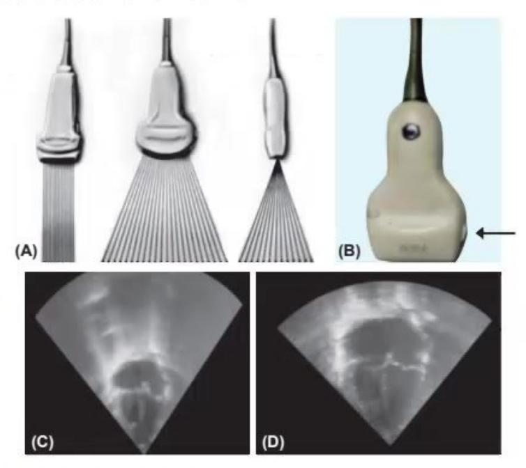
- What are the names of the 3 probes in Fig. A?
- What is the arrow-marked feature in Fig. B?
- In Fig. C and D, which depth is appropriate when you are doing ECHO, and how much depth is considered best?
Answer
- Linear, curved (convex), and phased array probes
- Probe orientation marker
- Figure D is of appropriate depth (the target organ / heart should occupy ~75% of the total screen).
MNJ03-002
What are the modalities in the given three images and write one use of each:
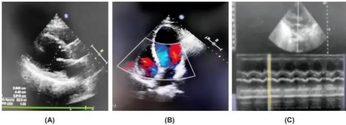
- Identify modality in Image A and state one use.
- Identify modality in Image B and state one use.
- Identify modality in Image C and state one use.
Answer
- 2D / B-mode (Grey scale): To visualize anatomical structure, spatial orientation, and morphology.
- Color Doppler: To assess blood flow presence, direction, turbulence, and velocity in chambers and vessels.
- M-Mode: To assess rapid movement of cardiac structures, chamber dimensions, and wall motion / contractility over time.
MNJ03-003
A preterm neonate (26 weeks) at day 3 became sick with tachycardia, desaturation, and feed intolerance. You observed the baby has bounding pulses and oxygen requirement has increased over the last 12 hours. Your unit in-charge advised ECHO screening in all preterm babies around day 3. Kindly answer the following questions:
- What is the possible diagnosis?
- Why is ECHO screening important in preterm babies at day 3?
- What is the importance of anterior cerebral artery Doppler in cases of PDA in neonates?
Answer
- Hemodynamically significant patent ductus arteriosus (hsPDA)
- To detect and monitor PDA, assess the degree of left-to-right shunt, detect systemic hypoperfusion, and guide targeted medical or surgical closure before complications arise (e.g., pulmonary hemorrhage, NEC, IVH).
- Ductal steal phenomenon: A large left-to-right shunt across the PDA steals blood during diastole, resulting in absent or reversed end-diastolic flow (AEDF/REDF) in the anterior cerebral artery, which indicates severe systemic/cerebral hypoperfusion.
MNJ03-004
In the given two images below, differentiate normal image and abnormal duct, and what is the diagnosis in the abnormal duct image? What are three important pathophysiological parameters of the abnormal duct?
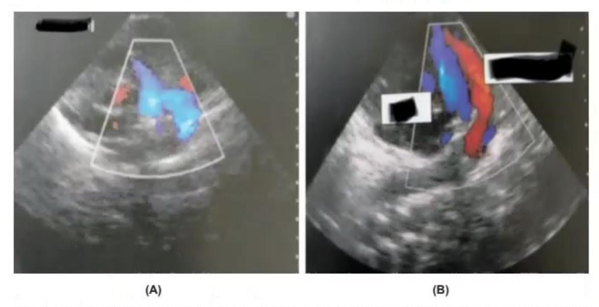
- Differentiate Image A and Image B.
- What are three important pathophysiological consequences of this abnormal duct?
Answer
- Image A: Closed ductus arteriosus (normal); Image B: Large patent ductus arteriosus (abnormal).
- Three pathophysiological consequences:
- Pulmonary overcirculation (pulmonary edema, respiratory deterioration)
- Systemic hypoperfusion / ductal steal (hypoperfusion of brain, gut, and kidneys)
- High-volume left-to-right shunt causing left ventricular volume overload
MNJ03-005
A neonate has issues related to his heart. A screening ECHO was planned by your unit head. While performing the ECHO, this is the first image you saw on screen. Observe this image and answer the following questions:
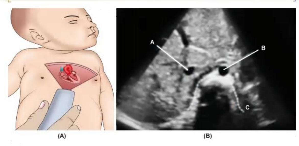
- Which view is this (Image A)?
- What are the marked structures in Image B?
- What does NeoPOCUS mean?
Answer
- Subcostal abdominal situs view (transverse abdominal view)
- A: Inferior Vena Cava (IVC), B: Abdominal Aorta, C: Spine / Vertebral body
- NeoPOCUS: Neonatal Point-Of-Care UltraSound
MNJ03-006
A 5 days old 26 weeks preterm baby started deteriorating in NICU with tachycardia, hyperactive precordium, and wide pulse pressure. You suspected a hemodynamically significant PDA. See the echocardiographic images below and answer the following questions:
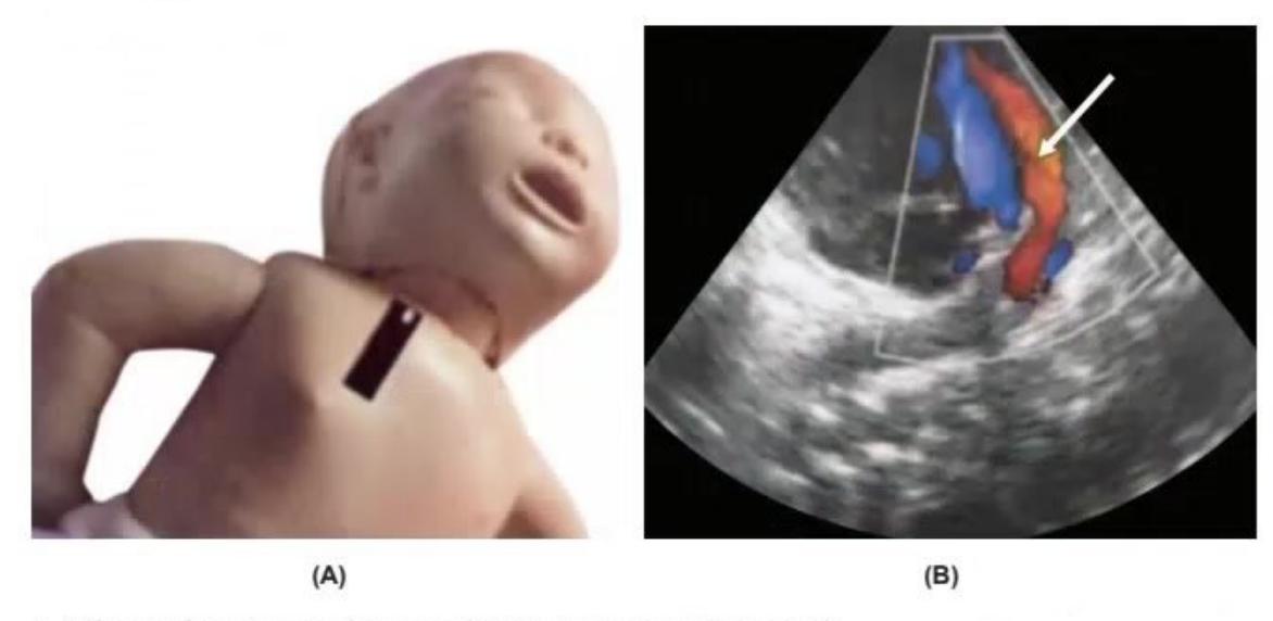
- What is this view used to diagnose PDA in a neonate (Image A)?
- What does the arrow marker show in Image B?
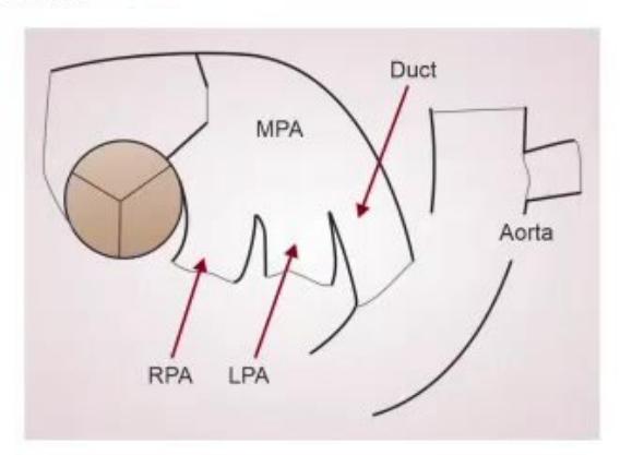
- What are the vessels that make up the characteristic "triple leg / high parasternal view"?
Answer
- High parasternal view (ductal view)
- Patent Ductus Arteriosus (PDA) with left-to-right shunt
- Main pulmonary artery (MPA), Left pulmonary artery (LPA), Right pulmonary artery (RPA), and the Patent Ductus Arteriosus (PDA) connecting MPA/LPA to the descending aorta.
MNJ03-007
A cranial ultrasound (CrUS) is performed on a preterm infant in the NICU. Review the ultrasound images below and answer the questions:

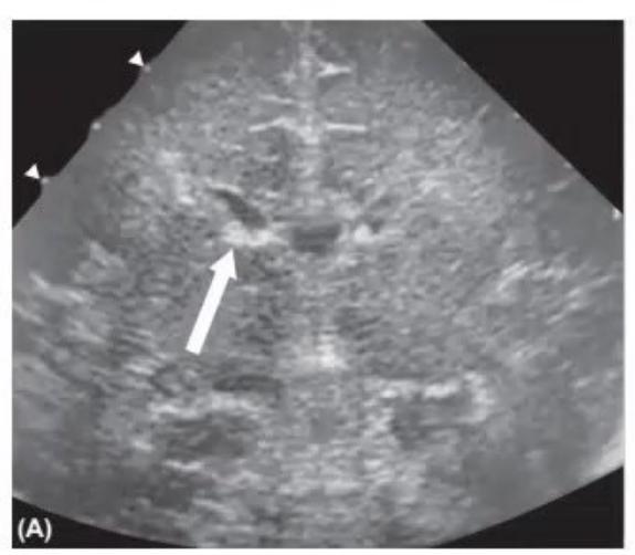
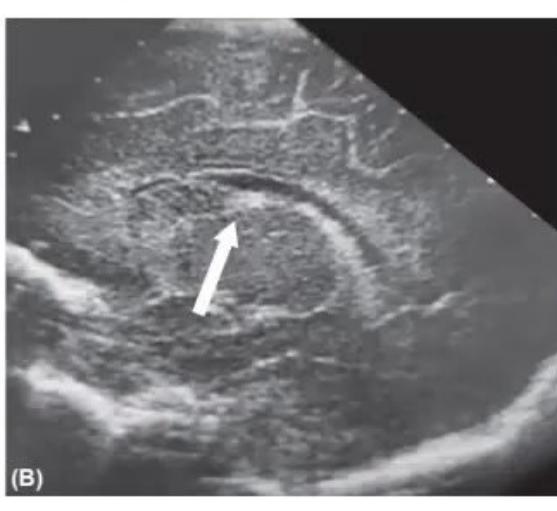
- What are the names of the two cranial ultrasound views shown (Image A and Image B)?
- What is the grade of intraventricular hemorrhage (IVH)?
- What is the anatomical structure shown at the arrow in both images?
- Why is this area important in preterm infants for long-term neurodevelopmental outcome?
Answer
- Image A: Coronal view (at the level of the foramen of Monro); Image B: Parasagittal view
- Grade I Intraventricular Hemorrhage (Germinal Matrix Hemorrhage / Subependymal Hemorrhage)
- Germinal matrix hemorrhage located at the caudothalamic groove
- The germinal matrix is a highly vascular, fragile structure with poor supporting tissue. Hemorrhage in this region can extend into the ventricles (Grade II/III) or cause venous periventricular hemorrhagic infarction (Grade IV), leading to increased risk of hydrocephalus, cerebral palsy, cognitive deficits, and neurodevelopmental impairment.