THG14-001
A 16-month-old boy presented with fever for 2 months. Fever episodes are associated with evanescent rashes (as shown in images) and no joint symptoms. Child playful between fever spikes. Temperatures went as high as 104°F and as low as 96°F once a day. Fever persisted in spite of antibiotics, infectious causes ruled out. Hemoglobin (Hb) 7.6 g/dL, white blood cell (WBC) 18,000/mm3 (N 78, L 10), platelet 7.7 lakh/mm3, erythrocyte sedimentation rate (ESR) 100 mm/hour, C-reactive protein (CRP) 56 mg/dL, serum glutamic oxaloacetic transaminase (SGOT) 38 IU/L, serum glutamate pyruvate transaminase (SGPT) 43 IU/L, serum creatinine 0.6 mg/dL, chest X-ray was normal, ultrasonography (USG) abdomen mild hepatomegaly, and ascites present.
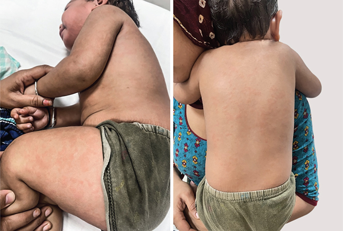
a. Identify the main diagnosis with likely complication described.
b. What are the International League of Associations for Rheumatology (ILAR) criteria for above condition and recent 2019 proposed criteria?
c. Is high CRP suggestive of sepsis in this disease?
d. What is the treatment of the disease?
Answer
- a. Systemic-onset juvenile idiopathic arthritis (sJIA / SOJIA) with Macrophage Activation Syndrome (MAS).
- b. Diagnostic criteria:
- ILAR Criteria: Arthritis in ≥1 joint with fever of at least 2 weeks duration (documented to be daily/quotidian for ≥3 days), accompanied by ≥1 of:
- Evanescent (non-fixed) salmon-pink rash
- Generalized lymphadenopathy
- Hepatomegaly and/or splenomegaly
- Serositis (pericarditis, pleuritis, or peritonitis)
- 2019 PRINTO Proposed Criteria: Daily fever documented for ≥3 consecutive days with quotidian pattern reoccurring over ≥2 weeks, plus 2 Major criteria OR 1 Major + 2 Minor criteria:
- Major Criteria: Evanescent rash, Arthritis
- Minor Criteria:
- Generalized lymphadenopathy and/or hepatomegaly and/or splenomegaly
- Serositis
- Arthralgia lasting ≥2 weeks (in absence of arthritis)
- Leukocytosis (≥15,000/mm³) with neutrophilia
- ILAR Criteria: Arthritis in ≥1 joint with fever of at least 2 weeks duration (documented to be daily/quotidian for ≥3 days), accompanied by ≥1 of:
- c. No. Children with sJIA inherently have marked elevation of CRP with neutrophilic leukocytosis due to high systemic IL-6 levels. Serum procalcitonin is useful to differentiate inflammatory flare from secondary bacterial sepsis.
- d. Treatment depends on predominant phenotype:
- Systemic features predominant: NSAIDs, systemic corticosteroids, IL-1 inhibitors (anakinra, canakinumab) or IL-6 inhibitors (tocilizumab).
- Arthritis predominant: Methotrexate with bridging steroids.
- Refractory/resource-constrained settings: Thalidomide, cyclosporine, leflunomide.
THG14-002
A 6-year-old boy with pain abdomen for 3 weeks extensively evaluated with all reports in normal range. Child was labelled as functional pain abdomen. After 4 weeks, he developed few purpuric lesions over lower limb and buttocks (as shown in images) with nonspecific joint pains. This was followed by scrotal swelling with one episode of blood in stool.
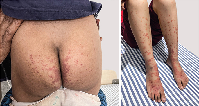
a. What is likely diagnosis and management?
b. Mention the classification criteria of this disease.
c. When are steroids indicated in this condition?
d. Which organ can be involved and needs monitoring after acute episode of this disease?
Answer
- a. IgA vasculitis (formerly Henoch–Schönlein purpura [HSP]).
- Management: Mostly supportive (hydration, analgesia). Short course of oral corticosteroids (prednisolone 1–2 mg/kg/day) for severe abdominal pain or joint symptoms. High-dose IV pulse methylprednisolone (10–30 mg/kg/day for 3 days) ± IV cyclophosphamide for rapidly progressive crescentic nephritis or life-threatening GI bleeding. Long-term DMARDs (mycophenolate mofetil or azathioprine) for chronic nephritis.
- b. EULAR/PReS/PRINTO Classification Criteria:
- Mandatory: Palpable purpura or petechiae (predominantly lower limbs and buttocks) not related to thrombocytopenia
- Plus at least 1 of the following 4:
- Acute diffuse colicky abdominal pain (may include intussusception or GI bleed)
- Histopathology showing leukocytoclastic vasculitis or proliferative glomerulonephritis with predominant IgA deposition
- Acute-onset arthralgia or arthritis
- Renal involvement (proteinuria >0.3 g/24 hr or hematuria / red cell casts)
- c. Indications for corticosteroids in IgA vasculitis:
- Severe colicky abdominal pain / GI bleeding
- Scrotal orchitis / epididymitis
- Pulmonary hemorrhage
- Central nervous system (cerebral) vasculitis
- Severe disabling arthralgia/myalgia or hemorrhagic bullous skin lesions
- d. Kidneys (renal involvement occurs in 20–80% of children). Requires regular blood pressure and urine analysis (protein and microscopy for active sediment) monitoring for at least 6–12 months.
THG14-003
A 1-year-old girl presented with fever for 10 days with no focus. She developed edema of hands and feet with irritability. She was on intravenous antibiotics for the last 6 days but fevers persisted.
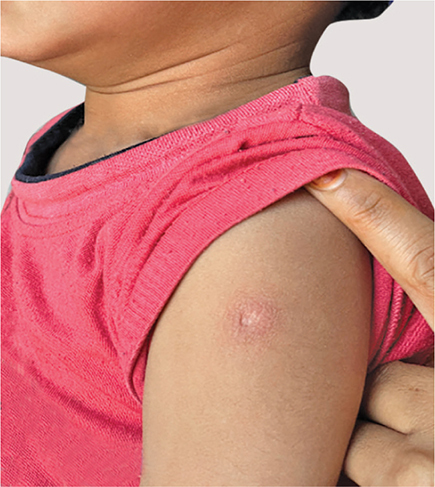
a. What is the likely diagnosis with pointers toward the diagnosis?
b. What is the next most important investigation?
c. Is this investigation needed to confirm the diagnosis?
d. Mention the diagnostic criteria for this disease.
e. What is the treatment for this condition?
Answer
- a. Incomplete Kawasaki disease. Pointers: prolonged unexplained fever >5 days unresponsive to antibiotics, extreme irritability, peripheral extremity edema, BCG site erythema/induration, thrombocytosis, and persistent elevated CRP.
- b. 2D echocardiography to evaluate for myocarditis, pericardial effusion, and coronary artery aneurysms (CAA).
- c. No. Kawasaki disease is primarily a clinical diagnosis; normal echocardiography does not rule out Kawasaki disease.
- d. Classical Diagnostic Criteria:
- Fever lasting ≥5 days
- Plus at least 4 of 5 principal clinical features:
- Bilateral non-exudative bulbar conjunctival injection
- Cervical lymphadenopathy (unilateral, usually >1.5 cm)
- Polymorphous rash (non-vesicular)
- Oral mucosal changes (strawberry tongue, erythema, lip cracking)
- Extremity changes (acute: erythema and indurative edema of hands/feet; subacute: periungual desquamation)
- Note: Can be diagnosed as Incomplete Kawasaki disease with <4 criteria if coronary artery dilation (Z-score ≥2.5) is present on echocardiogram or supportive laboratory criteria are met (AHA 2017 guidelines).
- e. Standard treatment:
- IVIg: 2 g/kg as a single IV infusion over 10–12 hours (within the first 10 days)
- Aspirin: Anti-inflammatory dose (30–50 mg/kg/day in 4 divided doses) until afebrile for 48 hours, followed by low-dose antiplatelet aspirin (3–5 mg/kg/day) for 6–8 weeks (or indefinitely if CAA persist).
- IVIg resistance/high-risk infants: Pulse methylprednisolone, infliximab, or cyclosporine.
THG14-004
A 10-year-old girl presented with prolonged fever, fatigue, and malar rash (as shown in image).
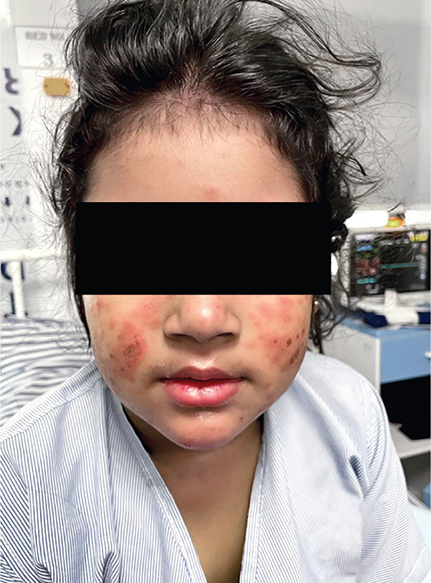
a. What goes against the diagnosis of active systemic lupus erythematosus (SLE)? Explain the reason to select your answer (multiple options can be selected).
b. Mention the 2019 Systemic Lupus International Collaborating Clinics (SLICC) classification criteria of SLE.
Answer
- a. Marked neutrophilic leukocytosis and markedly elevated CRP with normal ESR go against active uncomplicated SLE. Classically, active SLE presents with leukopenia, lymphopenia, thrombocytopenia, high ESR with normal/mildly elevated CRP (a very high CRP in SLE strongly suggests intercurrent infection or serositis).
- b. 2019 EULAR/ACR (SLICC-derived) SLE Classification Criteria:
- Entry Criterion: Positive ANA titer ≥1:80 by HEp-2 immunofluorescence
- Add weighted scores from clinical and immunological domains (Classification requires score ≥10, with ≥1 clinical criterion):
- Clinical Domains:
- Constitutional: Fever (>38.3°C) [2 pts]
- Hematologic: Leukopenia [3 pts], Thrombocytopenia [4 pts], Autoimmune hemolysis [4 pts]
- Neuropsychiatric: Delirium [2 pts], Psychosis [3 pts], Seizures [5 pts]
- Mucocutaneous: Non-scarring alopecia [2 pts], Oral ulcers [2 pts], Subacute cutaneous or discoid lupus [4 pts], Acute cutaneous lupus (malar rash) [6 pts]
- Serosal: Pleural or pericardial effusion [5 pts], Acute pericarditis [6 pts]
- Musculoskeletal: Joint involvement (synovitis / tenderness) [6 pts]
- Renal: Proteinuria >0.5 g/24 hr [4 pts], Class II/V lupus nephritis [8 pts], Class III/IV lupus nephritis [10 pts]
- Immunological Domains:
- Antiphospholipid antibodies (anticardiolipin, anti-β2GP1, lupus anticoagulant) [2 pts]
- Complement levels: Low C3 OR low C4 [3 pts], Low C3 AND low C4 [4 pts]
- SLE-specific antibodies: Anti-dsDNA OR Anti-Smith (anti-Sm) [6 pts]
- Clinical Domains:
THG14-005
A 5-year-old child presented with rashes around knuckles (image A) and muscle weakness for the last 2 years. Bloods reports show raised inflammatory markers and muscle enzymes with a total creatine phosphokinase (CPK) of 5,600 U/L, antinuclear antibody (ANA) by immunofluorescence assay (IFA) 1:320, and extractable nuclear antigen (ENA) profile negative with few hard nodules palpable under the skin of bilateral cubital fossa (image B).
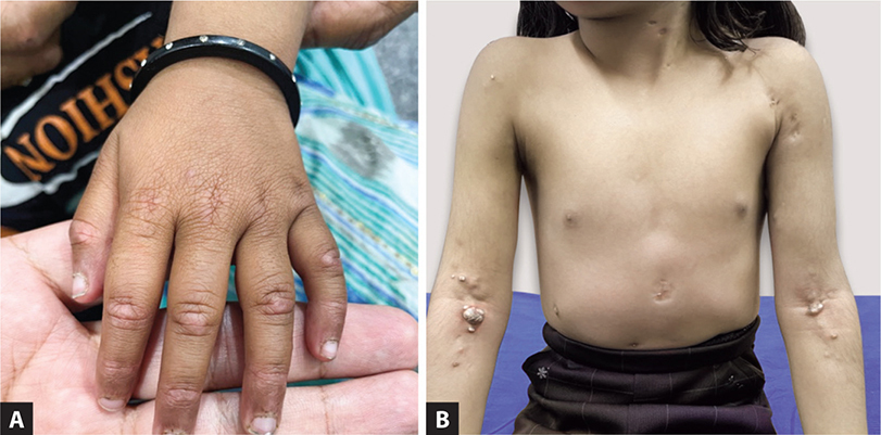
a. Name the disease and different type of rashes seen in this disease.
b. Mention the three commonest myositis antibodies seen in children with this disease and their clinical signifiance.
c. Is muscle biopsy indicated in all cases?
d. Name the complication shown in image B and the most common associated myositis antibody.
Answer
- a. Juvenile Dermatomyositis (JDM). Characteristic cutaneous lesions:
- Heliotrope rash: Violaceous periorbital erythema and edema of upper eyelids
- Gottron papules: Erythematous to violaceous papules/plaques over extensor surfaces of PIP and MCP joints, elbows, and knees
- Malar erythema: Facial flushing crossing the nasolabial folds
- Shawl sign: Erythema over posterior neck, upper back, and shoulders
- V-sign: Confluent erythema over anterior lower neck and upper anterior chest
- Holster sign: Violaceous rash over lateral hips/thighs
- Cuticular changes / periungual telangiectasia and inverse Gottron papules (palmar surface)
- b. Three major Myositis-Specific Antibodies (MSAs) in JDM:
- Anti-TIF1-γ (p155/140): Severe, extensive cutaneous disease, chronic disease course, lipodystrophy
- Anti-NXP-2 (anti-MJ): Severe myopathy, joint contractures, GI ulceration/perforation, high risk of severe dystrophic calcinosis
- Anti-MDA5: Cutaneous ulcerations, rapidly progressive interstitial lung disease (RP-ILD), arthritis, amyopathic/hypomyopathic phenotype
- c. No. While Bohan and Peter criteria (1975) required muscle biopsy, modern consensus and 2017 EULAR/ACR criteria allow non-invasive diagnosis via characteristic skin findings, high muscle enzymes (CPK, LDH, AST, aldolase), myositis antibodies, and muscle MRI (T2/STIR showing edema and fasciitis). Muscle biopsy is reserved for atypical or antibody-negative presentations.
- d. Calcinosis cutis / subcutaneous calcinosis. Most commonly associated with anti-NXP2 (anti-MJ) antibody.
THG14-006
A 2-year-old girl with swelling left knee for last 6 months and stiffness early morning hours. ANA by IFA titer is 1:640 other blood reports in normal range.
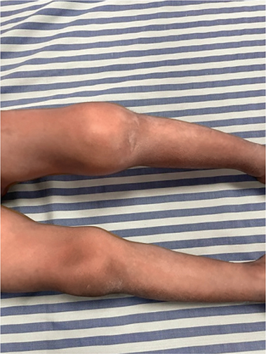
a. What is the relevance of ANA titer in a child with oligoarthritis?
b. What are the types of childhood arthritis as per the ILAR criteria?
c. When would you start oral bridging steroids and methotrexate in this case?
Answer
- a. A positive ANA (titer ≥1:80) in oligoarticular JIA is a major risk factor for asymptomatic, chronic anterior uveitis (which can cause silent blindness). High-risk children (oligoarticular JIA, ANA positive, age at onset <7 years, disease duration <4 years) require slit-lamp ophthalmologic screening every 3 months.
- b. Seven subtypes of JIA as per ILAR criteria:
- Oligoarticular JIA (Persistent or Extended)
- Polyarticular JIA (RF negative)
- Polyarticular JIA (RF positive)
- Systemic-onset JIA (sJIA)
- Enthesitis-related arthritis (ERA)
- Psoriatic arthritis (PsA)
- Undifferentiated JIA
- c. Initial treatment for oligoarticular JIA is intra-articular corticosteroid injections (triamcinolone hexacetonide) ± NSAIDs. Systemic therapy with methotrexate (along with short-course oral bridging corticosteroids) is indicated if there is:
- Recurrence after 2 intra-articular injections within 6 months
- Extension to polyarticular pattern (extended oligoarthritis)
- Severe refractory joint inflammation or high risk/active chronic uveitis