THG20-001
A 1-year-old infant presented with fever. Routine evaluation showed severe anemia with hemoglobin (Hb) 5 g/dL, MCV—50 fl, MCH—20 pg.
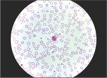
a. Comment on the smear picture.
b. Mention a few conditions which present with such smear.
c. What is Mentzer index?
d. How do you diagnose sideroblastic anemia?
e. What is reticulocyte index?
Answer
- a. Microcytic hypochromic anemia, marked anisopoikilocytosis, pencil/cigar-shaped cells, and target cells.
- b. Iron deficiency anemia (IDA), Thalassemia trait/syndromes, Sideroblastic anemia, Anemia of chronic disease, and Lead poisoning.
- c. Mentzer Index:
suggests Thalassemia trait. suggests Iron Deficiency Anemia.
- d. Increased serum iron and ferritin, transferrin saturation >50%, and presence of ringed sideroblasts (>15%) on Prussian blue iron staining of bone marrow erythroid precursors.
- e. Reticulocyte Production Index (RPI):
- RPI >2 indicates adequate erythropoietic response (hemolysis/blood loss).
- RPI <2 indicates inadequate erythropoietic response (hypoproliferative bone marrow).
THG20-002
A 3-year-old boy presented with fever, productive cough, and shortness of breath. He has had multiple episodes of hand and foot swelling with pain during episodes of fever. On arrival to emergency department (ED), he was febrile, tachycardic, and tachypneic with SpO2 of 90% at room air (RA). His investigation showed hemoglobin (Hb): 6 g/dL, TLC: 20 × 109/L (N—85% and L—15%), and platelets: 500 × 109/L.
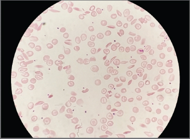
a. Describe the peripheral smear findings.
b. What is the diagnosis?
c. How do you manage?
d. Name new drugs approved for the above disease.
Answer
- a. Sickled RBCs (drepanocytes / crescent-shaped cells), target cells, polychromasia, nucleated RBCs, and Howell-Jolly bodies (autosplenectomy).
- b. Sickle Cell Anemia (HbSS) presenting with Acute Chest Syndrome (ACS).
- c. Management of Acute Chest Syndrome:
- Oxygen supplementation (maintain SpO2 ≥95%)
- Empiric IV antibiotics: 3rd generation Cephalosporin (Ceftriaxone) + Macrolide (Azithromycin)
- Judicious IV hydration (avoid pulmonary edema)
- Adequate analgesia (opioids) and incentive spirometry
- Simple blood transfusion or automated red cell exchange transfusion (erythrocytapheresis) for progressive hypoxemia or severe distress (target HbS <30%)
- Thromboprophylaxis
- d. Newer approved agents:
- Voxelotor (HbS polymerization inhibitor)
- Crizanlizumab (Anti-P-selectin monoclonal antibody)
- L-glutamine (reduces oxidative stress)
THG20-003
A 12-year-old patient regularly transfused β-thalassemia major with serum ferritin of 15,000 ng/mL came with acute onset shortness of breath of 2 days duration. On examination (O/E), pallor was present and jugular venous pressure (JVP) elevated. Systemic examination revealed muffled heart sounds with S3.
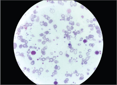
a. What is the significance of T2* cardiac magnetic resonance imaging (MRI)?
b. What is shuttle hypothesis?
c. Name the new drug approved for β-thalassemia.
d. What is the management of acute cardiac iron overload?
Answer
- a. Significance of T2* Cardiac MRI:
- Gold standard non-invasive method for quantifying myocardial iron overload (cardiac T2* <20 ms indicates cardiac iron overload; <10 ms indicates severe iron overload with high risk of heart failure/arrhythmias).
- Serum ferritin is a poor predictor of cardiac iron overload when ferritin >3,000 ng/mL.
- b. Shuttle Hypothesis (Chelator Synergy):
- Small, lipid-soluble Deferiprone (DFP) rapidly enters cardiomyocytes, removes intracellular iron, and shuttles it to extracellular Deferoxamine (DFO), which binds it tightly and promotes urinary/biliary excretion.
- c. Luspatercept (erythroid maturation agent / recombinant fusion protein that inhibits SMAD2/3 signaling, reducing transfusion burden).
- d. Management of acute cardiac siderosis:
- Continuous 24-hour IV Deferoxamine (50–60 mg/kg/day) combined with oral Deferiprone (75–100 mg/kg/day).
- Inotropes, ACE inhibitors, diuretics, and antiarrhythmics for heart failure.
THG20-004
A 4-year-old child was treated with cotrimoxazole for urinary infection. He presented with acute fatigue and high colored urine.
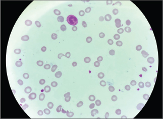
a. Describe the peripheral smear.
b. What is the likely diagnosis?
c. What is the recent classification?
d. What is the mode of inheritance?
Answer
- a. "Bite cells" (degmacytes), "blister cells", spherocytes, polychromasia, and helmet-shaped schistocytes (Heinz bodies seen on supravital stain).
- b. Glucose-6-Phosphate Dehydrogenase (G6PD) Deficiency with drug-induced acute intravascular hemolytic anemia.
- c. WHO Classification of G6PD Deficiency:
- Class I: Severe enzyme deficiency (<10% activity) with chronic non-spherocytic hemolytic anemia
- Class II: Severe deficiency (<10% activity) with acute intermittent hemolysis
- Class III: Moderate deficiency (10–60% activity) with episodic hemolysis triggered by drugs/infections
- Class IV: Normal enzyme activity (60–150%)
- Class V: Increased enzyme activity (>150%)
- d. X-linked recessive.
THG20-005
A 4-year-girl with lethargy and poor weight gain. Examination showed knuckle hyperpigmentation.
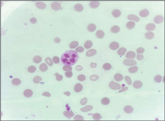
a. Identify the above smear.
b. What is the diagnosis?
c. What are the additional findings expected in morphology?
d. Name an infection causing it.
Answer
- a. Oval macrocytes (macro-ovalocytes) and hypersegmented neutrophils (≥6 lobes in one neutrophil or ≥5% neutrophils with 5 lobes).
- b. Megaloblastic Anemia (Vitamin B12 or Folate deficiency).
- c. Additional findings:
- Smear: Howell-Jolly bodies, Cabot rings, basophilic stippling, leukopenia, thrombocytopenia.
- Bone marrow: Hypercellular marrow with megaloblastic erythroid hyperplasia, nuclear-cytoplasmic asynchrony, giant metamyelocytes, and giant band forms.
- d. Diphyllobothrium latum (fish tapeworm causing Vitamin B12 malabsorption).
THG20-006
A 4-year-old boy presented with acute epistaxis and gum bleed with platelet count of 10,000/mm3. He had history of fever 1 week ago. His peripheral smear is given below.
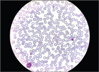
a. What is the diagnosis?
b. What is immature platelet fraction (IPF)?
c. What are the new drugs approved in this condition?
d. What is the management?
Answer
- a. Immune Thrombocytopenia (ITP).
- b. Immature Platelet Fraction (IPF): Percentage of newly released, RNA-rich reticulated platelets reflecting active megakaryocyte thrombopoiesis; elevated in peripheral platelet destruction (ITP) and low in marrow aplasia.
- c. Approved Second-line / Thrombopoietin Receptor Agonists (TPO-RAs):
- Eltrombopag
- Romiplostim
- Avatrombopag
- Rituximab (Anti-CD20)
- d. Management:
- Asymptomatic / mild cutaneous bleeding: Observation and follow-up.
- Active mucosal bleeding or severe thrombocytopenia: Oral Prednisolone (2–4 mg/kg/day for 4–7 days) OR IV Immunoglobulin (IVIG 0.8–1 g/kg single dose) OR IV Anti-D (in Rh-positive, non-splenectomized children).
THG20-007
A 3-year-old child presented with fever, epistaxis, generalized lymphadenopathy, and hepatosplenomegaly.
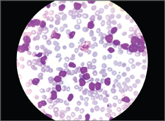
a. Describe the smear findings.
b. What are the drugs commonly employed in treatment?
c. What are the features of tumor lysis syndrome?
d. What is the prognostic indicator by cytogenetics?
Answer
- a. Smear shows high cellularity with replacement by monomorphic lymphoblasts (high N:C ratio, immature condensed-to-fine chromatin, indistinct nucleoli, scant agranular cytoplasm) and smudge cells -> Acute Lymphoblastic Leukemia (ALL).
- b. Induction chemotherapy: Glucocorticoids (Prednisolone/Dexamethasone), Vincristine, L-Asparaginase / PEG-asparaginase, and Daunorubicin (4-drug induction).
- c. Tumor Lysis Syndrome (Cairo-Bishop criteria): Hyperkalemia, Hyperuricemia, Hyperphosphatemia, and Hypocalcemia.
- d. Cytogenetic prognostic factors:
- Favorable / Good: Hyperdiploidy (>50 chromosomes / DNA index >1.16), t(12;21) [ETV6-RUNX1].
- Unfavorable / Poor: Hypodiploidy (<44 chromosomes), t(9;22) [BCR-ABL1 / Philadelphia chromosome], KMT2A (MLL) rearrangements / t(4;11), TP53 mutations.
THG20-008
A 6-year-old child presented with anuria and seizures with hypertension. His investigations showed urea: 100 mg/dL and creatinine: 6 mg/dL. Urine microscopy shows protein 2+. He has past history of diarrhea with abdominal pain present.
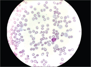
a. Identify the cells.
b. What is the diagnosis?
c. What is the etiology?
d. What is the treatment?
Answer
- a. Helmet cells, triangular cells, and fragmented red blood cells (Schistocytes / Microangiopathic hemolytic anemia).
- b. Hemolytic Uremic Syndrome (HUS) (typical / D+ HUS).
- c. Etiology: Shiga toxin-producing Escherichia coli (STEC serotype O157:H7) or Shigella dysenteriae type 1.
- d. Treatment:
- Supportive therapy: Fluid and electrolyte management, control of hypertension.
- Renal replacement therapy (hemodialysis / peritoneal dialysis) for anuria/uremia/fluid overload.
- Packed RBC transfusion for severe anemia (avoid platelet transfusion unless life-threatening hemorrhage).
- Avoid antimotility agents and antibiotics (may increase toxin release).
THG20-009
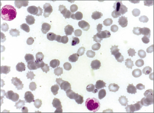
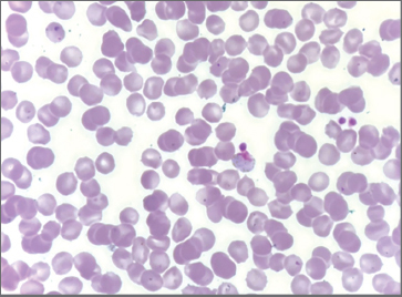
A 4-year-old male child with fever and seizures was brought in unconscious state. His peripheral smear is given below.
a. What do you see in the smear?
b. What is the drug of choice?
c. Which underlying condition is to be ruled out before starting antimalarials?
Answer
- a. Smear A: Intraerythrocytic delicate ring forms (trophozoites) of Plasmodium falciparum (multiple rings per RBC, applique forms). Smear B: Banana/crescent-shaped gametocyte of P. falciparum (or amoeboid trophozoite of P. vivax).
- b. Drug of choice for severe / cerebral malaria: IV Artesunate (2.4 mg/kg IV at 0, 12, 24 hours, then daily) followed by a complete oral course of Artemisinin-based Combination Therapy (ACT).
- c. G6PD Deficiency (mandatory before administering Primaquine or 8-aminoquinolines to avoid severe acute intravascular hemolysis).
THG20-010
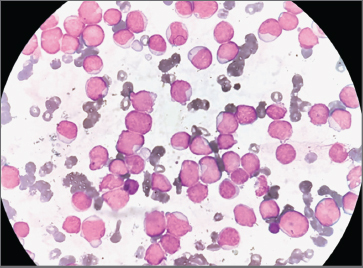
A 10-year-old child presented with fever, lethargy, and epistaxis. Examination shows gum hypertrophy and splenomegaly.
a. What is the diagnosis?
b. What are the common genetic translocations identified?
c. What are the drugs used in above condition?
d. What are the syndromes associated?
Answer
- a. Acute Myeloid Leukemia (AML) (specifically acute monocytic/myelomonocytic leukemia presenting with gingival hyperplasia; blasts with open chromatin, prominent nucleoli, and Auer rods).
- b. Cytogenetic translocations:
- t(8;21)(q22;q22) [RUNX1-RUNX1T1]
- inv(16)(p13.1q22) / t(16;16) [CBFB-MYH11]
- t(15;17)(q22;q12) [PML-RARA - Acute Promyelocytic Leukemia]
- 11q23 (KMT2A) translocations
- c. Chemotherapy drugs: Cytarabine (Ara-C) and Daunorubicin (3+7 regimen); ATRA + Arsenic trioxide for APL.
- d. Associated syndromes: Down syndrome (Trisomy 21), Fanconi anemia, Shwachman-Diamond syndrome, Severe congenital neutropenia (Kostmann), Bloom syndrome, Neurofibromatosis type 1.
THG20-011
A 10-year-old girl with chronic kidney disease (CKD) on maintenance hemodialysis presented with easy fatigability, lethargy, and poor weight. She has no history of chronic transfusion, jaundice, and blood loss.
a. What is functional iron deficiency?
b. What are the new RBC parameters helpful in this case which can guide therapy?
c. What is the target Hb level in CKD patients?
d. How do you manage?
Answer
- a. Functional Iron Deficiency: Condition where total body storage iron is normal or elevated, but iron cannot be mobilized rapidly enough from macrophages and hepatocytes to supply the demands of enhanced bone marrow erythropoiesis (due to hepcidin excess).
- b. Reticulocyte Hemoglobin Content (Ret-He / CHr <29 pg) and percentage of hypochromic red blood cells (%Hypo >6%).
- c. Target Hb level: 10.0 to 11.5 g/dL (avoid Hb >12 g/dL due to thrombosis/cardiovascular risks).
- d. Management:
- IV Iron therapy (Iron sucrose / Ferric carboxymaltose)
- Erythropoiesis-Stimulating Agents (ESA - Recombinant Human Erythropoietin-alpha / Darbepoetin-alpha)
THG20-012
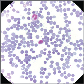
A 5-year-old boy presented with lethargy, persistent jaundice, and abdominal distension. O/E, he had Hb: 5 g/dL, MCV: 80 fl, MCH: 30 pg, and MCHC: 38 g/dL. His smear is shown below.
a. Comment on the smear.
b. What are the conditions associated with the above image?
c. What is the indication of splenectomy in the above condition?
d. What are the common genetic mutations with the condition?
Answer
- a. Numerous uniform, dense, small microspherocytes lacking central pallor with polychromasia.
- b. Hereditary Spherocytosis, Autoimmune Hemolytic Anemia (AIHA), ABO incompatibility of newborn, thermal burns, and Clostridium welchii sepsis.
- c. Indications for splenectomy:
- Severe anemia requiring frequent blood transfusions
- Growth failure, skeletal deformities, or failure to thrive
- Symptomatic splenomegaly / symptomatic cholelithiasis (perform after 5–6 years of age following complete encapsulation vaccination).
- d. Mutations in RBC membrane skeleton proteins: Ankyrin (ANK1), Beta-spectrin (SPTB), Alpha-spectrin (SPTA1), Band 3 (SLC4A1), and Protein 4.2 (EPB42).
THG20-013
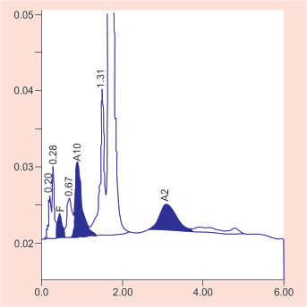
An 11-year-old girl presented with refractory iron deficiency anemia.
a. What is the likely diagnosis?
b. What are the conditions associated with low HbA2?
c. What are the common α variants in India?
d. What is the principle of high-performance liquid chromatography (HPLC)?
Answer
- a. Beta-Thalassemia Trait / Minor (elevated HbA2 >3.5–4.0%).
- b. Conditions associated with low HbA2 (<2%):
- Severe iron deficiency anemia
- Alpha-thalassemia (e.g., HbH disease)
- Delta-thalassemia
- Sideroblastic anemia
- c. Common alpha-thalassemia deletions in India:
single-gene deletion and deletion. - d. Principle of HPLC: Automated cation-exchange high-performance liquid chromatography separates hemoglobin fractions based on charge differences as they bind to negatively charged resin cartridges and elute with gradients of ionic buffer at characteristic retention times.
THG20-014
A 10-year-old girl came with lethargy, jaundice, and high-colored urine. She has no other significant history. Investigations revealed Hb: 5 g/dL, MCV: 110 fl, MCH: 40 pg, MCHC: 40 g/dL, and reticulocyte count: 8%. Smear shows normochromic normocytic red cells with microspherocytes and 25 nRBC/100 white blood cell (WBC). Direct Coombs test showed 4+. Monospecific Coombs test showed immunoglobulin G (IgG): 4+, C3b = 4+.
a. What is the significance of the monospecific Coombs test?
b. What is the first-line treatment for the above condition?
c. What are the causes associated with the above condition?
d. In which condition Donath–Landsteiner antibody is detected?
Answer
- a. Significance: Distinguishes Warm AIHA (IgG-positive ± C3d-positive) from Cold AIHA / Cold Agglutinin Disease (C3d-positive only) and mixed-type AIHA.
- b. First-line treatment: Systemic glucocorticoids (Oral Prednisolone 1–2 mg/kg/day or IV Methylprednisolone pulses).
- c. Secondary causes: Systemic Lupus Erythematosus (SLE), Common Variable Immunodeficiency (CVID), Autoimmune Lymphoproliferative Syndrome (ALPS), viral infections (EBV, CMV, HIV, Hepatitis), lymphoma, drugs.
- d. Paroxysmal Cold Hemoglobinuria (PCH) (biphasic IgG Donath-Landsteiner hemolysin).
THG20-015
A 6-year-old child came with lethargy and bleeding gums. O/E, child was short with generalized hyperpigmentation and bifid thumb.
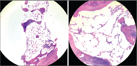
a. What is the likely diagnosis?
b. What are the other tests routinely performed?
c. What are the hematological and nonhematological complications of the above condition?
d. What is the treatment?
Answer
- a. Fanconi Anemia (Constitutional Aplastic Anemia).
- b. Diagnostic tests: Chromosomal breakage analysis on peripheral blood lymphocytes using DNA-crosslinking clastogenic agents (Diepoxybutane [DEB] or Mitomycin C [MMC]), followed by FANCA-FANCV multigene next-generation sequencing.
- c. Complications:
- Hematological: Progressive bone marrow failure (aplastic anemia), Myelodysplastic Syndrome (MDS), and Acute Myeloid Leukemia (AML).
- Non-hematological: Solid tumors (squamous cell carcinoma of oral cavity, esophagus, and anogenital tract; hepatic adenoma/carcinoma), endocrine dysfunction (hypothyroidism, GH deficiency), renal abnormalities.
- d. Definitive cure: Allogeneic Hematopoietic Stem Cell Transplantation (HSCT). Androgens (Oxymetholone) and G-CSF used as bridging therapy.
THG20-016
Bone marrow aspirate of an infant with isolated hepatosplenomegaly.
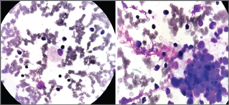
a. What is the diagnosis?
b. What is the enzyme deficiency associated with this disease?
c. Describe the classical bone marrow picture.
Answer
- a. Gaucher Disease (Type 1 / Type 2 / Type 3).
- b. Deficiency of lysosomal Acid beta-glucosidase (Glucocerebrosidase).
- c. Gaucher cells: Large, lipid-laden macrophages/histiocytes (20–100 µm) with eccentric nuclei and abundant fibrillary, pale blue-gray cytoplasm resembling "wrinkled tissue paper" or "crinkled silk".
THG20-017
A 1-year-boy presented with fever of 2 weeks duration. He had hemoglobin of 5 g/dL, total white cell counts of 2000/mm3 and platelets of 20 ×109/L with hepatosplenomegaly.
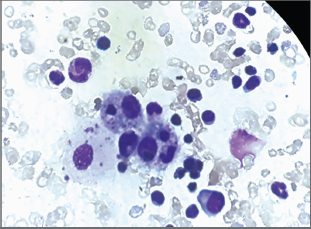
a. What is the diagnosis?
b. What are the mutation defects that can be associated?
c. What are the laboratory abnormalities specific to the disease?
d. Name some drugs used to treat the disease.
Answer
- a. Hemophagocytic Lymphohistiocytosis (HLH) (bone marrow showing activated macrophage engulfing RBCs, platelets, and leukocytes—hemophagocytosis).
- b. Genetic mutations (Primary/Familial HLH): PRF1 (Perforin), UNC13D (Munc13-4), STX11 (Syntaxin 11), STXBP2 (Munc18-2), RAB27A (Griscelli 2), LYST (Chédiak-Higashi).
- c. HLH-2004 diagnostic criteria abnormalities:
- Cytopenias affecting ≥2 lineages
- Hyperferritinemia (>500 µg/L, often >3,000–10,000)
- Hypertriglyceridemia (fasting triglycerides ≥265 mg/dL)
- Hypofibrinogenemia (≤150 mg/dL)
- Elevated soluble CD25 (sIL-2 receptor)
- Low/absent NK cell activity
- d. Treatment (HLH-94/2004 protocol): Dexamethasone, Etoposide (VP-16), Cyclosporine A, IVIG, and Emapalumab (Anti-IFN-gamma).
THG20-018
A 10-year-old acute myeloid leukemia (AML) child on induction phase chemotherapy presented with cough, breathlessness, and pleuritic chest pain. High-resolution computed tomography (HRCT) is shown below showed multiple nodules in both lung field with halo sign.
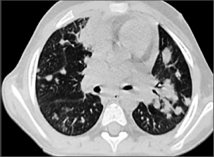
a. What is the likely diagnosis?
b. What are the etiological factors?
c. What are the other additional tests for confirmation?
d. What are the treatments available for the above condition?
Answer
- a. Invasive Pulmonary Aspergillosis (IPA) (CT showing macronodules surrounded by a perimeter of ground-glass attenuation—CT Halo Sign indicating angioinvasion and hemorrhage).
- b. Aspergillus fumigatus, Aspergillus flavus, Aspergillus niger, Aspergillus terreus; Mucorales (Rhizopus).
- c. Confirmatory tests:
- Serum and Bronchoalveolar Lavage (BAL) Galactomannan antigen assay
- BAL fungal smear (calcofluor white, 45° acute-angle branching septate hyphae) and fungal culture
- Aspergillus PCR
- d. Antifungal therapy:
- First-line: IV/Oral Voriconazole (or Isavuconazole)
- Alternative: Liposomal Amphotericin B
THG20-019
An 18-year-old girl presented with menorrhagia with history of similar menorrhagia in mother.
a. What are the next investigations to be done?
b. What is the probable diagnosis?
c. What is the mode of inheritance?
d. What is the available management?
Answer
- a. Investigations:
- CBC and platelet count (usually normal)
- Coagulation screen: Bleeding time / PFA-100, PT, and aPTT (may be prolonged)
- Specific assays: von Willebrand Factor Antigen (VWF:Ag), Ristocetin Cofactor activity (VWF:RCo), and Factor VIII coagulant activity (FVIII:C)
- b. von Willebrand Disease (VWD) (most common inherited bleeding disorder).
- c. Mode of inheritance:
- Autosomal dominant: Type 1, Type 2A, Type 2B, Type 2M
- Autosomal recessive: Type 2N, Type 3
- d. Management:
- Antifibrinolytics: Oral/IV Tranexamic acid
- Combined oral contraceptive pills (COCP) or Levonorgestrel IUD for menorrhagia
- Desmopressin (DDAVP) nasal spray/IV (for Type 1)
- Plasma-derived VWF/FVIII concentrates or recombinant VWF (Vonvendi)
THG20-020
A 1-year-old boy presented with spontaneous skin ecchymosis and epistaxis. She has no history of hematoma or hemarthrosis. There is no family history of bleeding.
a. Identify the diagnosis.
b. What is the mode of inheritance?
c. What is the defect?
d. What are the treatment options available?
Answer
- a. Glanzmann Thrombasthenia (GT).
- b. Autosomal recessive.
- c. Deficiency or dysfunction of platelet membrane Integrin alpha-IIb/beta-3 (Glycoprotein IIb/IIIa / CD41/CD61) receptor complex, leading to failure of platelet aggregation with all agonists except ristocetin.
- d. Treatment options:
- Local hemostatic measures, pressure packing, and oral Tranexamic acid
- HLA-matched / leukodepleted Platelet transfusions (reserved for refractory severe bleeding)
- Recombinant activated Factor VII (rFVIIa)
- Hematopoietic Stem Cell Transplantation (curative for severe phenotype)
THG20-021
A 1-year-old boy presented with acute right hemarthrosis.
a. What is the likely diagnosis?
b. What is the mode of inheritance?
c. What is aged and adsorbed plasma?
d. How you do classify the above condition?
Answer
- a. Hemophilia A (Factor VIII deficiency) or Hemophilia B (Factor IX deficiency).
- b. X-linked recessive.
- c. Aged vs. Adsorbed Plasma in mixing studies:
- Aged plasma: Contains all coagulation factors EXCEPT labile factors (Deficient in Factor V and Factor VIII).
- Adsorbed plasma: Vitamin K-dependent factors are adsorbed by barium sulfate/aluminum hydroxide (Deficient in Factors II, VII, IX, and X).
- d. Severity Classification:
- Severe: Factor VIII/IX level <1% (<0.01 IU/mL) — spontaneous hemarthroses and muscle bleeds.
- Moderate: Factor VIII/IX level 1–5% (0.01–0.05 IU/mL) — bleeding with minor trauma.
- Mild: Factor VIII/IX level 5–40% (0.05–0.40 IU/mL) — bleeding only with major trauma or surgery.
THG20-022
Blood Bank Equipment.
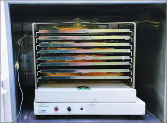
a. Identify the above image.
b. What is the blood product used in this?
c. What is the shelf-life of platelet?
d. What is the temperature storage?
Answer
- a. Platelet Incubator with Agitator (flatbed continuous platelet shaker).
- b. Platelet concentrates (Random Donor Platelets / Single Donor Apheresis Platelets).
- c. 5 days (up to 7 days with approved bacterial screening/pathogen reduction).
- d. 20°C to 24°C (with continuous gentle horizontal agitation to prevent aggregation and maintain gas exchange).
THG20-023
A 1-month-old neonate with Down syndrome presented with abdominal distension. Investigations showed Hb: 11 g/dL, total platelet counts: 50 × 109/L, and peripheral smear: 50% large atypical cells with high nuclear–cytoplasmic ratio, with deep basophilic cytoplasm with cytoplasmic blebs.
a. What is the likely diagnosis?
b. What is the natural course of disease?
c. Name gene involved.
d. What is the management?
Answer
- a. Transient Abnormal Myelopoiesis (TAM) / Transient Myeloproliferative Disorder (TMD) of Down Syndrome.
- b. Natural course: Spontaneous resolution occurs in >80–90% of infants within the first 3 months of life; however, ~20–30% develop true Myeloid Leukemia of Down Syndrome (ML-DS / AML-M7) within the subsequent 1–4 years.
- c. Somatic GATA1 gene mutation (in addition to constitutional Trisomy 21).
- d. Management:
- Asymptomatic / mild cases: Close observation and monitoring of blood counts.
- Life-threatening symptoms (hydrops, severe hepatic dysfunction, extreme leukocytosis, organ failure): Low-dose Cytarabine (Ara-C) (0.5–1.5 mg/kg/day for 3–7 days).
THG20-024
A 5-year-old child presented with acute onset shortness of breath after taking over the counter medicine chloroquine. SpO2 is 80% not improving with oxygen and PaO2 is 90 mm Hg.
a. What is the diagnosis?
b. What is the etiology?
c. What are the confirmatory tests?
d. What is the management?
Answer
- a. Acquired Methemoglobinemia ("saturation gap": low pulse oximetry SpO2 refractory to 100% O2 with normal arterial PaO2 on ABG, chocolate brown blood).
- b. Etiology: Oxidizing agents—Antimalarials (Chloroquine, Primaquine), Dapsone, Sulfonamides, Local anesthetics (Benzocaine, Prilocaine), Nitrates/Nitrites.
- c. Confirmatory test: Multiple-wavelength CO-oximetry directly measuring MetHb percentage (MetHb level >1.5–3%).
- d. Management:
- Discontinue offending drug; high-flow 100% supplemental oxygen.
- First-line antidote: IV Methylene Blue (1–2 mg/kg of 1% solution IV over 5 minutes) (Contraindicated in G6PD deficiency due to risk of severe hemolysis).
- Alternative in G6PD deficiency / refractory cases: High-dose IV Ascorbic acid (Vitamin C), hyperbaric oxygen, or red cell exchange transfusion.
THG20-025
A 3-day-old newborn, born to parent with third degree consanguinity, presented with blackish discoloration of toes and fingers.
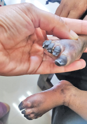
a. What is the diagnosis?
b. How to confirm diagnosis?
c. What is the treatment option?
d. What is the differential diagnosis?
Answer
- a. Neonatal Purpura Fulminans (Homozygous Protein C or Protein S Deficiency).
- b. Markedly reduced or undetectable plasma Protein C / Protein S activity and antigen levels; confirmatory PROC / PROS1 gene sequencing.
- c. Treatment:
- Acute phase: Purified Protein C concentrate (or Fresh Frozen Plasma 10–15 mL/kg q 12h) + therapeutic systemic anticoagulation (Unfractionated / LMW Heparin).
- Long-term: Lifelong Protein C replacement or oral anticoagulants; Liver transplantation is curative.
- d. Differential diagnosis: Sepsis-induced DIC with purpura fulminans (Meningococcemia), catastrophic antiphospholipid syndrome, severe congenital antithrombin deficiency, warfarin-induced skin necrosis.
THG20-026
A 3-month-old child is brought with the complaints of severe anemia and noisy breathing. He has moderate splenomegaly.
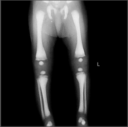
a. What is the diagnosis?
b. What is the characteristic peripheral smear finding seen in these children?
c. What visual complication happens in this disease?
d. What is the curative treatment for this disease?
Answer
- a. Malignant Infantile Osteopetrosis (Albers-Schönberg disease / Marble bone disease) (diffuse skeletal sclerosis, "bone within a bone" appearance).
- b. Leukoerythroblastic blood picture (nucleated RBCs, myelocytes, metamyelocytes, and teardrop RBCs due to myelophthisis / marrow space obliteration).
- c. Progressive bilateral optic atrophy and blindness due to bony narrowing and compression of the optic foramina / optic nerve canal.
- d. Allogeneic Hematopoietic Stem Cell Transplantation (HSCT) (provides donor monocytic precursors that differentiate into functional osteoclasts).
THG20-027
A 4-year-old boy has presented to the emergency room with acute pain abdomen. He has a doughy abdomen and an ultrasound of the abdomen reveals an ileocecal mass. His renal functions are deranged with following ECG changes.
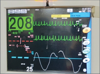
a. What is the ECG finding and the etiology beyond the ECG change?
b. What is the metabolic complication leading to this emergency?
c. Which tumor has a high predisposition to develop this metabolic syndrome?
d. What are the treatment options?
Answer
- a. ECG finding: Tall, peaked, symmetrical tented T waves caused by acute severe Hyperkalemia.
- b. Acute Tumor Lysis Syndrome (TLS) with secondary acute kidney injury.
- c. Burkitt Lymphoma (and T-cell ALL) due to extremely rapid cellular turnover and high tumor burden.
- d. Treatment:
- Emergency hyperkalemia stabilization: IV Calcium gluconate (10%), regular insulin + dextrose infusion, nebulized salbutamol, IV Sodium bicarbonate.
- Hyperhydration (2.5–3 L/m²/day without added potassium).
- Hypouricemic therapy: Recombinant urate oxidase (Rasburicase) or Allopurinol.
- Urgent Renal Replacement Therapy (Hemodialysis / Continuous RRT) for refractory hyperkalemia, severe hyperphosphatemia, or fluid overload.
THG20-028
A 5-year-old boy presented with recurrent pain in joints for past 10 months—initially left shoulder, then left ankle, then right hip pain. He has mild hepatomegaly. His blood smear finding and MRI pelvis picture are shown below.
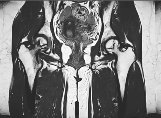
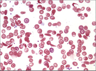
a. Comment on this smear and what is your diagnosis?
b. What is the complication noted in the MRI?
c. How do you confirm the diagnosis of this condition?
d. What are the curative treatment options?
Answer
- a. Smear: Crescentic sickle cells, target cells, nucleated RBCs -> Sickle Cell Disease (HbSS).
- b. Bilateral Avascular Necrosis (Aseptic necrosis / osteonecrosis) of the femoral heads (Right > Left).
- c. Confirmatory tests: High-Performance Liquid Chromatography (HPLC) / Hemoglobin Electrophoresis (demonstrating HbS band, absent HbA in homozygous state) and beta-globin gene sequencing.
- d. Curative therapies: Allogeneic Hematopoietic Stem Cell Transplantation (HSCT) and Gene Therapy (Lentiviral vector gene addition / CRISPR-Cas9 gene editing - Exagamglogene autotemcel).
THG20-029
An 8-year-old boy presented with a right-sided scalp swelling. X-ray skull reveals a lytic skull lesion corresponding to this scalp swelling. He has no other lytic lesions. He has no lymphadenopathy or hepatosplenomegaly. His blood counts are normal.
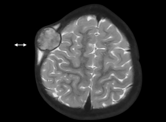
a. What is the probable diagnosis?
b. What is the expected histopathological finding and electron microscopy finding?
c. What are the treatment options?
d. What is the anticipated endocrine abnormality?
Answer
- a. Langerhans Cell Histiocytosis (LCH) (Single-system unifocal bone disease / Eosinophilic granuloma with "punched-out" calvarial lytic lesion).
- b.
- Histopathology: Infiltration by Langerhans histiocytes (grooved "coffee-bean" nuclei) mixed with abundant eosinophils, lymphocytes, and osteoclast-like giant cells; positive for CD1a, CD207 (Langerin), and S100.
- Electron Microscopy: Tennis-racket shaped Birbeck granules in cytoplasm.
- c. Treatment: Curettage / surgical excision, intralesional steroid injection (Methylprednisolone), or systemic Vinblastine + Prednisolone for multifocal/systemic disease.
- d. Central Diabetes Insipidus (due to hypothalamic-pituitary axis involvement).
THG20-030
A 5-month-old child, product of third-degree consanguineous marriage, is admitted with second episode of pneumonia. He has no reaction at the BCG scar site. There is family history of previous sibling death at 10 months of age due to severe pneumonia.
a. What is the possible inborn error of immunity in this child?
b. Which newborn screening test if performed can detect the disease in the latent phase?
c. Which subset of this disease has an enzyme replacement available? Name the enzyme available.
d. What is the precaution to be taken when transfusing these children?
Answer
- a. Severe Combined Immunodeficiency (SCID) (T-cell negative SCID).
- b. TREC Assay (T-cell Receptor Excision Circles quantitation via RT-PCR on dried blood spots).
- c. Adenosine Deaminase (ADA) Deficiency SCID; Enzyme replacement therapy: Pegademase bovine / Elapegademase (PEG-ADA).
- d. All blood products must be Leukodepleted and Gamma-Irradiated (to prevent fatal Transfusion-Associated Graft-versus-Host Disease [TA-GvHD]) and CMV-seronegative.
THG20-031
A 6-month-old infant is brought with complaints of recurrent transfusions from birth. He has received four red cell transfusions till date. He has no history of neonatal jaundice and has no organomegaly. He is noted to have macrocytic anemia with thrombocytosis. Bone marrow studies reveal normal marrow cellularity with paucity of erythroid precursors.
a. What is the diagnosis?
b. What is the mode of inheritance?
c. What are the differential diagnoses?
d. Name a drug which will improve hematopoiesis in 80% of affected children.
Answer
- a. Diamond-Blackfan Anemia (DBA) (Congenital pure red cell aplasia).
- b. Autosomal dominant (with variable penetrance; ribosomal protein gene mutations like RPS19).
- c. Differential diagnoses:
- Transient Erythroblastopenia of Childhood (TEC)
- Congenital dyserythropoietic anemia (CDA)
- Pearson marrow-pancreas syndrome
- Parvovirus B19 pure red cell aplasia
- Fanconi anemia
- d. Oral Corticosteroids (Prednisolone) (initial dose 2 mg/kg/day, tapered to lowest effective alternate-day dose).
THG20-032
A 1-year-old boy is brought with complaints of fever for 1 week. He has anemia, petechiae, and moderate splenohepatomegaly. He also has multiple café au lait spots. His complete blood count (CBC) reveals Hb: 8 g/dL, WBC count of 33,000/µL, and platelet count of 15,000/µL. Peripheral smear shows monocytosis with no blasts. He has elevated fetal hemoglobin.
a. What is the diagnosis?
b. Name two predisposing conditions.
c. Which germline/somatic genetic mutation is seen in 90% of patients?
d. Name two drugs given as a bridge to a curative hematopoietic stem cell transplantation.
Answer
- a. Juvenile Myelomonocytic Leukemia (JMML).
- b. Predisposing conditions: Neurofibromatosis type 1 (NF1), Noonan syndrome, CBL syndrome.
- c. Mutations hyperactivating the RAS-MAPK signaling pathway (PTPN11, NRAS, KRAS, NF1, CBL).
- d. Bridging therapies: Oral 6-Mercaptopurine (6-MP), Azacitidine (hypomethylating agent), or low-dose Cytarabine prior to allogeneic HSCT.
THG20-033
A 2-year-old male child is brought with complaints of recurrent pneumonia since 6 months of life. He has history of cervical lymphadenitis evolving into abscess requiring an incision and drainage at 1 year of age. Pus grew methicillin resistant Staphylococcus aureus. The below mentioned flow cytometry based diagnostic test yielded the diagnosis.
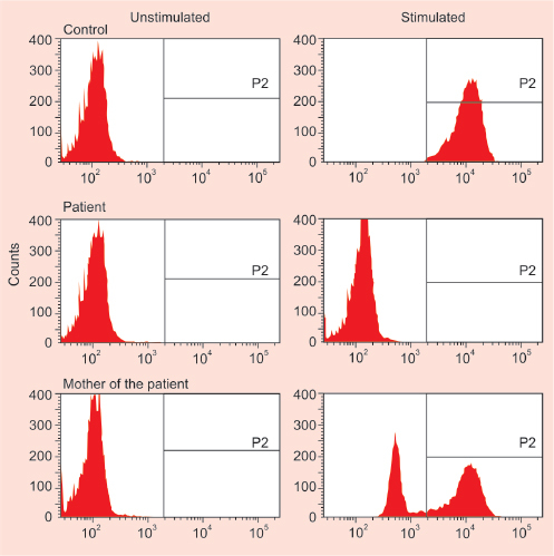
a. What is the diagnostic test depicted?
b. What is the inheritance pattern arrived based on the results?
c. What antimicrobial prophylaxis is initiated for these patients?
d. What is the drug of choice for obstructive lesions of gastrointestinal tract?
Answer
- a. Dihydrorhodamine 123 (DHR) flow cytometric oxidation assay for Chronic Granulomatous Disease (CGD).
- b. X-linked recessive CGD (CYBB gene mutation encoding gp91-phox).
- c. Antimicrobial prophylaxis:
- Antibacterial: Daily Cotrimoxazole (Trimethoprim-Sulfamethoxazole)
- Antifungal: Daily Itraconazole
- Immunomodulator: Subcutaneous Interferon-gamma (IFN-gamma)
- d. Systemic Corticosteroids (Prednisolone) for non-infectious granulomatous inflammatory obstructions (e.g., gastric outlet or ureteral obstruction).
THG20-034
A 2-year-old child has been brought with complaints of recurrent fever episodes. He has hypopigmented hair, organomegaly, and blood counts reveal anemia with thrombocytopenia. He has lost an elder sibling with similar illness. His hair microscopy reveals irregular melanin granules.
a. What is the possible diagnosis?
b. What are the biochemical investigations which can be done to support the diagnosis?
c. What are the pigment disorders which can have the same disease predisposition?
d. What are the bridging drugs which can be used prior to a curative treatment?
Answer
- a. Griscelli Syndrome type 2 (RAB27A mutation) presenting with Hemophagocytic Lymphohistiocytosis (HLH).
- b. Serum Ferritin, Fasting Triglycerides, Fibrinogen, Soluble CD25 / sIL-2R levels, and liver function tests.
- c. Chédiak-Higashi Syndrome (LYST) and Hermansky-Pudlak Syndrome type II (AP3B1).
- d. Dexamethasone, Etoposide (VP-16), and Cyclosporine A (HLH-2004 protocol) prior to HSCT.
THG20-035
A 5-year-old previously well girl is brought with low-grade fever and listlessness. At a local hospital, she was told to be anemic on evaluation but the hospital was unable to arrange compatible blood for her.
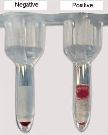
a. What is the most likely diagnosis?
b. What is the principle behind the diagnostic test in the picture?
c. What is the first-line treatment of choice?
d. List four conditions that can have this as a secondary complication in children.
e. Mention characteristic peripheral smear findings in this disease besides polychromasia.
Answer
- a. Autoimmune Hemolytic Anemia (AIHA) (Warm AIHA).
- b. Direct Antiglobulin (Coombs) Test Principle: Antihuman globulin (polyspecific Coombs reagent) binds to and cross-links RBCs coated in vivo with autoantibodies (IgG) and/or complement (C3d), producing visible agglutination.
- c. Oral Glucocorticoids (Prednisolone 2–4 mg/kg/day).
- d. Secondary causes: Systemic Lupus Erythematosus (SLE), Common Variable Immunodeficiency (CVID), Autoimmune Lymphoproliferative Syndrome (ALPS), Epstein-Barr Virus / Mycoplasma infection.
- e. Prominent Microspherocytes, nucleated RBCs, and red cell agglutination.
THG20-036
A 12-year-old boy came with 1-week history of fever, dyspnea, unable to lie flat for 2 days.
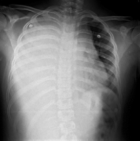
a. Identify the syndrome.
b. List two specific malignant etiologies.
c. Urgent biopsy recommended here, True or False.
d. List four immediate measures for treatment.
Answer
- a. Superior Mediastinal Syndrome (SMS) / Superior Vena Cava (SVC) Syndrome.
- b. Malignant causes:
- T-cell Acute Lymphoblastic Leukemia (T-ALL)
- T-cell Lymphoblastic Lymphoma
- Hodgkin Lymphoma
- Primary Mediastinal Large B-Cell Lymphoma (PMBCL)
- c. False (General anesthesia/invasive biopsy carries extreme risk of fatal cardiopulmonary arrest and tracheal collapse; perform least invasive diagnostic procedures under local anesthesia or upright position first).
- d. Immediate measures:
-
- Maintain upright / propped-up sitting position
-
- Supplemental oxygen support
-
- Avoid sedation and intravenous access in upper extremities (place lower limb IV cannula)
-
- Empiric IV Dexamethasone / Methylprednisolone (± emergency mediastinal radiotherapy if refractory airway compression).
-
THG20-037
Bone Marrow Aspiration Needle.
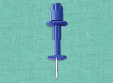
a. Name the instrument.
b. List two sites where it can be used for the test.
c. List four causes for which the test is needed for diagnosis.
d. The test is contraindicated in children with platelet count of <10,000/mm3, True or False.
e. If four draws are made, which draw by order is preferred to make the smears?
f. Mention usual gauge used in children.
Answer
- a. Salah / Jamshidi Bone Marrow Aspiration Needle (with stylet and adjustable guard).
- b. Aspiration sites in children:
- Posterior Superior Iliac Spine (PSIS) (preferred in children >1–2 years)
- Anteromedial surface of proximal tibia (infants <1 year to 18 months)
- Anterior Superior Iliac Spine (ASIS)
- c. Indications: Acute leukemias, unexplained pancytopenia / aplastic anemia, staging of neuroblastoma/lymphoma, lysosomal storage diseases (Gaucher, Niemann-Pick), pyrexia of unknown origin (marrow culture).
- d. False (Thrombocytopenia is NOT a contraindication; apply local pressure for 5–10 minutes post-procedure).
- e. The first draw (0.5–1.0 mL) is preferred for preparing morphology smears to avoid dilution with sinusoidal peripheral blood.
- f. Gauges: Aspiration needles: 15G–18G; Trephine biopsy needles: 11G–13G.
THG20-038
A 3-year-old happy and playful boy was brought by parents since they could feel a lump in right abdomen since 2 weeks. O/E, per abdomen: distended, firm mass in right flank, ballotable and bimanually palpable.
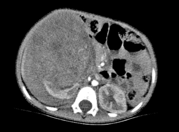
a. List two neoplasms which can present as flank mass in children.
b. Describe finding in CT.
c. What is the likely diagnosis?
d. Name the two principal modalities of treatment.
Answer
- a. Wilms Tumor (Nephroblastoma) and Neuroblastoma.
- b. Large, heterogeneous, well-circumscribed intrarenal mass arising from the right kidney showing the "Claw sign" (beak of compressed normal renal parenchyma wrapping around the tumor) with normal left kidney.
- c. Wilms Tumor (Nephroblastoma).
- d. Neoadjuvant/adjuvant Chemotherapy (Vincristine, Dactinomycin ± Doxorubicin) and Radical Nephrectomy (Radiotherapy indicated for Stage III/IV or unfavorable histology).
THG20-039
An 8-year-old boy presented with fever and fatigue since 2 weeks. O/E, he is 25 kg of weight. General examination shows pallor and generalized lymphadenopathy and systemic examination shows hepatosplenomegaly.
a. Identify the oncological emergencies in this child.
b. List two measures to prevent this.
c. Which drug can be used to decrease the uric acid and mention its mechanism of action?
d. Comment on treatment of the calcium value.
e. List the interventions for potassium value.
Answer
- a. Tumor Lysis Syndrome (TLS) and Hyperleukocytosis / Leukostasis.
- b. Aggressive IV Hyperhydration (
) and prophylactic hypouricemic agent (Allopurinol / Rasburicase). - c. Rasburicase (Recombinant Urate Oxidase enzyme): Catalyzes enzymatic conversion of insoluble uric acid into highly soluble allantoin.
- d. Asymptomatic hypocalcemia should NOT be treated with IV calcium, as calcium administration precipitates calcium phosphate in renal tubules and tissues. Treat only symptomatic hypocalcemia (tetany/seizures).
- e. Interventions for severe hyperkalemia:
- 10% Calcium gluconate IV (membrane stabilizer)
- Regular insulin with 25% dextrose infusion
- Nebulized Salbutamol / Albuterol
- Potassium binders (Sodium polystyrene sulfonate / Patiromer)
- Emergency hemodialysis if refractory
THG20-040
A 1.5-month-old baby was brought with coalescing purplish lesions on skin. The previous sibling died in early infancy with similar lesions.
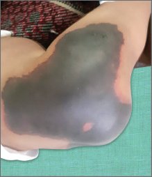
a. What is the most likely diagnosis with underlying defect?
b. What is the inheritance of the condition?
c. List steps of management.
d. List three more inherited causes and three acquired causes of thrombosis.
e. Name two direct-acting oral anticoagulants approved in children.
Answer
- a. Neonatal Purpura Fulminans due to severe congenital Protein C (or Protein S) Deficiency.
- b. Autosomal recessive.
- c. Management:
- Acute replacement: Protein C concentrate (or FFP 15–20 mL/kg q 12h)
- Therapeutic anticoagulation: IV Unfractionated Heparin or subcutaneous LMWH
- Transition to oral anticoagulation or maintenance PC replacement; definitive cure: Liver transplantation.
- d. Thrombophilia causes:
- Inherited: Factor V Leiden mutation, Prothrombin G20210A mutation, Antithrombin deficiency, Protein S deficiency.
- Acquired: Central venous catheters, Nephrotic syndrome, Antiphospholipid Syndrome (APS), Sepsis/DIC, malignancy, severe dehydration.
- e. DOACs approved in pediatrics: Rivaroxaban and Dabigatran (also Apixaban).
THG20-041
This is the peripheral smear of a 4-year-old boy who came with pallor and severe body pains. O/E, spleen was 3 cm below the costal margin.
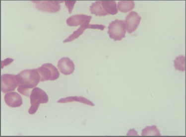
a. Mention the genetic defect here.
b. Describe in one and two sentences the pathophysiology of pain.
c. Name the conventional drug used with dose for management.
d. Name the monoclonal antibody recently introduced for treatment.
e. What is the test done and its cut-off used to decide risk of stroke in the child?
f. List four acute complications.
Answer
- a. Point mutation (single nucleotide substitution GAG -> GTG / codon 6) in the
-globin gene (HBB) resulting in the substitution of valine for glutamic acid at position 6 of the -globin chain. - b. Deoxygenation causes polymerization of HbS into long fibers, deforming RBCs into rigid sickled shapes. These non-deformable sickled erythrocytes adhere to endothelium and occlude post-capillary venules, causing tissue ischemia, infarction, and excruciating inflammatory pain (Vaso-occlusive Crisis).
- c. Hydroxyurea (starting dose 15–20 mg/kg/day, titrated up to 30–35 mg/kg/day; increases HbF synthesis).
- d. Crizanlizumab (humanized anti-P-selectin monoclonal antibody).
- e. Transcranial Doppler (TCD) ultrasonography; Time-averaged mean maximum velocity (TAMMV)
in distal internal carotid/middle cerebral artery indicates high risk of ischemic stroke. - f. Acute complications: Vaso-occlusive painful crisis, Acute Chest Syndrome, Splenic sequestration crisis, Aplastic crisis (Parvovirus B19), Priapism, and Stroke.
THG20-042
Below images are clinical picture and peripheral blood smear shown of a day 2 newborn who has developed respiratory distress and abdominal distention.
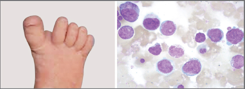
a. What are the findings in this image?
b. What is the likely diagnosis?
c. Is bone marrow testing mandatory?
d. Likely mutation leading to this complication.
e. List any four complications.
f. What is the treatment for this life-threatening disease?
Answer
- a. Wide sandal gap between first and second toes (characteristic of Down Syndrome / Trisomy 21) and circulating megakaryoblastic blasts in peripheral blood smear.
- b. Transient Abnormal Myelopoiesis (TAM) / Transient Myeloproliferative Disorder (TMD) in a newborn with Down Syndrome.
- c. No, bone marrow examination is not mandatory as blasts originate from the fetal liver; diagnosis is established from peripheral blood showing
megakaryoblasts in a neonate with Down syndrome. - d. Somatic mutation in the transcription factor gene GATA1.
- e. Complications: Hepatic fibrosis / fulminant hepatic failure, severe cholestasis, hydrops fetalis, pericardial/pleural effusions, disseminated intravascular coagulation (DIC), multi-organ failure.
- f. Low-dose Cytarabine (Ara-C) (
for 3–7 days).
THG20-043
A 2-year-old girl is brought with 3 weeks history of irritability, abnormal eye movements, and frequent falls leading to inability to walk. Her CT abdomen picture is shown below.
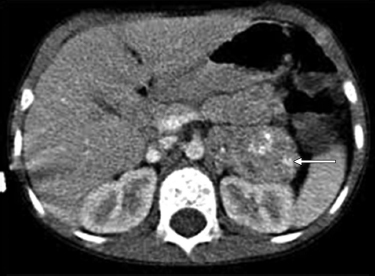
a. Describe the imaging finding.
b. What is the most likely diagnosis?
c. What are the four criteria for diagnosis?
d. List three drugs used for immunological therapy.
e. What is the long-term complication?
Answer
- a. Heterogeneous, calcified suprarenal retroperitoneal mass in the left adrenal gland engulfing retroperitoneal vessels.
- b. Neuroblastoma presenting with paraneoplastic Opsoclonus-Myoclonus-Ataxia Syndrome (OMAS / "Dancing eyes-dancing feet" syndrome).
- c. Diagnostic criteria for OMAS (at least 3 of 4 required):
-
- Opsoclonus (rapid, chaotic, conjugate involuntary saccades)
-
- Myoclonus and/or Ataxia
-
- Behavioral changes / sleep disturbance / irritability
-
- Identification of underlying Neuroblastoma
-
- d. Immunotherapy / Immunomodulators: Systemic Corticosteroids / ACTH, IV Immunoglobulin (IVIG), and Rituximab (Anti-CD20).
- e. Long-term neurocognitive, behavioral, and motor sequelae.
THG20-044
A term and well-baby body weight (BW) of 2.8 kg with no adverse perinatal events is found to have few petechial spots and ecchymoses on day 3 of life. No pallor/organomegaly was found.
a. What is the most possible diagnosis?
b. What is the risk of intracranial bleed?
c. What are the two types of platelet transfusions recommended?
d. What is the treatment of choice?
e. Name four differentials for neonatal thrombocytopenia besides this condition.
Answer
- a. Neonatal Alloimmune Thrombocytopenia (NAIT / FNAIT) (maternal alloimmunization against fetal platelet antigens, most commonly HPA-1a or HPA-5b).
- b. 10% to 20% (frequently occurs in utero).
- c.
- HPA-compatible (antigen-negative) donor platelets
- Maternal platelets (washed and irradiated to remove maternal anti-HPA antibodies)
- d. IV Immunoglobulin (IVIG 1 g/kg/day for 2 days) ± compatible platelet transfusion.
- e. Differentials:
- Increased destruction: Maternal ITP (autoimmune), neonatal sepsis/TORCH infections, necrotizing enterocolitis (NEC), Kasabach-Merritt phenomenon, perinatal asphyxia.
- Decreased production: Congenital amegakaryocytic thrombocytopenia (CAMT), Thrombocytopenia-absent radius (TAR) syndrome, Wiskott-Aldrich syndrome, congenital leukemia.
THG20-045
A 5-day-old boy with no adverse perinatal events and BW of 3.2 kg, active and well, is brought with multiple ecchymoses on trunk and limbs.
a. What is the most likely diagnosis?
b. Which protein level can be used to confirm diagnosis?
c. List three types with the age of presentation.
d. List four predisposing factors.
e. List two other differentials which can present with similar laboratory parameters.
Answer
- a. Vitamin K Deficiency Bleeding (VKDB) / Hemorrhagic Disease of the Newborn (HDN) (Classic form).
- b. PIVKA-II (Protein Induced by Vitamin K Absence / Incomplete des-carboxy prothrombin).
- c. Classification by onset:
- Early: <24 hours of life (maternal drugs)
- Classic: 2 to 7 days of life (inadequate vitamin K prophylaxis/exclusive breastfeeding)
- Late: 2 to 24 weeks of age (cholestatic liver disease, malabsorption, cystic fibrosis)
- d. Predisposing factors: Omission of neonatal vitamin K prophylaxis at birth, exclusive breastfeeding, maternal antiepileptics/ATT during pregnancy, biliary atresia, cystic fibrosis, celiac disease, and diarrhea.
- e. Common pathway coagulation factor deficiencies (congenital Factor X, V, II deficiency), severe hepatic failure/neonatal liver disease, and DIC (differentiated by severe thrombocytopenia in DIC).
THG20-046
A 5-year-old boy comes with yellow eye discoloration noticed since 1 year. There is history of neonatal jaundice at birth.
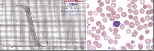
a. Identify the peripheral smear finding and the test done.
b. What is the likely defect?
c. Name any three proteins that could be defective.
d. Mention the characteristic red blood cell (RBC) index abnormality expected.
e. Name two surgeries that patients may require for this disease.
f. Describe inheritance.
Answer
- a. Smear: Microspherocytes (lacking central pallor) with polychromasia. Diagnostic test: Osmotic Fragility Test (or Eosin-5-Maleimide [EMA] binding flow cytometry).
- b. Hereditary Spherocytosis (HS) (erythrocyte membrane cytoskeleton defect causing membrane loss, spheroidal remodeling, and premature splenic destruction).
- c. Defective membrane proteins: Ankyrin,
-Spectrin, -Spectrin, Band 3, and Protein 4.2. - d. Elevated Mean Corpuscular Hemoglobin Concentration (MCHC >36 g/dL) and elevated RDW.
- e. Surgeries: Splenectomy (total or subtotal) and Cholecystectomy (for pigmented gallstones).
- f. Autosomal dominant (~75% of cases; autosomal recessive in ~25%).
THG20-047
This 11-month-old girl needed five transfusions in last 6 months. No organomegaly on examination.
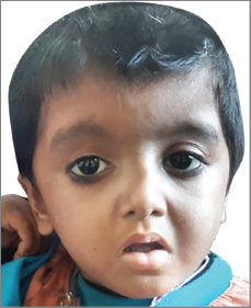
a. What are the anomalies seen in the above picture?
b. What is the likely diagnosis?
c. Mention four criteria for diagnosis.
d. Which are the two options for treatment?
e. What proportion of patients responds to the frontline drug?
Answer
- a. Craniofacial dysmorphism (hypertelorism, flat nasal bridge, long philtrum) and upper limb anomalies (triphalangeal / bifid thumb).
- b. Diamond-Blackfan Anemia (DBA).
- c. Diagnostic criteria:
- Normochromic macrocytic anemia presenting in the first year of life
- Reticulocytopenia
- Normocellular bone marrow with selective erythroid hypoplasia (<5% erythroid precursors)
- Normal platelet and leukocyte counts
- Supporting criteria: Elevated erythrocyte adenosine deaminase (eADA), elevated HbF, characteristic congenital anomalies, and identified ribosomal gene mutation (RPS19, RPL5, etc.).
- d. Treatment options: Oral Prednisolone and Hematopoietic Stem Cell Transplantation (HSCT) (with chronic transfusion + iron chelation for steroid non-responders).
- e. ~75% to 80% of patients respond to initial corticosteroid therapy.
THG20-048
A 4-year-old boy posted for closure of ventricular septal defect was referred for hematological clearance due to mild anemia. His CBC reveals Hb: 9 g/dL, MCV: 64, MCH: 19, pMCHC: 22, RBC count: 5.5, red cell distribution width (RDW): 12.5, and smear showed occasional target cells.
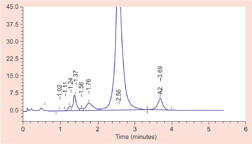
a. Name the test and interpret.
b. Calculate Mentzer index and comment.
c. Iron supplementation is contraindicated in this child, True or False.
d. What is the counseling essential for the child’s family based on report?
e. In India, what is the approximate rate of this condition?
f. Name two conditions which can reduce A2 concentration.
Answer
- a. High-Performance Liquid Chromatography (HPLC): Demonstrates elevated
(typically 4.0–6.0%), confirming -Thalassemia Trait (Minor). - b. Mentzer Index:
- Value
strongly supports Thalassemia trait rather than iron deficiency anemia.
- Value
- c. False (Iron is not contraindicated if co-existing nutritional iron deficiency is documented by low serum ferritin; however, empiric iron without deficiency must be avoided).
- d. Premarital/Preconceptional Genetic Counseling: Partner screening for beta-thalassemia trait is essential; if both parents are carriers, there is a 25% risk of having a child with Thalassemia Major in each pregnancy, which can be diagnosed via prenatal chorionic villus sampling/amniocentesis.
- e. Carrier frequency in India is approximately 3% to 4% (higher in certain ethnic communities like Sindhis, Punjabis, Gujaratis, Bengalis).
- f. Co-existing severe iron deficiency anemia and
-thalassemia (or -thalassemia).
THG20-049
Clinical image and fine needle aspiration cytology (FNAC) slide image of an 11-year-old boy are shown.
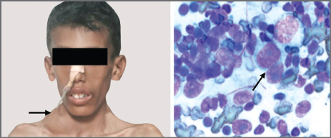
a. Identify the findings marked by arrows.
b. What is the likely diagnosis?
c. List B symptoms in context of this case.
d. List four differences in clinical presentation between Hodgkin and non-Hodgkin lymphoma.
e. Name two modalities used in management of condition.
Answer
- a. Clinical: Asymmetric, non-tender, firm, matted right cervical/supraclavicular lymphadenopathy. Cytology: Classic binucleated Reed-Sternberg cell with prominent eosinophilic inclusion-like nucleoli surrounded by a clear halo ("owl-eye" appearance).
- b. Hodgkin Lymphoma.
- c. B Symptoms:
- Unexplained recurrent fever
- Drenching night sweats
- Unexplained loss of
body weight within preceding 6 months
- Unexplained recurrent fever
- d. Hodgkin vs. Non-Hodgkin Lymphoma:
| Feature | Hodgkin Lymphoma | Non-Hodgkin Lymphoma |
|---|---|---|
| Nodal spread | Contiguous, orderly spread to adjacent node chains | Non-contiguous, unpredictable nodal spread |
| Extranodal involvement | Rare initially (bone marrow/CNS rare) | Common (GI tract, bone marrow, CNS) |
| Mediastinal involvement | Common (anterior-superior mediastinum) | Common (anterior mediastinum in T-ALL/LBL) |
| Constitutional B symptoms | Frequent (~30–40%) | Less common / variable |
| Waldeyer's ring / Mesenteric | Rarely involved | Frequently involved |
- e. Combination Multiagent Chemotherapy (e.g., ABVD / OEPA-COPDAC) and Involved-Field / Involved-Site Radiation Therapy (ISRT).
THG20-050
Vincristine Chemotherapy.
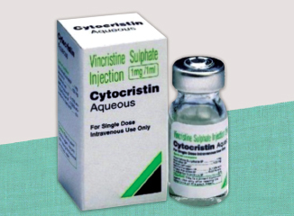
a. What is the dose limiting toxicity of the drug?
b. What is the mechanism of action?
c. List two precautions to take while administering.
d. Mention two interventions for extravasation.
Answer
- a. Peripheral Neurotoxicity (sensorimotor peripheral neuropathy, loss of deep tendon reflexes, foot drop, paralytic ileus, constipation, cranial nerve palsies).
- b. Binds to tubulin dimers and inhibits microtubule polymerization and mitotic spindle assembly, arresting cells in the metaphase of mitosis.
- c. Administration precautions:
- FATAL IF GIVEN INTRATHECALLY — administer strictly by intravenous route only with distinct labeling.
- Administer through a fresh, free-flowing IV cannula/central line and monitor for extravasation.
- d. Extravasation management (Vinca alkaloid is a vesicant):
- Stop infusion immediately; aspirate remaining drug; do NOT apply cold.
- Apply warm dry compresses (to promote local vasodilation and drug dispersion).
- Subcutaneous injection of Hyaluronidase (150–300 units locally around extravasation site).
THG20-051
L-Asparaginase Chemotherapy.
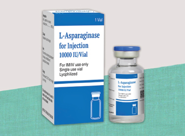
a. Mention the cancer in which it is most commonly used in children.
b. List any two adverse reactions.
c. List two routes of administration.
d. List two chemotherapy drugs in children that cause minimal/no emesis.
Answer
- a. Acute Lymphoblastic Leukemia (ALL).
- b. Adverse reactions:
- Anaphylaxis and hypersensitivity reactions
- Acute pancreatitis
- Thrombosis (due to antithrombin III deficiency) or hemorrhage (hypofibrinogenemia)
- Hyperglycemia / transient diabetes mellitus (inhibition of insulin synthesis)
- Hepatic dysfunction / hyperbilirubinemia
- c. Intramuscular (IM) and Intravenous (IV).
- d. Vincristine, L-Asparaginase, oral 6-Mercaptopurine, and oral Methotrexate.