MNJ29-001
A 9-year-old child Rampal presented with malaise, anorexia, vomiting, muscle weakness since one month. On examination, you find orthostatic hypotension. He had history of a febrile illness with purpuric rash a few days before:
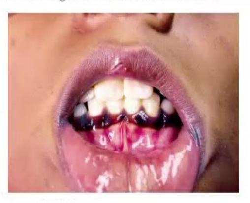
- What is the significant finding in the picture?
- What is the cause of this finding?
- What is the possible infection preceding it and what is it called?
- What are the possible electrolyte abnormalities?
- What is the definitive test for diagnosis of this condition?
Answer
- Hyperpigmentation of the gingival and buccal mucosa (and skin creases).
- Primary adrenal insufficiency (Addison disease): Cortisol deficiency removes negative feedback on the pituitary-hypothalamic axis, leading to increased ACTH and
-MSH production from the common precursor pro-opiomelanocortin (POMC). - Meningococcemia (Neisseria meningitidis) causing acute bilateral adrenal hemorrhage; known as Waterhouse-Friderichsen syndrome.
- Electrolyte abnormalities:
- Hyponatremia
- Hyperkalemia
- Hypoglycemia
- Metabolic acidosis (normal anion gap)
- Prerenal azotemia / elevated BUN
- Standard high-dose ACTH (Synacthen/Cosyntropin) stimulation test (measuring baseline and 30/60-minute serum cortisol levels).
MNJ29-002
This is the blood glucose profile of an 8-year-old boy with diabetes. His
| Insulin | Morning | Evening |
|---|---|---|
| Regular | 6 U | 3 U |
| NPH | 8 U | 5 U |
- What changes would you institute in insulin dosage?
- Considering his
, over what period is his glycemic control likely to be unsatisfactory? - When do you start screening for diabetic retinopathy?
- What is the preferred method for screening for diabetic retinopathy?
Answer
- Increase the morning regular insulin dose by 1–2 units (and adjust overall daily insulin dose by 10–20% based on pre- and post-prandial glucose logs).
- Over the preceding 3 months (8–12 weeks).
- Retinopathy screening initiation:
- After 5 years of diabetes duration in prepubertal children, OR
- Starting at age 10–11 years / onset of puberty (whichever is earlier), then annually.
- Preferred screening method: Dilated pupillary digital fundal photography (or dilated indirect ophthalmoscopy by an ophthalmologist).
Reference: Screening guidelines in pediatric diabetes:
- Retinopathy: After 5 years duration (or from puberty), annually by fundal photography.
- Nephropathy: After 5 years duration (or from puberty), annually by spot urine albumin-to-creatinine ratio (ACR).
- Thyroid disease: At diagnosis, then every 1–2 years (TSH, anti-TPO).
- Celiac disease: At diagnosis, then every 2–5 years (anti-tTG IgA).
MNJ29-003
Answer the following questions about
- What does its testing indicate?
- What are the normal values in non-diabetics?
- What are the target goals of therapy in pediatric type 1 diabetes?
Answer
- Glycated hemoglobin reflects mean ambient glycemic control over the preceding 2–3 months (erythrocyte lifespan ~120 days).
- Normal non-diabetic range:
( ). - Target
in pediatric Type 1 Diabetes (ISPAD / ADA guidelines): ( ) for most children if achievable without significant hypoglycemia. where continuous glucose monitoring / advanced technology is unavailable.
MNJ29-004
Thyroid function reports in an 8-year-old girl Shivani revealed Total
- What is this girl suffering from according to these lab results?
- What is the commonest etiology in children?
- What medical treatment can be given?
Answer
- Hyperthyroidism / Thyrotoxicosis.
- Graves' disease (autoimmune stimulation of TSH receptors by TRAb / TSI).
- Medical management:
- Antithyroid drugs (Thionamides): Methimazole / Carbimazole (
in 1–2 divided doses). (Propylthiouracil is avoided in children due to severe hepatotoxicity risk). -blockers: Propranolol ( in 3 divided doses) for symptomatic control of adrenergic symptoms (tachycardia, tremors). - Definitive therapy if refractory: Radioactive iodine (
) ablation or near-total thyroidectomy.
- Antithyroid drugs (Thionamides): Methimazole / Carbimazole (
MNJ29-005
A 5-year-old girl Bhawna was brought with complaints of obesity. On examination, she had weight of 38 kg and height of 122 cm. She had hyperpigmentation of axilla, groin and neck regions:
- What is the Body Mass Index (BMI) of the child?
- What does the pigmentation denote? What investigation would you do for the same?
- What musculoskeletal problems can this girl suffer from?
Answer
(well above the 95th percentile for age and sex, indicating obesity). - Acanthosis nigricans, which denotes severe insulin resistance / hyperinsulinemia. Investigations: Fasting lipid profile, Oral Glucose Tolerance Test (OGTT) / fasting plasma glucose, and fasting serum insulin (
). - Musculoskeletal complications of childhood obesity:
- Slipped Capital Femoral Epiphysis (SCFE)
- Blount disease (Tibia vara)
- Genu valgum ("knock knees")
- Pes planus (flat feet)
MNJ29-006
A 10-year-old boy Manish was brought with history of episodic flushing for the last three months. His BP was 160/110 mmHg and heart rate 150/min. His adrenals were enlarged on USG:
- What is the likely diagnosis?
- How will you confirm the diagnosis?
- What is the medical treatment before surgery?
Answer
- Pheochromocytoma (chromaffin tissue catecholamine-secreting tumor).
- Diagnostic confirmation:
- 24-hour urinary fractionated metanephrines and normetanephrines (or plasma free metanephrines).
- 24-hour urinary vanillylmandelic acid (VMA).
- Contrast CT / MRI abdomen (adrenal protocol) and
-MIBG scintigraphy or -DOTATATE PET-CT.
- Preoperative medical management:
-adrenergic blockade first: Phenoxybenzamine ( titrated) or Prazosin for 10–14 days to control BP and expand intravascular volume. -adrenergic blockade second: Propranolol or Atenolol started only AFTER adequate -blockade (to avoid unopposed -vasoconstriction and hypertensive crisis).
MNJ29-007
A 16-year-old short girl Sagarika was brought with history of delayed puberty. On examination she had a short 4th metacarpal, absent lower limb pulses and hypertension:
- What is the likely diagnosis?
- What cardiovascular abnormality does she have, and what other CVS abnormalities are seen in this disorder?
- What endocrine problems can you get in this condition?
- What medication can you give for her delayed puberty?
Answer
- Turner syndrome (45,X / Gonadal dysgenesis).
- Cardiovascular abnormalities:
- Present finding: Coarctation of the Aorta (causing upper extremity hypertension and absent/delayed lower limb femoral pulses).
- Other CVS anomalies: Bicuspid aortic valve (most common), aortic root dilatation / dissection, partial anomalous pulmonary venous connection (PAPVC).
- Associated endocrine disorders:
- Hypergonadotropic hypogonadism (streak gonads / ovarian failure)
- Autoimmune thyroiditis (Hashimoto thyroiditis / hypothyroidism)
- Type 2 diabetes / Impaired glucose tolerance with insulin resistance
- Pubertal induction therapy:
- Low-dose oral or transdermal Estrogen (e.g.,
-estradiol) initiated around 11–12 years of age, gradually titrated over 2–3 years. - Cyclic Progesterone added after 2 years of estrogen therapy or when breakthrough bleeding occurs to achieve regular menstrual withdrawal bleeding and protect endometrium.
- Low-dose oral or transdermal Estrogen (e.g.,
MNJ29-008
A 5-year-old female child Rajini is brought with recurrent bleeding per vaginam for the past 1 year. On examination she has large hyperpigmented patches with irregular "coast of Maine" borders over the neck and back, and breast development Tanner stage 2. X-ray shows advanced bone age:
- What is your diagnosis?
- What is the classic triad of this syndrome?
- Name two other endocrine associations.
- How will you treat the peripheral precocious puberty?
Answer
- McCune-Albright Syndrome (MAS) (due to postzygotic somatic activating mutation in GNAS gene).
- Classic triad:
- Peripheral (GnRH-independent) precocious puberty (recurrent autonomous ovarian follicular cysts)
- Polyostotic fibrous dysplasia of bones
- Large, irregular café-au-lait skin macules with "coast of Maine" jagged borders
- Other hyperfunctioning endocrinopathies:
- Hyperthyroidism (toxic nodular goiter)
- GH excess / Gigantism / Acromegaly
- Cushing syndrome (autonomous bilateral adrenal hyperplasia)
- Renal phosphate wasting / hypophosphatemic rickets (FGF-23 excess)
- Treatment:
- Aromatase inhibitors (e.g., Letrozole or Anastrozole) to block estrogen synthesis
- Estrogen receptor antagonists (e.g., Tamoxifen or Fulvestrant)
- Bisphosphonates (Pamidronate/Zoledronic acid) for painful polyostotic fibrous dysplasia.
MNJ29-009
A 12-year-old female Ritu weighing 31 kg, a diagnosed case of Type 1 Diabetes on split-mix insulin therapy (Morning: Regular 10 U + NPH 30 U; Evening: Regular 10 U + NPH 12 U), presents with high fasting early-morning blood glucose (7:30 AM blood glucose 270 mg/dL):
- What are the Somogyi phenomenon and Dawn phenomenon?
- How will you differentiate these two in this child?
- How will you treat each condition?
Answer
- Definitions:
- Somogyi Phenomenon (Rebound Hyperglycemia): Nocturnal hypoglycemia (around 2–3 AM) triggers a surge of counter-regulatory hormones (epinephrine, glucagon, cortisol, GH) producing early-morning rebound hyperglycemia.
- Dawn Phenomenon: Early-morning (4–8 AM) hyperglycemia without preceding nocturnal hypoglycemia, caused by circadian physiological rise in Growth Hormone and cortisol together with declining nighttime insulin levels.
- Differentiation:
- Check blood glucose at 2:00 AM – 3:00 AM:
- If 3:00 AM glucose is low (< 60–70 mg/dL)
Somogyi phenomenon. - If 3:00 AM glucose is normal or elevated (> 80–100 mg/dL)
Dawn phenomenon.
- If 3:00 AM glucose is low (< 60–70 mg/dL)
- Check blood glucose at 2:00 AM – 3:00 AM:
- Treatment:
- For Somogyi phenomenon: Decrease the evening intermediate/long-acting (NPH) insulin dose by 10–15% or provide a bedtime complex carbohydrate snack.
- For Dawn phenomenon: Increase the evening/nighttime insulin dose by 10–15% or shift the evening NPH injection from dinnertime to bedtime (around 10:00 PM).
MNJ29-010
An infant is being evaluated for ambiguous genitalia. You find clitoral hypertrophy and other signs of virilization. On investigations: serum cortisol levels are very low, ACTH and PRA are markedly elevated, ACTH stimulation test reveals markedly increased 17-hydroxyprogesterone (17-OHP), serum testosterone is elevated, and the child has severe hyponatremia and hyperkalemia:
- What is your diagnosis?
- What is the mode of inheritance?
- What is the method for prenatal diagnosis?
- What advice will you give for the mother's next pregnancy?
- What medical treatment will you offer for this baby?
Answer
- Congenital Adrenal Hyperplasia (CAH) due to 21-Hydroxylase deficiency (Salt-wasting classic form, 46,XX virilized female).
- Autosomal recessive (CYP21A2 gene mutation on chr 6p21.3).
- Prenatal diagnosis: Chorionic Villus Sampling (CVS) at 10–12 weeks or Amniocentesis at 15–18 weeks for CYP21A2 molecular genetic analysis (MLPA/Sanger sequencing).
- Management in next pregnancy:
- Start maternal oral Dexamethasone (
in 3 divided doses, max 1.5 mg/day) as soon as pregnancy is confirmed (before 6–7 weeks) to suppress fetal pituitary ACTH and prevent virilization of female genitalia. - Perform CVS for fetal sex determination and CYP21A2 mutation: If fetus is male or an unaffected female, stop dexamethasone; if affected 46,XX female, continue dexamethasone until term.
- Start maternal oral Dexamethasone (
- Treatment:
- Glucocorticoid replacement: Oral Hydrocortisone (
in 3 divided doses; doubled/tripled during stress/illness). - Mineralocorticoid replacement: Oral Fludrocortisone (
). - Sodium chloride supplementation:
(17–51 mEq/day) in infants. - Surgical feminizing genitoplasty (clitoroplasty/vaginoplasty) at 6–12 months of age.
- Glucocorticoid replacement: Oral Hydrocortisone (
MNJ29-011
An infant with seizures is being investigated. The following are the lab reports:
- Serum calcium: 6.6 mg/dL (Low)
- Serum phosphate: 9.0 mg/dL (High)
- Serum alkaline phosphatase (SAP): 500 IU/L (Normal)
- Serum magnesium: 3.0 mg/dL (Normal)
- What is the probable diagnosis?
- What will be the levels of PTH and
? - If the same infant is noted to have hyperpigmentation and chronic mucocutaneous candidiasis, what is your diagnosis?
- What finding do you expect on CT brain?
Answer
- Primary Hypoparathyroidism (or Pseudohypoparathyroidism).
- Serum intact PTH is low / undetectable in primary hypoparathyroidism (elevated in pseudohypoparathyroidism);
(Calcitriol) is low in both. - Autoimmune Polyendocrinopathy-Candidiasis-Ectodermal Dystrophy (APECED / Autoimmune Polyglandular Syndrome Type 1 [APS-1], due to AIRE gene mutation).
- Bilateral symmetrical basal ganglia calcifications (and subcortical white matter calcifications).
MNJ29-012
A child with meningoencephalitis is comatose. His serum sodium is 114 mmol/L and you suspect a diagnosis of SIADH:
- Which of the following lab values will be present?
a. Urine Osmolality < 100 mOsm/kg
b. Intravascular / plasma volume normal or mildly increased
c. Urine Sodium > 20–40 mEq/L
d. Serum uric acid > 10 mg/dL - Name 2 drugs that can increase vasopressin (ADH) levels.
- What is the main principle of fluid management in SIADH?
- Which pharmacological agent may be given for chronic SIADH?
- Name two acute respiratory illnesses that can cause SIADH.
- Which anticonvulsant is known to decrease ADH secretion/action?
Answer
- Lab findings in SIADH:
- a. False (Urine Osmolality is inappropriately concentrated, typically
). - b. True (Euvolemic / mildly increased plasma volume without clinical edema).
- c. True (Urine
despite hyponatremia due to natriuresis). - d. False (Serum uric acid and BUN are low due to dilution and increased urate clearance).
- a. False (Urine Osmolality is inappropriately concentrated, typically
- Drugs increasing ADH: Carbamazepine, Vincristine, Cyclophosphamide, Tricyclic antidepressants (TCAs), SSRIs.
- Fluid restriction: Restrict total fluid intake to
of maintenance ( ). - Pharmacotherapy: Demeclocycline (causes nephrogenic DI), Tolvaptan / Conivaptan (vasopressin V2 receptor antagonists), or oral urea / salt tablets.
- Respiratory illnesses: Acute bronchiolitis (RSV), bacterial pneumonia, tuberculosis, status asthmaticus.
- Phenytoin (inhibits ADH release).
MNJ29-013
- What is premature thelarche?
- Up to what age is it considered a benign variant?
- What is exaggerated or atypical thelarche?
- What will be the levels of FSH, LH, and estradiol in benign premature thelarche?
- What will be the pelvic USG findings?
Answer
- Isolated unilateral or bilateral breast development in young girls without other signs of sexual maturation (no pubic/axillary hair, no growth spurt, no bone age advancement).
- Benign variant when presenting before 2–3 years of age (often self-limiting with spontaneous regression).
- Exaggerated / Atypical thelarche: Breast development accompanied by mild acceleration of bone age or fluctuating estradiol levels, requiring close surveillance for progression to central precocious puberty.
- Hormone profile in benign premature thelarche:
- Basal and GnRH-stimulated LH: Prepubertal / suppressed (
) - FSH: Normal to mildly elevated (FSH-predominant response to GnRH)
- Serum Estradiol: Prepubertal / low (
)
- Basal and GnRH-stimulated LH: Prepubertal / suppressed (
- Pelvic USG findings: Prepubertal tubular uterus (length
, uterine volume , endometrial stripe absent); normal small ovaries ( ) with small non-dominant follicles ( ).
MNJ29-014
A girl child Dipika presented with palpitations, diarrhea, and weight loss for 3 months:
- What is the diagnosis?
- The thyroid swelling is non-tender and diffuse. She has exophthalmos. What is the cause?
- List 4 other causes of childhood thyrotoxicosis.
- What are the key diagnostic investigations?
- Name 2 drug classes used in initial medical management.
Answer
- Hyperthyroidism / Thyrotoxicosis.
- Graves' disease (autoimmune stimulation by TSH-receptor antibodies [TRAb/TSI] leading to diffuse goiter and thyroid-associated orbitopathy/exophthalmos).
- Other causes of thyrotoxicosis:
- Toxic solitary adenoma
- Toxic multinodular goiter (Plummer disease)
- Subacute granulomatous thyroiditis (de Quervain thyroiditis)
- Chronic autoimmune thyroiditis (Hashitoxicosis - early phase)
- Exogenous thyroid hormone ingestion (factitious)
- TSH-secreting pituitary adenoma
- Investigations:
- Free
, Free (both elevated), TSH (suppressed ) - Serum TSH receptor antibodies (TRAb / TSI) and anti-TPO antibodies
- Technetium-99m (
) or thyroid scintigraphy (diffuse increased uptake) - Thyroid USG with color Doppler (increased vascular flow - "thyroid inferno").
- Free
- Drug classes:
- Antithyroid drugs (Thionamides): Methimazole or Carbimazole
-blockers: Propranolol or Atenolol.
MNJ29-015
Please determine the Tanner pubertal stage (Sexual Maturity Rating [SMR]) for the following patients:
- Girl: Breast and papilla elevated as a small mound; areolar diameter enlarged ("breast bud" stage).
- Boy: Pubic hair: scanty, long, slightly pigmented at base of penis; Penis: minimal enlargement; Scrotum: enlarged, reddened, with altered texture.
- Name the three chronological sub-stages of adolescence with their respective age ranges.
Answer
- Tanner Stage B2 (Breast Stage 2 / Thelarche).
- Tanner Stage G2 / P2 (Genital Stage 2, Pubic Hair Stage 2).
- Sub-stages of adolescence:
- Early adolescence: 10–13 years
- Middle adolescence: 14–16 years
- Late adolescence: 17–20 years
MNJ29-016
A 3-year-old girl Richa is brought with pubic hair and clitoromegaly, increased irritability gradually worsening over 2 months. No history of drug ingestion or hyperpigmentation. On examination: BMI > 95th percentile, BP: 110/82 mmHg, HR: 90/min, RR: 22/min. She has hirsutism and acne on the face, chubby "moon" cheeks, and pink striae over the abdomen. Genital examination shows pubic hair Tanner stage 3 and clitoris measuring 1.9 cm:
- What is the clinical diagnosis?
- What is the most likely etiology at this age?
- Enlist 2 important first-line screening tests used for this condition.
- Name 2 drugs used for the medical management of hypercortisolemia.
Answer
- Cushing Syndrome (with virilization / adrenal tumor).
- Autonomous adrenal neoplasm (Adrenocortical Adenoma or Adrenocortical Carcinoma). (In children
years, adrenal tumors are the commonest cause of endogenous Cushing syndrome; in children years, ACTH-secreting pituitary adenoma [Cushing disease] predominates). - First-line screening tests:
- 24-hour urinary free cortisol (UFC) excretion (at least 2 collections)
- Overnight low-dose Dexamethasone Suppression Test (1 mg DST or
DST) - Late-night salivary cortisol (at 11:00 PM)
- Loss of normal diurnal plasma cortisol rhythm
- Adrenolytic / steroidogenesis-inhibiting drugs:
- Ketoconazole (inhibits CYP17 and 11
-hydroxylase) - Metyrapone (inhibits 11
-hydroxylase) - Mitotane (adrenolytic agent)
- Aminoglutethimide / Etomidate
- Ketoconazole (inhibits CYP17 and 11
MNJ29-017
- Give the differential diagnosis of short 4th metacarpal (brachydactyly type E).
- Name syndromes associated with "radial ray defect" (absent/hypoplastic radius).
- Identify the condition in the radiographic findings showing extensive soft-tissue heterotopic ossification in back/extremities.
Answer
- Differential diagnosis of short 4th metacarpal:
- Pseudohypoparathyroidism (Albright Hereditary Osteodystrophy)
- Pseudo-pseudohypoparathyroidism (PPHP)
- Turner syndrome (45,X)
- Klinefelter syndrome (47,XXY)
- Ellis-van Creveld syndrome
- Idiopathic / hereditary brachydactyly
- Syndromes with Radial Ray Defects:
- Fanconi Anemia (FA)
- Thrombocytopenia-Absent Radius (TAR) syndrome
- Holt-Oram syndrome (cardiac defects + radial ray)
- VACTERL association
- Baller-Gerold syndrome
- Diamond-Blackfan anemia
- Fibrodysplasia Ossificans Progressiva (FOP / Myositis ossificans progressiva) characterized by congenital hallux valgus (microdactyly of great toes) and progressive heterotopic extraskeletal endochondral ossification.
MNJ29-018
A 4-year-old girl Mona is brought with short stature. She was noticed to have short 4th and 5th metacarpals, round facies, subcutaneous ossifications, hypocalcemia, hyperphosphatemia, and elevated PTH levels:
- What is the diagnosis in this girl?
- What biochemical response will you see in this child on exogenous PTH administration (Ellsworth-Howard test)?
- Mention the inheritance pattern and genetic basis.
- Which of the following diseases shares the same mode of inheritance? (Incontinentia pigmenti / Tuberous sclerosis / Prader-Willi syndrome / Duchenne muscular dystrophy).
Answer
- Pseudohypoparathyroidism Type 1a (PHP-1a / Albright Hereditary Osteodystrophy [AHO]).
- Ellsworth-Howard test (PTH infusion test): Blunted or absent increase in urinary cyclic AMP (cAMP) and urinary phosphate excretion (confirming end-organ PTH receptor resistance).
- Autosomal dominant with genomic imprinting (maternally inherited inactivating mutation of the GNAS gene encoding the
protein subunit). - Tuberous sclerosis (Autosomal dominant).
MNJ29-019
- What test is performed as newborn screening for Congenital Adrenal Hyperplasia?
- Define micropenis in a term male newborn by measurement.
- List 5 causes / syndromes associated with micropenis.
- What is paraphimosis?
- How is paraphimosis managed?
Answer
- Dried blood spot 17-hydroxyprogesterone (17-OHP) assay by immunoassay / tandem mass spectrometry (LC-MS/MS).
- Definition: Stretched penile length (SPL)
below the mean for age ( in a full-term neonate; normal term mean SPL ). - Causes / syndromes:
- Hypogonadotropic hypogonadism: Kallmann syndrome, Prader-Willi syndrome, CHARGE syndrome, congenital hypopituitarism (GH/LH/FSH deficiency)
- Hypergonadotropic hypogonadism: Klinefelter syndrome, testicular dysgenesis/anorchia
- Robinow syndrome (fetal face syndrome)
- Idiopathic micropenis
- Paraphimosis: A urological emergency where a retracted, tight foreskin becomes trapped behind the coronal sulcus, leading to venous and lymphatic congestion, glans edema, and arterial compromise.
- Management:
- Manual reduction: Apply manual compression to reduce glans edema (with topical ice/osmotic agent like 50% dextrose), followed by gentle distal traction on the prepuce and pushing the glans proximally.
- Puncture technique / dorsal slit of the constricting band under local anesthesia if manual reduction fails.
- Elective circumcision after resolution of acute inflammation.
MNJ29-020
A 4-year-old girl Manisha has been brought with history of red staining of diapers for one month. Her height is 102 cm (1 year back it was 92 cm), weight is 17.5 kg (1 year back it was 13.5 kg). She has been observed to be more quiet than usual with intermittent episodes of laughing (gelastic seizures). Abdominal and neurological exam is normal:
- What is the likely complete diagnosis?
- What diagnostic investigations would you ask for?
- What is the first-line medical treatment?
- If medical treatment fails, what intervention is indicated?
Answer
- Central (GnRH-dependent) Precocious Puberty secondary to Hypothalamic Hamartoma (presenting with gelastic seizures and accelerated linear growth).
- Diagnostic investigations:
- High-resolution MRI Brain (with thin cuts through the hypothalamus and pituitary to identify sessile or pedunculated tuber cinereum hamartoma)
- Video-EEG (to capture gelastic seizures)
- Baseline and GnRH agonist stimulation test (showing pubertal LH surge
) - Serum estradiol and pelvic USG (uterine and ovarian enlargement)
- Bone age radiograph (advanced bone age).
- Medical treatment:
- Long-acting GnRH agonists (e.g., Leuprolide acetate depot
every 4–12 weeks, or Triptorelin) to desensitize pituitary GnRH receptors and arrest pubertal progression. - Antiseizure medications (e.g., Carbamazepine, Lamotrigine, Levetiracetam) for seizures.
- Long-acting GnRH agonists (e.g., Leuprolide acetate depot
- Refractory cases: Stereotactic radiofrequency ablation, Gamma Knife radiosurgery, MRI-guided laser interstitial thermal therapy (LITT), or endoscopic transsphenoidal/microsurgical resection.
MNJ29-021
Interpret the following diagnostic hormone profiles:
Table A: Thyroid Disorders
| Clinical Scenario | TSH | TSH Response to TRH | Diagnosis | ||
|---|---|---|---|---|---|
| 1 | Low | Low | Very High | Exaggerated | ? |
| 2 | Low | Low | Low / Normal | Absent / Blunted | ? |
| 3 | Normal Free |
Normal Free |
Normal | Normal | ? |
| 4 | Normal | Normal / Low | High | Exaggerated | ? |
Table B: Metabolic Bone Disorders
| Condition | Serum Calcium | Serum Phosphate | Serum Alk Phos | Urinary Amino Acids | ||
|---|---|---|---|---|---|---|
| Cystinosis / Fanconi syndrome | Normal / Low | Low | High | Normal | Variable | Generalized aminoaciduria (1) |
| Chronic Renal Failure (CRF) | Low | High | High | Normal | Low | Normal |
| Hypophosphatasia | Normal / High | Normal / High | Markedly Low (3) | Normal | Normal | Normal (High urinary PEA) |
| VDDR Type II (End-organ resistance) | Low | Low | High | Normal | Markedly High (4) | Normal |
| VDDR Type I ( |
Low | Low | High | Normal | Markedly Low (5) | Normal |
Answer
Table A Answers:
- Primary Hypothyroidism (Hashimoto / Thyroid dysgenesis).
- Secondary (Central / Pituitary) Hypothyroidism.
- Thyroxine-Binding Globulin (TBG) deficiency (Total
is low, but Free and TSH are normal). - Subclinical / Compensated Hypothyroidism.
Table B Answers:
- 1
Generalized aminoaciduria (Renal Fanconi syndrome in Cystinosis). - 3
Markedly Decreased Serum Alkaline Phosphatase (hallmark of Hypophosphatasia due to ALPL gene mutation). - 4
Markedly Increased (due to vitamin D receptor [VDR] mutation in VDDR Type II). - 5
Markedly Decreased (due to -hydroxylase deficiency in VDDR Type I).
MNJ29-022
An 11-year-old boy Ranveer presents with complaints of secondary enuresis and increased frequency of urination and thirst for the last 3 months. Match the diagnoses A, B, and C with the test profiles below:
| Parameter | Patient A | Patient B | Patient C |
|---|---|---|---|
| Serum Sodium | Low ( |
Low ( |
High ( |
| Urine Output | Normal / Low | High (Polyuria) | High (Polyuria) |
| Urine Sodium | Moderate ( |
Very High ( |
Low ( |
| Intravascular Volume | Euvolemic / Normal | Hypovolemic / Low | Hypovolemic / Low |
| Serum Vasopressin (ADH) | Inappropriately High | Suppressed / Low | Suppressed / Absent |
Answer
- Patient A: Syndrome of Inappropriate Antidiuretic Hormone Secretion (SIADH) (Euvolemic hyponatremia with inappropriately high ADH).
- Patient B: Cerebral Salt Wasting (CSW) (Hypovolemic hyponatremia with massive natriuresis).
- Patient C: Central Diabetes Insipidus (CDI) (Hypernatremic polyuria with low/absent ADH).
MNJ29-023
A 7-year-old girl Akansha presents with breast development Tanner stage 3, pubic hair stage 1, and delayed bone age. She has constipation and has gained weight rapidly over the last 12 months. Laboratory reports show serum
- What is this clinical condition and its underlying cause?
- What other investigations will you perform?
- What are causes of combined central and peripheral precocious puberty?
- What is the clinical definition of precocious puberty in boys and girls?
- What is the treatment for this girl?
Answer
- Van Wyk-Grumbach Syndrome (severe primary juvenile hypothyroidism presenting with precocious thelarche, multicystic ovaries, uterine bleeding, and delayed bone age). Underlying mechanism: Cross-reactivity of extremely high TSH at ovarian FSH receptors (and hyperprolactinemia due to high TRH).
- Investigations:
- Free
(markedly low), Free , anti-TPO and anti-thyroglobulin antibodies - Serum Prolactin (elevated)
- Pelvic USG (shows bilateral multicystic enlarged ovaries and enlarged uterus)
- MRI Brain / Pituitary (shows pituitary hyperplasia/macroadenoma reversible with
).
- Free
- Causes of secondary central activation after peripheral precocious puberty:
- Congenital Adrenal Hyperplasia (after initiating glucocorticoid therapy)
- McCune-Albright syndrome
- Familial male-limited precocious puberty (testotoxicosis).
- Clinical definitions:
- Girls: Onset of secondary sexual characteristics (breast budding) before 8 years of age (or menarche before 9 years).
- Boys: Testicular enlargement (
volume or length ) before 9 years of age.
- Treatment: Levothyroxine replacement therapy (
or ); causes prompt regression of breast enlargement, resolution of ovarian cysts, normalization of pituitary size, and resumption of linear growth.
MNJ29-024
An 8-year-old female child presents with weight gain, constipation, cold intolerance, and excessive sleepiness. Her bone age is significantly delayed:
- What is the most likely diagnosis and its most common etiology?
- What investigations will you order?
- She is studying in 4th standard. What is the expected school performance in acquired childhood hypothyroidism?
- What transient behavioral side effects can occur in the first year after starting thyroid replacement?
- What is the long-term effect on final adult height if treatment was delayed?
Answer
- Acquired Hypothyroidism; most common cause is Hashimoto thyroiditis (Chronic Autoimmune Lymphocytic Thyroiditis).
- Investigations:
- Serum Free
(low) and TSH (elevated) - Anti-Thyroid Peroxidase (anti-TPO) and Anti-Thyroglobulin (anti-Tg) antibodies
- Thyroid USG (heterogeneous, hypoechoic parenchyma with micronodularity)
- Bone age radiograph (epiphyseal dysgenesis, marked delay in skeletal maturation).
- Serum Free
- School performance: Academic performance is surprisingly well-preserved in acquired juvenile hypothyroidism (unlike congenital hypothyroidism), though processing speed and alertness may be slowed.
- Post-treatment behavioral changes: Restlessness, decreased attention span, hyperactive behavior, sleep difficulties, and transient decline in school grades as metabolic rate normalizes.
- Long-term height effect: Although significant catch-up growth occurs, persistent untreated long-standing juvenile hypothyroidism may lead to a permanent deficit of
in final adult height.
MNJ29-025
A 35-week preterm newborn baby is admitted in NICU for voracious feeding, excessive crying, and irritability for 28 hours. On examination: anxious, irritable, microcephaly, hepatosplenomegaly, hypertension, tachycardia (HR 200/min), tachypnea, and warm moist skin. Bone age is advanced. History of fetal tachycardia was noted:
- What is the probable diagnosis?
- What is the diagnostic test and expected results?
- What is the treatment of this condition?
Answer
- Neonatal Graves' Disease / Congenital Thyrotoxicosis (due to transplacental transfer of maternal thyroid-stimulating immunoglobulin [TSI / TRAb]).
- Diagnostic tests:
- Serum Free
and Free (markedly elevated), TSH (suppressed ). - Neonatal and maternal TSH-Receptor Antibodies (TRAb / TSI)
of control.
- Serum Free
- Treatment:
- Methimazole:
orally in 2–3 divided doses (or Propylthiouracil ). - Propranolol:
in 3 divided doses to control tachycardia and sympathetic hyperactivity. - Lugol's iodine (5% iodine + 10% KI): 1 drop orally TDS to acutely block thyroid hormone release (given 1 hour after antithyroid drug).
- Prednisolone (
) in severe refractory cases.
(Self-limiting disorder resolving in 2–4 months as maternal IgG antibodies are cleared from infant's circulation).
- Methimazole:
MNJ29-026
A 13-year-old boy presents with growth failure. Match the short stature syndromes A, B, C, and D based on the evaluation table below:
| Feature | A | B | C | D |
|---|---|---|---|---|
| Family History of Short Stature | Absent | Absent | Present (Delayed puberty) | Present (Short parents) |
| Birth Length | Normal | Normal | Normal | Low / Normal |
| Bone Age | Markedly Delayed | Markedly Delayed | Delayed ( |
Normal ( |
| Pubertal Timing | Delayed | Delayed | Delayed | Normal |
| GH Stimulation Peak | Low ( |
Normal | Normal | Normal |
| Gonadotropins | Low / Absent | Variable | Normal for bone age | Normal |
Answer
- Diagnosis A: Growth Hormone Deficiency / Hypopituitarism
- Diagnosis B: Hypothyroidism
- Diagnosis C: Constitutional Delay of Growth and Puberty (CDGP)
- Diagnosis D: Familial (Genetic) Short Stature (FSS)
MNJ29-027
An 8-month-old boy presents with recurrent vomiting, constipation, failure to thrive, and polyuria. Investigations: BUN/Creatinine: 60/0.7 mg/dL, Serum Calcium: 12.5 mg/dL (High), Phosphate: 2.9 mg/dL (Low), pH: 7.45,
- What is the probable diagnosis?
- What is the characteristic radiological finding?
- What is an important differential diagnosis and how will you rule it out?
- Following curative parathyroidectomy, the child develops generalized seizures. What is the cause?
Answer
- Primary Hyperparathyroidism (solitary parathyroid adenoma or parathyroid hyperplasia).
- Subperiosteal bone resorption (particularly at the radial margins of the middle phalanges, distal clavicles, and "salt-and-pepper" skull appearance / osteitis fibrosa cystica).
- Familial Hypocalciuric Hypercalcemia (FHH / CASR mutation); ruled out by urinary calcium excretion (urinary calcium is low in FHH with Calcium-to-Creatinine clearance ratio
, whereas urinary calcium is elevated in primary hyperparathyroidism with ratio ). - Severe hypocalcemia ("Hungry Bone Syndrome" / acute transient hypoparathyroidism) leading to tetany and hypocalcemic seizures following surgical excision of the hyperactive parathyroid tissue.
MNJ29-028
A 13-year-old boy Nitin presented with viral URI. On examination you noticed hyperpigmented, velvety plaques on the neck and axillae. In the family history, both parents and siblings are obese:
- What is this clinical sign called, and what other anatomical sites can it involve?
- What does this sign indicate in children and adolescents?
- What lifestyle and clinical advice would you give for Nitin?
Answer
- Acanthosis Nigricans (Obesity-associated / Pseudoacanthosis nigricans). Other anatomical sites: Flexural creases, antecubital fossae, popliteal fossae, groin, knuckles, umbilicus, lips, and mucosal surfaces.
- Severe hyperinsulinemia, peripheral insulin resistance, and increased risk for Metabolic Syndrome / Type 2 Diabetes Mellitus.
- Clinical recommendations:
- Comprehensive screening: Fasting lipid panel, fasting glucose and OGTT, liver transaminases (NAFLD screening), and blood pressure monitoring.
- Structured weight management: Caloric restriction with whole foods, elimination of sugar-sweetened beverages.
- Physical activity: At least 60 minutes of moderate-to-vigorous aerobic exercise daily with reduction of recreational screen time to
. - Family-centered lifestyle modifications.