THG17-001
A 5-year-old boy brought with complaints of vomiting; vomitus contained fresh blood along with clots. No history of similar episodes in the past. No history of hospital admissions in the past.
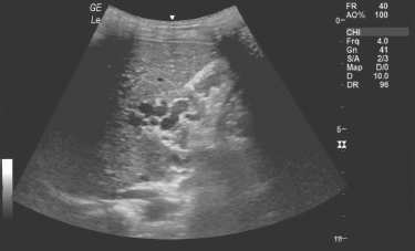
a. Identify the finding on ultrasound abdomen in given image.
b. What is the diagnosis?
c. What is the common clinical presentation?
d. How will you manage the child if he comes with this presentation?
Answer
- a. Cavernomatous transformation of portal vein—portal cavernoma.
- b. Extrahepatic portal venous obstruction (EHPVO).
- c. Sudden onset of unprovoked painless recurrent gastrointestinal (GI) bleeding, usually a major bleed and is well tolerated without signs of decompensation like ascites or encephalopathy.
- d. Management of acute variceal bleed with hemodynamic stabilization, use of vasopressors (octreotide/terlipressin), and endotherapy namely endoscopic variceal band ligation (EVL) or sclerotherapy depending upon the size of varices.
THG17-002
A 10-year-old boy presented with intermittent episodes of nocturnal regurgitation and difficulty in swallowing for 1 year duration. Clinical examination was unremarkable. Esophageal manometry (as shown in image) was done.
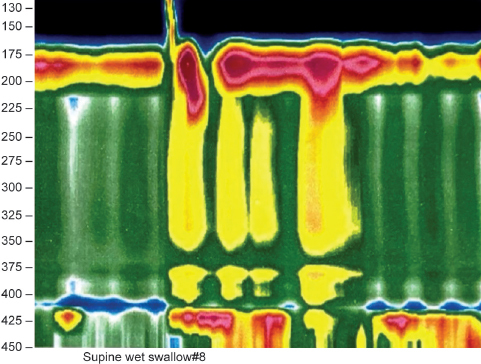
a. What are the findings?
b. What is the diagnosis?
c. Name the modalities of treatment.
Answer
- a. Hypertensive lower esophageal sphincter (LES) pressure with absent or inadequate relaxation and failed esophageal body peristalsis.
- b. The diagnosis is achalasia cardia.
- c. Modalities of treatment:
- Drugs: Calcium channel blockers, nitrates
- Endotherapy: Pneumatic balloon dilatation, botulinum toxin injection, peroral endoscopic myotomy (POEM)
- Surgery: Heller's myotomy (laparoscopic/open)
THG17-003
A 14-year-old boy presented in OPD with abdominal distension, bilateral pedal edema, and firm hepatomegaly and splenomegaly. Examination of the eye showed the finding (as shown in image).
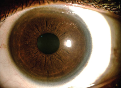
a. What is the finding in image given?
b. What is the likely diagnosis?
c. What are the other tests done to diagnosis this condition?
d. What are the drugs used to treat this?
Answer
- a. Pigmented golden-brown/greenish ring around the cornea—Kayser–Fleischer (KF) ring.
- b. Likely diagnosis: Wilson’s disease.
- c. Other tests: Serum ceruloplasmin, 24-hour urinary copper estimation, liver biopsy with quantitative copper assay, and slit-lamp examination.
- d. Medical treatment: D-penicillamine, trientine, and zinc along with avoidance of copper-rich diet (organ meats, shellfish, chocolate, nuts, mushrooms).
THG17-004
A 12-year-old male presented with recurrent episodes of upper abdominal pain, aggravated by food and radiation to back of 4 years duration. Clinically, he was thin built and the abdominal plain X-ray (as shown in image) showed the finding.
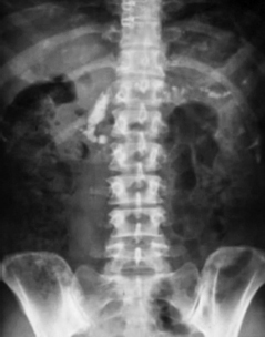
a. What is the diagnosis?
b. Write the X-ray finding.
c. What are the important complications?
Answer
- a. Chronic pancreatitis (tropical/calcific pancreatitis).
- b. Plain abdominal X-ray shows radiopaque calcification crossing the midline in the pancreatic region (pancreatic ductal calculi).
- c. Important complications:
- Exocrine insufficiency: Steatorrhea, malabsorption, malnutrition
- Endocrine insufficiency: Diabetes mellitus (pancreatogenic/Type 3c)
- Pseudocyst formation, splenic vein thrombosis, and rarely pancreatic cancer
THG17-005
A 14-year-old boy from Rajasthan presented with recurrent episodes of loose stools with fatigability of 2 years duration. His endoscopy showed scalloping of duodenal folds.
a. What is the likely diagnosis?
b. What are the serological tests done?
c. What will be findings on histology?
d. How will you treat?
Answer
- a. Celiac disease (gluten-sensitive enteropathy).
- b. Serological tests: Anti-tissue transglutaminase (anti-tTG IgA), anti-endomysial antibody (EMA IgA), and total serum IgA (to rule out IgA deficiency).
- c. Histopathology findings (Marsh classification):
- Flattening and blunting/atrophy of villi
- Crypt hyperplasia
- Increased intraepithelial lymphocytes (IELs)
- Chronic inflammatory infiltrate in lamina propria
- d. Lifelong strict adherence to a gluten-free diet (avoid wheat, rye, barley).
THG17-006
A 15-year-old girl presented with passing hard stools associated with bleeding per rectum. She also gave history of digital evacuation.
a. What is the probable diagnosis?
b. What will the finding on colonoscopy?
c. How will you treat?
Answer
- a. Solitary rectal ulcer syndrome (SRUS).
- b. Colonoscopy finding: Single or multiple shallow ulcers, usually on the anterior rectal wall within 10 cm of the anal verge; occasionally polypoid or erythematous lesions.
- c. Management:
- High-fiber diet, bulk laxatives, and behavioral therapy (avoid straining/digital evacuation)
- Sucralfate or corticosteroid enemas for active bleeding
- Argon plasma coagulation (APC)
- Biofeedback therapy and surgery (rectopexy) for refractory cases
THG17-007
A 42-day-old male infant who had an uneventful natal and postnatal period, now brought with jaundice, high-colored urine staining the diaper and passing clay-colored stools. On examination, child is alert, hepatomegaly present, no ascites, and no features of coagulopathy.
a. What is the most likely diagnosis?
b. When to suspect it on a USG abdomen?
c. What is the gold standard test?
d. How will you manage?
Answer
- a. Neonatal cholestasis syndrome—most likely Biliary Atresia.
- b. USG abdomen findings suggestive of biliary atresia:
- Absent or small, contracted gallbladder after fasting (triangular cord sign)
- Triangular cord sign (>4 mm echogenic fibrous tissue anterior to the portal vein bifurcation)
- Non-visualization of common bile duct
- Associated anomalies: Polysplenia/asplenia, situs inversus (biliary atresia splenic malformation syndrome)
- c. Intraoperative cholangiogram (IOC) with liver biopsy.
- d. Management:
- Surgical: Kasai portoenterostomy (ideally before 60 days of life)
- Nutritional & supportive: Fat-soluble vitamin supplementation (A, D, E, K), MCT oil, ursodeoxycholic acid (UDCA)
- Definitive for end-stage: Pediatric liver transplantation
THG17-008
A 9-month-old infant brought to emergency room (ER) with incessant cry, irritability, and passage of blood in stool of 1 day duration. Ultrasonography (USG) of abdomen has been shown this image.
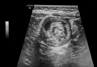
a. What is the diagnosis and clinical triad?
b. What is the USG finding shown in the above image?
c. What is the management?
d. What are the important complications?
Answer
- a. Diagnosis: Intussusception (ileocolic). Classical triad: Paroxysmal colicky abdominal pain (incessant cry), vomiting, and red "currant jelly" stools.
- b. Target sign (doughnut sign) in transverse view / pseudokidney sign in longitudinal view.
- c. Management:
- Resuscitation: IV fluids, NG tube decompression, IV antibiotics
- Non-operative reduction: Pneumatic (air) or hydrostatic (saline/contrast) enema reduction under fluoroscopy/ultrasound
- Operative reduction / bowel resection: Indicated if peritonitis, bowel perforation, shock, or failed non-operative reduction
- d. Bowel ischemia, gangrene, perforation, and peritonitis.
THG17-009
A 2-year-old child presented who has a history of delayed passage of meconium, now brought with constipation intermittently with occasional blood in the stools and failure to thrive.
a. What is the probable diagnosis?
b. How will you investigate?
c. How will you treat?
Answer
- a. Hirschsprung disease (congenital aganglionic megacolon).
- b. Investigations:
- Digital rectal examination: Empty rectum with explosive release of liquid stool and gas upon withdrawal of finger.
- Barium enema: Narrow aganglionic segment, transition zone (cone-shaped), and dilated proximal colon.
- Anorectal manometry: Absence of rectoanal inhibitory reflex (RAIR).
- Gold standard: Rectal suction biopsy showing absence of ganglion cells and presence of hypertrophied nerve trunks with positive acetylcholinesterase (AChE) staining.
- c. Definitive surgical management: Pull-through procedures (Duhamel, Soave, or Swenson procedure / transanal endorectal pull-through).
THG17-010
An 8-year-old boy presented with recurrent episodes of colicky abdominal pain with passage of loose stools and vomiting and weight loss of 6 months duration.
a. Name at least two differential diagnoses.
b. When to suspect Crohn’s disease on magnetic resonance enterography?
c. What are the extraintestinal manifestations (EIMs) in Crohn’s disease?
d. When to use biological agents in Crohn’s disease?
Answer
- a. Differential diagnoses:
- Intestinal tuberculosis
- Crohn's disease
- Gastrointestinal lymphoma
- b. MR Enterography features of Crohn's disease:
- Segmental mural thickening and hyperenhancement with "skip lesions"
- Luminal narrowing and strictures
- Comb sign (engorged vasa recta) and fibrofatty proliferation ("creeping fat")
- Fistulae and abscess formation
- c. Extraintestinal manifestations (EIMs):
- Aphthous oral ulcers
- Ocular: Episcleritis, anterior uveitis
- Cutaneous: Erythema nodosum, pyoderma gangrenosum, perianal skin tags/fissures
- Musculoskeletal: Peripheral arthritis, ankylosing spondylitis
- d. Indications for biologicals (Anti-TNF agents, e.g., Infliximab, Adalimumab):
- Fistulizing or penetrating Crohn's disease
- Severe active disease refractory to steroids and immunomodulators
- Extensive disease with high risk of poor outcome
THG17-011
A 4-year-old child was brought with recurrent episodes of abdominal pain. On examination, he had a palpable mass in the right hypochondrium.
a. What is the most probable diagnosis?
b. What are the clinical presentations?
c. How will you classify?
d. What is the treatment?
Answer
- a. Choledochal cyst (congenital cystic dilation of biliary tree).
- b. Clinical presentations:
- Classic triad: Abdominal pain, right upper quadrant mass, and jaundice (present in <20% of cases)
- Recurrent cholangitis, fever, pancreatitis, or asymptomatic incidental finding
- c. Todani classification:
- Type IA: Cystic dilatation of common bile duct
- Type IB: Focal segmental dilatation of extrahepatic duct
- Type IC: Fusiform dilatation of entire extrahepatic duct
- Type II: Diverticulum of common bile duct
- Type III (Choledochocele): Dilatation of intramural duodenal portion of CBD
- Type IVA: Multiple intrahepatic and extrahepatic cysts
- Type IVB: Multiple extrahepatic cysts only
- Type V (Caroli disease): Dilatation limited to intrahepatic bile ducts
- d. Complete surgical excision of cyst with Roux-en-Y hepaticojejunostomy.
THG17-012
A 2-year-old child born of second-degree consanguineous marriage was brought with loose stools on and off from 6 months of age. His birth weight was 2.7 kg. Clinically, the child was weighing 7.5 kg with bilateral pedal edema.
a. What is the role of peripheral smear?
b. How clinical examination is important.
c. What will be the finding in small bowel biopsy?
d. How will you manage?
Answer
- a. Role of peripheral smear:
- Microcytic or macrocytic anemia (malnutrition/blood loss)
- Thrombocytosis (inflammatory bowel disease)
- Eosinophilia (eosinophilic gastroenteritis)
- Lymphopenia (intestinal lymphangiectasia)
- Acanthocytes (abetalipoproteinemia)
- b. Examination clues: Look for hemihypertrophy, vascular malformations (Klippel-Trenaunay-Weber syndrome), facial dysmorphism (syndromic lymphangiectasia like Hennekam syndrome), or signs of protein-losing enteropathy.
- c. Small bowel biopsy finding: Dilated mucosal and submucosal lymphatic channels (lacteals) with distortion of villi, diagnostic of primary intestinal lymphangiectasia (Waldmann disease).
- d. Management:
- High-protein, low-fat diet enriched with medium-chain triglycerides (MCT)
- Fat-soluble vitamin supplementation (A, D, E, K)
- Correction of hypoalbuminemia with IV albumin if severe
- Octreotide (long-acting somatostatin analog) for refractory cases
- Surgical resection if localized to a short segment
THG17-013
A 2-year-old child was brought with history of foreign body (FB) (one rupee coin) ingestion an hour ago.
a. What investigation is to be advised?
b. What are dangerous foreign bodies?
c. What are the clinical signs which warrant immediate removal?
d. How will you manage?
Answer
- a. Plain biplane radiograph (X-ray neck, chest, and abdomen: AP and lateral views).
- b. Dangerous foreign bodies:
- Button batteries (risk of liquefaction necrosis, tracheoesophageal fistula, perforation within 2 hours)
- Multiple magnets or magnet plus metal object (bowel wall entrapment, necrosis, perforation)
- Sharp and pointed foreign bodies (needles, bones, pins)
- Superabsorbent expandable objects / water beads
- Large/long objects (>2 cm width, >5 cm length in small children)
- c. Red flag clinical signs for emergent endoscopy:
- Stridor, respiratory distress, wheezing, or drooling of saliva (airway compromise)
- Severe dysphagia, odynophagia, inability to swallow secretions, neck/chest tenderness, subcutaneous crepitus (impaction/perforation)
- d. Management:
- If coin in esophagus and symptomatic or >24 hours: Urgent endoscopic removal.
- If coin in stomach and asymptomatic: Conservative management; check stools and repeat X-ray in 1–2 weeks if not passed.
THG17-014
An 8-year-old boy brought with intermittent bleeding per rectum (PR) of 6-month duration. Colonoscopy was done. Identify the finding given in image here.
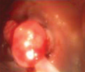
a. Identify the finding?
b. What are the findings expected to be seen in head-to-foot examination?
c. How will you manage this patient?
d. What are the complications?
Answer
- a. Colonic polyp (pedunculated juvenile polyp).
- b. Head-to-foot examination findings:
- Pallor due to chronic blood loss anemia
- Mucocutaneous pigmentation around lips, buccal mucosa, digits (Peutz-Jeghers syndrome)
- Dental anomalies, osteomas, epidermal cysts (Gardner syndrome / FAP)
- Macrocephaly, trichilemmomas (Cowden syndrome)
- c. Endoscopic snare polypectomy with histopathological examination.
- d. Complications: Severe anemia, acute lower GI bleed, colocolonic intussusception, and malignant transformation (in polyposis syndromes).
THG17-015
A 5-year-old female child was brought with recurrent bouts of vomiting of 2 years duration. Clinical examination showed some dehydration.
a. When will you suspect cyclical vomiting syndrome?
b. Name some triggers.
c. How will you treat?
Answer
- a. CVS suspicion criteria (Rome IV criteria):
- Stereotypical episodes of intense nausea and unremitting vomiting
- At least 2 acute episodes in the past 6 months, lasting 1 hour to 10 days, occurring at least 1 week apart
- Complete return to baseline health between episodes
- Absence of other identifiable causes
- b. Triggers:
- Psychological stress, excitement, sleep deprivation, physical exhaustion
- Intercurrent infections (URI, sinusitis)
- Foods: Chocolate, cheese, MSG, caffeine
- Motion sickness, hot weather, menses in adolescents
- c. Management:
- Evaluation: Rule out malrotation, hydronephrosis, metabolic disorders, and space-occupying CNS lesions.
- Acute attack: Quiet dark room, IV hydration, dextrose, antiemetics (ondansetron), sedatives (lorazepam), triptans (sumatriptan).
- Prophylaxis (if frequent episodes): Cyproheptadine (<5 years), propranolol, amitriptyline (>5 years), or coenzyme Q10/L-carnitine.
THG17-016
An 8-month-old infant was brought with streaks of blood in the stools for 1 month duration. Child was introduced to formula feeds and regular milk used in the household since 6 months of age.
a. What is the probable diagnosis?
b. How will you manage?
c. What are the different formulations available?
d. What is FPIES?
Answer
- a. Cow's milk protein allergy (CMPA / allergic proctocolitis).
- b. Management:
- Strict elimination of cow's milk protein from the infant's diet.
- If breastfed, eliminate dairy from the mother's diet with maternal calcium supplementation.
- Re-evaluate / food challenge after 6–12 months of age (most outgrow by 1–3 years).
- c. Therapeutic formulations:
- Extensively hydrolyzed formula (eHF) — first line
- Amino acid-based formula (AAF) — for severe cases / anaphylaxis / failure of eHF
- Soy formula (for infants >6 months if eHF not tolerated/accepted)
- d. FPIES (Food Protein-Induced Enterocolitis Syndrome): A non-IgE mediated food allergy presenting with severe repetitive vomiting, lethargy, dehydration, and hypovolemic shock 1–4 hours after ingestion of trigger food (commonly cow's milk or soy).
THG17-017
A 48-hour-old male infant was brought with ER with respiratory distress and drooling of saliva.
a. What is the most probable diagnosis?
b. What is the next step of investigation?
c. How will you treat?
d. What is the long-term complication?
Answer
- a. Tracheoesophageal fistula (TEF) with Esophageal Atresia (EA) (most commonly Type C: proximal EA with distal TEF).
- b. Next step of investigation: Insertion of a wide-bore radiopaque catheter (10 Fr) through the mouth/nose into the esophagus followed by plain chest and abdominal radiograph (shows coiling of catheter in upper esophageal pouch at T4-T5 and air in GI tract).
- c. Treatment:
- Preoperative: Upright positioning, continuous suction of upper pouch (Replogle tube), IV fluids, withhold feeds, antibiotics.
- Definitive surgery: Primary thoracoscopic or open ligation of fistula and end-to-end esophageal anastomosis.
- d. Long-term complications: Esophageal anastomotic stricture requiring dilatation, tracheomalacia, gastroesophageal reflux disease (GERD), and recurrent TEF.
THG17-018
An 8-year-old female child presented with history of ataxia and visual disturbances. Her growth chart shows height and weight below 3rd centile. Her peripheral smear is given below. Identify the finding on peripheral smear (as shown in image).
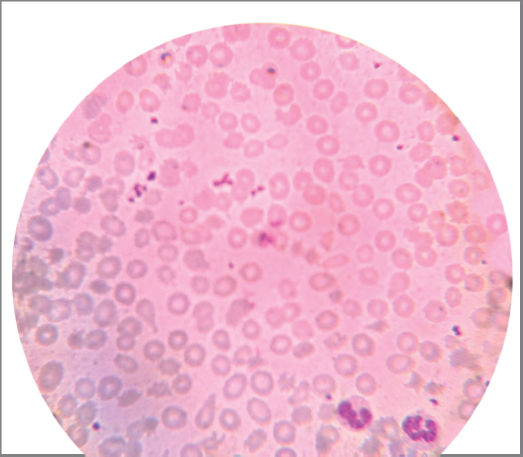
a. What will be the clinical picture?
b. What will be the finding on intestinal biopsy?
c. What are the long-term complications of the disease?
Answer
- a. Clinical picture of Abetalipoproteinemia (Bassen-Kornzweig syndrome):
- Peripheral smear shows thorny red blood cells (acanthocytes).
- Infancy: Severe fat malabsorption, steatorrhea, failure to thrive.
- Later childhood: Spinocerebellar ataxia, peripheral neuropathy, loss of deep tendon reflexes, retinitis pigmentosa (atypical), and visual impairment.
- b. Intestinal biopsy: Enterocytes packed with clear lipid vacuoles / fat droplets (lipid-laden enterocytes) with normal villous architecture.
- c. Long-term complications: Progressive neurological disability (ataxia, loss of proprioception), blindness due to retinitis pigmentosa, severe hypocholesterolemia, hypovitaminosis A, D, E, K, hepatic steatosis/cirrhosis, and cardiomyopathy.
THG17-019
Helicobacter pylori infection.
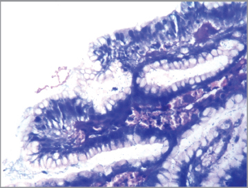
a. Identify the organism?
b. Who discovered it?
c. Name any two factors responsible for virulence in the organism.
d. How will you test?
Answer
- a. Gastric biopsy showing Helicobacter pylori (spiral/curved gram-negative bacilli).
- b. Robin Warren and Barry Marshall (Nobel Prize 2005).
- c. Virulence factors:
- Cytotoxin-associated gene A (CagA) protein
- Vacuolating cytotoxin A (VacA)
- Urease enzyme production
- Flagellar motility and BabA/SabA adhesins
- d. Diagnostic tests:
- Invasive (Endoscopy): Rapid urease test (RUT), histopathology (Giemsa/Warthin-Starry stain), gastric culture, biopsy PCR.
- Non-invasive: Urea breath test (13C-UBT), stool antigen test (monoclonal EIA), serology (IgG).
THG17-020
A 4-year-old boy was brought with history of recurrent seizures since infancy. Clinical examination showed hepatomegaly with liver span of 10 cm.
a. When will you suspect glycogen storage disease?
b. How will you differentiate type 1 and 3?
c. How will you manage?
Answer
- a. Suspicion of Glycogen Storage Disease (GSD):
- Doll-like facies with chubby cheeks, thin extremities, protuberant abdomen with massive hepatomegaly (normal spleen).
- Recurrent severe fasting hypoglycemia and morning seizures, lactic acidosis, failure to thrive.
- Differential for massive hepatomegaly: GSD, lysosomal storage disorders (Gaucher, Niemann-Pick), congestive hepatopathy, Budd-Chiari syndrome, and hepatoblastoma.
- b. Differentiating Type 1 (von Gierke) vs. Type 3 (Cori/Forbes):
- Type 1 (GSD I): Severe fasting hypoglycemia, profound lactic acidosis, marked hyperuricemia, hypertriglyceridemia, renomegaly; normal transaminases initially; spleen normal.
- Type 3 (GSD III): Mild hypoglycemia, normal lactate and uric acid, markedly elevated transaminases (AST/ALT), muscle weakness/cardiomyopathy, progressive hepatic fibrosis/cirrhosis; spleen can be enlarged due to portal hypertension.
- c. Management:
- Prevent hypoglycemia: Frequent feeds with uncooked cornstarch (1.5–2 g/kg every 4–6 hours) or continuous nocturnal nasogastric glucose infusion.
- Diet: High complex carbohydrate, restriction of simple sugars (fructose/galactose in Type 1; high protein in Type 3).
- Allopurinol for hyperuricemia, statins/fibrates for hyperlipidemia, ACE inhibitors for microalbuminuria.