MNJ18-001
A 10-year-old male is referred for autoimmune hemolytic anemia, frequent diarrhea, normal tonsils; father is a diagnosed case having low IgA levels. His IgM level is normal but IgA and IgG levels are low:
- What is the immunodeficiency type you will suspect?
- How will you treat the patient?
- What is the mode of inheritance?
- What is the future risk for these children?
Answer
- Suspected type: Common Variable Immunodeficiency (CVID) (Note: IgA deficiency occurs commonly in pedigrees having CVID).
- Treatment: Regular IVIG replacement therapy, co-trimoxazole (Septran) prophylaxis.
- Mode of inheritance: Autosomal Dominant (AD) (or complex/polygenic in many cases).
- Future risk: Up to 38-fold increased risk of malignancies, particularly lymphomas and gastric carcinoma.
MNJ18-002
A 6-year-old child was admitted for treatment of LRTI. This was his 6th episode of severe pneumonia apart from 4 episodes of severe diarrhea in past. He was transfused blood in view of anemia (Hb: 8.5 gm%) but developed severe transfusion reactions:
- What immunological defect is likely?
- What is the critical serum level to make the diagnosis?
- Discontinuation of which drug leads to resolution of drug-induced deficiency?
- True/False:
- a. IVIG administration is not useful as treatment.
- b. These patients have antibodies against cow's milk.
- c. This deficiency may evolve into CVID.
- How will you manage the same?
- What is the common GI organism associated with this defect?
Answer
- Immunological defect: Selective IgA Deficiency.
- Diagnostic serum level: Serum
with normal IgG and IgM levels. - Drug association: Phenytoin (also sulfasalazine, penicillamine, gold).
- True/False:
- a. True (IVIG is not indicated and contains trace IgA which can cause fatal anaphylaxis due to anti-IgA antibodies).
- b. True (frequent antibodies to dietary proteins/cow's milk proteins).
- c. True (can evolve into CVID over time).
- Management:
- Prompt treatment of infections with antibiotics; prophylactic antibiotics if recurrent infections.
- Avoid routine blood/plasma transfusions; if blood transfusion is essential, use washed RBCs or blood products from IgA-deficient donors.
- Common GI organism: Giardia lamblia.
MNJ18-003
A 1.5-year-old male came with breathlessness, fever, and poor feeding. He was a wasted and stunted child with absent tonsils and thymus.
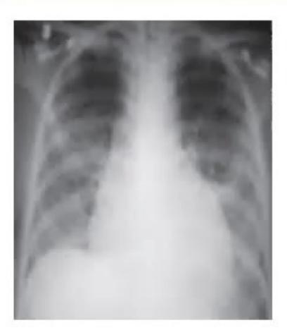
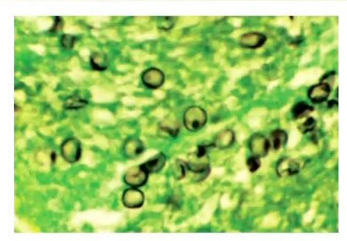
- Identify the organism.
- What is the likely diagnosis?
- Mention the 4 molecular defects identified.
Answer
- Organism: Pneumocystis jirovecii (formerly P. carinii).
- Likely diagnosis: Hyper-IgM Syndrome.
- Molecular defects identified:
- Mutations in CD40 ligand (CD40L / CD154) on X-chromosome (X-linked, most common)
- Mutations in NEMO (IKBKG) on X-chromosome
- Mutations in Activation-Induced Cytidine Deaminase (AID) gene on chromosome 12
- Mutations in CD40 gene on chromosome 20 (or UNG deficiency)
MNJ18-004
A dysmorphic newborn with receding chin was admitted with hypocalcemic seizures, congestive cardiac failure with cardiac murmur, and bifid uvula. X-ray revealed absent thymus:
- Identify the syndrome.
- Identify the defective embryologic stage.
- Which chromosome is involved?
- What is the treatment?
Answer
- Syndrome: DiGeorge syndrome (22q11.2 deletion syndrome / Velocardiofacial syndrome).
- Defective embryologic stage: Dysmorphogenesis / maldevelopment of the 3rd and 4th pharyngeal pouches and arches.
- Chromosome involved: Microdeletion of chromosome 22q11.2.
- Treatment:
- Management of hypocalcemia (IV calcium gluconate, oral calcium, active vitamin D/calcitriol).
- Correct congenital cardiac defects.
- For complete DiGeorge syndrome: Cultured thymic tissue transplant or HLA-matched bone marrow transplant (BMT).
- Use irradiated, CMV-negative blood products.
MNJ18-005
A 4-month-old infant exclusively breastfed presented with severe illness. He had severe oral thrush, perianal dermatitis, and fever. He was diagnosed to have Klebsiella septicemia. During the course of admission he developed brain abscesses. He was transfused platelets for treatment of DIC. He later developed skin rash, bloody diarrhea, and elevated liver enzymes. Patient succumbed to fungal meningitis. Lymph nodes/tonsils were absent. On admission his Hb: 9 gm%, WBC: 2400, P: 85, L: 10, M: 2, E: 3, CXR: absent thymus:
- Identify the phenotype/condition.
- What care is needed in transfusing these infants?
- How are they cured?
- What advice will you give to parents about vaccination?
Answer
- Condition: Severe Combined Immunodeficiency (SCID) (complicated by transfusion-associated Graft-versus-Host Disease [GVHD]).
- Care in transfusion: All blood and blood products must be leukodepleted, irradiated, and CMV-negative to prevent transfusion-associated GVHD.
- Definitive cure: Hematopoietic stem cell transplantation (HSCT / Bone marrow transplant) or gene therapy.
- Vaccination advice: Strictly avoid all live vaccines (e.g., BCG, OPV, rotavirus, MMR, varicella) as they can cause fatal disseminated vaccine-strain infections.
MNJ18-006
An 8-year-old female had difficulty in walking, had 5 episodes of sinusitis, and 2 episodes of pneumonias; skin and eyes showed abnormal vessels:
- What is the likely diagnosis?
- What is the genetic defect?
- Which immunoglobulin level will you find low?
- What are 5 systems involved in this disease?
Answer
- Likely diagnosis: Ataxia-Telangiectasia.
- Genetic defect: Mutations in the ATM (Ataxia-Telangiectasia Mutated) gene on the long arm of chromosome 11 (11q22.3).
- Immunoglobulin level: Low IgA (and commonly IgE and
). - Systems involved:
- Central Nervous System (cerebellar ataxia, oculomotor apraxia)
- Cutaneous/Vascular (oculocutaneous telangiectasias, premature aging)
- Immune system (humoral and cellular immunodeficiency, recurrent sinopulmonary infections)
- Endocrine system (insulin resistance/diabetes, hypogonadism)
- Hepatic/Malignancy/Hematologic predisposition (high
-fetoprotein, elevated risk of leukemias and lymphomas).
MNJ18-007
A 6-month-old male had skin lesions and gum bleeds, WBC: 9500, P: 40, L: 45, E: 15, Plt: 56,000/
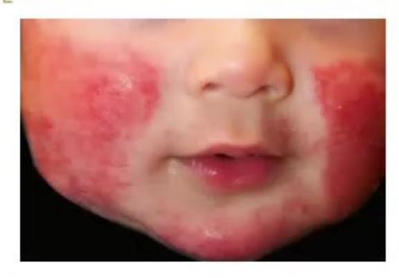
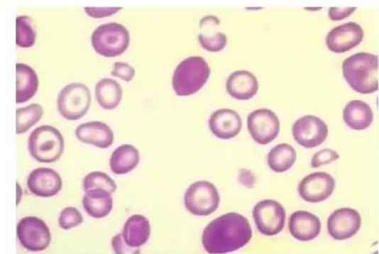
- What diagnosis will you suspect?
- What are the typical serum immunoglobulin levels?
- How will you treat the patient?
Answer
- Diagnosis: Wiskott-Aldrich Syndrome (WAS) (characterized by microthrombocytopenia with low MPV, eczema, and immunodeficiency).
- Serum Ig levels: Typically low IgM, elevated IgA and IgE, and normal to decreased IgG.
- Treatment:
- Definitive cure: Hematopoietic stem cell transplantation (HSCT) or gene therapy.
- Supportive/Medical care: IVIG replacement therapy, antibiotic prophylaxis (co-trimoxazole) and prompt treatment of infections, platelet transfusions (irradiated/leukodepleted) for severe bleeding, topical corticosteroids for eczema, avoid NSAIDs/antiplatelet agents.
MNJ18-008
A 3-month-old male is referred for fever, umbilical sepsis, and persistently elevated WBC counts (WBC: 75,000, P: 92, L: 6, M: 2), not responding to therapy. Child has history of delayed umbilical cord detachment:
- Which immunodeficiency will you suspect?
- How will you prove the same?
- What is the definitive therapy?
Answer
- Suspected immunodeficiency: Leukocyte Adhesion Deficiency type 1 (LAD-1).
- Proof/Diagnostic test: Flow cytometry demonstrating deficiency or absence of
(integrin subunit) on leukocytes, and Rebuck skin window test (absence of neutrophil migration). - Definitive therapy: Allogeneic Hematopoietic Stem Cell Transplantation (HSCT / BMT).
MNJ18-009
A fair-appearing child with intellectual disability was brought with recurrent infections (recurrent fevers, otitis media, gingivitis, and septicemia). His peripheral smear led to diagnosis of the condition. Skin biopsy showed giant melanosomes:
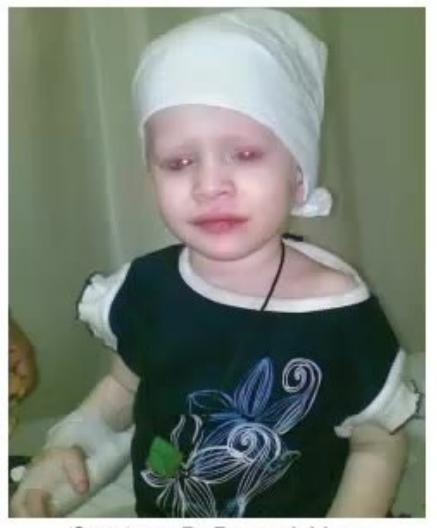
Courtesy: Dr Pravesh Vyas
- Name the disorder.
- Name the genetic defect.
Answer
- Disorder: Chédiak-Higashi Syndrome.
- Genetic defect: Autosomal recessive mutations in the Lysosomal Trafficking Regulator (LYST / CHS1) gene located on chromosome 1 (1q42-q43).
MNJ18-010
A 6-year-old boy is evaluated for recurrent pneumonia and osteomyelitis of multiple sites. On examination, he had anemia, malnutrition, and cervical lymphadenitis. He also had folliculitis and multiple cutaneous granulomas:
- What is the possible diagnosis?
- What is the closest differential diagnosis?
- What are the gastrointestinal complications of this condition?
- What is the recent diagnostic test for this condition?
- Name 3 drugs used in the management.
Answer
- Possible diagnosis: Chronic Granulomatous Disease (CGD).
- Closest differential diagnosis: Severe G6PD deficiency (or Hyper-IgE syndrome / Crohn's disease / Mycobacterial infection).
- Gastrointestinal complications: Pyloric/antral outlet obstruction, granulomatous colitis/enteritis (simulating Crohn's disease), rectal/perianal fistulae, esophageal strictures, and hepatic abscesses.
- Recent diagnostic test: Flow cytometry using Dihydrorhodamine 123 (DHR) oxidation test (evaluates neutrophil respiratory burst).
- Drugs in management:
- Daily co-trimoxazole (trimethoprim-sulfamethoxazole) for antibacterial prophylaxis
- Itraconazole for antifungal prophylaxis
- Interferon-gamma (
) to enhance phagocytic oxidative burst and reduce serious infections - Corticosteroids for inflammatory granulomatous complications (e.g., gastric outlet or urinary tract obstruction).
MNJ18-011
A 2-year-old child presents with episodes of otitis media, eczematous dermatitis, and thrombocytopenia with bloody stool:
- What is the diagnosis of this condition and what is the size of megakaryocytes/platelets in this?
- What is the inheritance pattern?
- What are the common organisms causing infection in this condition?
Answer
- Diagnosis & size: Wiskott-Aldrich Syndrome (WAS). Megakaryocytes are normal in number and morphology in bone marrow, but circulating platelets are characteristically small (microthrombocytopenia / low MPV).
- Inheritance pattern: X-linked recessive (mutation in WAS gene on Xp11.22).
- Common organisms: Encapsulated bacteria (Streptococcus pneumoniae, Neisseria meningitidis, Haemophilus influenzae), Pneumocystis jirovecii, and viral pathogens (Herpes simplex, CMV, EBV, Varicella).
MNJ18-012
A 2-year-old child presents with the following:
- 5 episodes of abscesses in 6 months
- Photosensitivity
- Light skin and silvery hair
- Peripheral smear shows large inclusions in all nucleated blood cells
- What is the diagnosis?
- What is the cause for the lightened hair?
- What is the mode of inheritance?
- Name one life-threatening hematological complication.
- What is the neurological manifestation?
- Which drug/therapy is indicated?
Answer
- Diagnosis: Chédiak-Higashi Syndrome.
- Cause of lightened hair: Failure to properly disperse giant melanosomes from melanocytes to keratinocytes and hair shafts.
- Mode of inheritance: Autosomal recessive.
- Life-threatening hematological complication: Accelerated phase (hemophagocytic lymphohistiocytosis [HLH]-like syndrome with pancytopenia, hepatosplenomegaly, and multiorgan failure).
- Neurological manifestation: Peripheral neuropathy, cranial neuropathies, cerebellar ataxia, motor and sensory deficits, intellectual disability/dementia.
- Drug/Treatment: High-dose ascorbic acid (Vitamin C: 200 mg/day in infants, up to 2000 mg/day in older children) in the stable phase; chemotherapy/immunotherapy protocols for accelerated phase; definitive cure is Hematopoietic Stem Cell Transplantation (HSCT).
MNJ18-013
Answer the following rapid-fire clinical questions:
- A 6-month-old boy with bloody stools, draining ears, eczema, and normal IgA: What is the diagnosis?
- A 2-year-old with recurrent cutaneous and/or systemic staphylococcal abscesses and coarse facial features: What is the diagnosis?
- Oculocutaneous albinism and primary immunodeficiency: What condition is this?
- Failure to thrive, protracted diarrhea, and extensive mucocutaneous candidiasis: What cell line is likely compromised?
- Absolute lymphocyte count on screening for immunodeficiency rules out which cell line defect?
- What is the first screening test for complement deficiency?
- What are the screening tests for B-cell deficiency?
- What is the inheritance pattern of Bruton syndrome (agammaglobulinemia)?
- What is the difference between allogeneic and autologous hematopoietic stem cells?
- Mention two fungal and two viral infections common post stem cell transplant.
Answer
- Wiskott-Aldrich Syndrome
- Hyper-IgE Syndrome (Job syndrome)
- Chédiak-Higashi Syndrome (or Griscelli syndrome)
- Predominant T-cell defect (Cell-Mediated Immunity / SCID)
- T-cell defect (normal absolute lymphocyte count rules against severe T-cell lymphopenia/SCID)
- Total hemolytic complement assay (
) (and for alternative pathway) - Screening tests for B-cell deficiency: Quantitative serum immunoglobulin levels (IgG, IgA, IgM, IgE), isohemagglutinin titers (anti-A, anti-B), and specific antibody response to protein/polysaccharide vaccine antigens (e.g., tetanus, diphtheria, Haemophilus influenzae, pneumococcal).
- X-linked recessive
- Difference: Allogeneic stem cells are collected from a genetically matched related or unrelated donor, whereas autologous stem cells are harvested from the patient's own body.
- Infections:
- Fungal: Candida, Aspergillus
- Viral: Cytomegalovirus (CMV), Epstein-Barr virus (EBV) / Adenovirus / HSV
MNJ18-014
- Write (any two) screening tests for B-cell primary immunodeficiency.
- Write screening tests for complement deficiency.
- Give (any 4) indications for hematopoietic stem cell transplant in treatment of congenital diseases.
Answer
- Screening tests for B-cell primary immunodeficiency:
- Quantitative serum immunoglobulin levels (IgG, IgM, IgA)
- Isohemagglutinin titers
- Specific antibody titers to vaccine antigens (e.g., tetanus, diphtheria, H. influenzae, pneumococcus)
- B-cell enumeration by flow cytometry (CD19/CD20).
- Screening tests for complement deficiency:
(classical pathway activity) (alternative pathway activity) - Individual complement component assays (
, ).
- Indications for HSCT in congenital diseases:
- Severe Combined Immunodeficiency (SCID)
- Wiskott-Aldrich Syndrome / Hyper-IgM Syndrome
- Thalassemia Major / Sickle Cell Disease
- Diamond-Blackfan Anemia / Fanconi Anemia / Severe Congenital Neutropenia
- Inborn errors of metabolism (e.g., Hurler syndrome, adrenoleukodystrophy).
MNJ18-015
A 14-month-old male child presents with fever with acute flaccid paralysis (AFP) following polio vaccine. CSF was done to rule out the possibility of GBS, but report showed positive enterovirus from CSF. On physical examination, there is absence of tonsils and no palpable lymph nodes:
- What is the possible diagnosis?
- What is the mode of inheritance?
- What will you find on flow cytometry?
Answer
- Possible diagnosis: X-Linked Agammaglobulinemia (XLA / Bruton Agammaglobulinemia / BTK deficiency) presenting with vaccine-associated paralytic poliomyelitis (VAPP).
- Mode of inheritance: X-linked recessive (mutation in the Bruton tyrosine kinase [BTK] gene).
- Flow cytometry findings: Profound absence / near-total lack of circulating mature B-cells (
), with normal numbers and proportions of T-lymphocytes ( ).