VVK09-001
Write eight distinguishing features between gray and white matter diseases of the brain:
Answer
| Feature | Gray Matter Disease | White Matter Disease |
|---|---|---|
| Dementia / Cognitive decline | Early and prominent | Late |
| Seizures | Early, prominent, and frequent | Late or uncommon |
| Motor signs (Tone, Reflexes, Ataxia, Spasticity) | Late / uncommon until end-stage | Early and prominent (spasticity, hyperreflexia, ataxia) |
| Basal ganglia / Extrapyramidal signs | Often present early (chorea, dystonia, tremor) | Usually absent |
| Retinal changes / Retinitis pigmentosa | May be present (macular cherry-red spot, retinitis pigmentosa) | Absent (may have optic atrophy) |
| Neuroimaging (MRI Brain) | Cortical atrophy, basal ganglia signal changes | Diffuse symmetrical white matter T2/FLAIR hyperintensities |
| Electroretinogram (ERG) | May be abnormal | Normal |
| Evoked potentials (VER / BERA) | Usually normal | Characteristically abnormal (prolonged latencies / conduction delay) |
VVK09-002
A 4-year-old boy is brought by the parents with history that the school has complained that he is aggressive, cannot sit in one place, is forgetful, restless and constantly getting into fights with his friends. At home, the parents say that he is constantly on the move and even does not watch television continuously for more than a few minutes. The child had a normal birth and developmental milestones.
a. What is the diagnosis?
b. Give three cardinal features of this condition.
c. Name two treatment options.
Answer
- a. Attention-Deficit / Hyperactivity Disorder (ADHD).
- b. Cardinal Features (Triad):
-
- Inattention
-
- Hyperactivity
-
- Impulsivity
-
- c.
- Pharmacotherapy: CNS stimulants (Methylphenidate, Dextroamphetamine) or non-stimulants (Atomoxetine, Clonidine, Guanfacine).
- Behavioral Therapy / Psychosocial Intervention: Parent training, classroom behavioral interventions, positive reinforcement.
VVK09-003
A three-year-old male child is brought to you by the mother with the concern that he has been having trouble running and keeping up with his peers. She also states that he had been slow attaining other major motor milestones like walking and climbing stairs. The child is in the exam room sitting on the floor. You ask him to get up and he proceeds to arise by using his arms to climb up his legs and body.
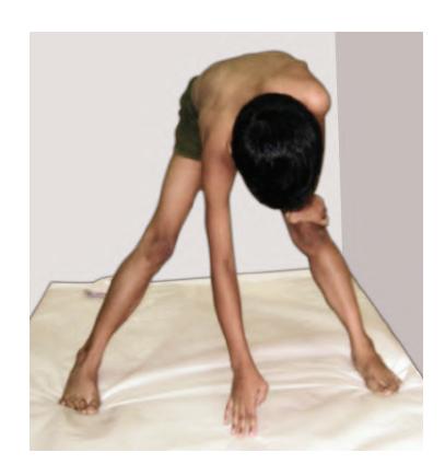
a. Name the sign.
b. Name three conditions in which this sign can be seen.
c. Name three investigations in sequence of importance.
d. What is the mode of inheritance and the locus of abnormal gene in the most common cause for this condition?
Answer
- a. Gower's sign (indicates proximal pelvic girdle muscle weakness).
- b.
- Duchenne Muscular Dystrophy (DMD)
- Becker Muscular Dystrophy (BMD)
- Spinal Muscular Atrophy (SMA type III / Kugelberg-Welander)
- Limb-Girdle Muscular Dystrophy (LGMD)
- Congenital or inflammatory myopathies (Polymyositis, Dermatomyositis)
- c.
-
- Serum Creatine Phosphokinase (CPK / CK) levels (massively elevated in DMD, > 10–50 times normal).
-
- Genetic testing: DMD gene deletion/duplication analysis (MLPA / Next-Generation Sequencing).
-
- Muscle biopsy with dystrophin immunohistochemistry / Electromyography (EMG).
-
- d. X-linked recessive; dystrophin gene locus at chromosome Xp21.2.
VVK09-004
Parents bring a 1½-year-old infant for routine evaluation. He is thriving well and gross and fine motor milestones are normal for his age. The parents are concerned that the child has not started speaking as yet. A previously consulted pediatrician diagnosed the child to be autistic. On further questioning and examination, the infant is not using bisyllables like 'mama' and 'dada'. He started cooing, laughing, and babbling at the appropriate ages, but the babbling is disappearing now. He responds to a command with gestures. The child interacts with his parents and asks for objects by pointing to them. He was born at 32 weeks of gestation and required care in the NICU, during which he received a course of antibiotics and phototherapy.
a. What is the likely diagnosis?
b. Do you agree with the diagnosis of autism? Justify your answer.
c. What is your assessment? Name two significant points in the history that could suggest an etiology for your diagnosis.
d. What investigations will you do on the child?
Answer
- a. Sensorineural Hearing Loss (SNHL) / Congenital/Prelingual Deafness.
- b. No, disagree with autism. The child exhibits normal non-verbal social interaction, makes eye contact, interacts appropriately with parents, and uses communicative pointing and gestures to request objects (which are preserved in isolated hearing loss but impaired in autism spectrum disorder).
- c. The infant has prelingual hearing loss causing speech regression/delay.
- Etiological risk factors in history:
-
- Ototoxic antibiotic exposure in NICU (aminoglycosides like gentamicin/amikacin).
-
- Hyperbilirubinemia requiring phototherapy (auditory neuropathy / kernicterus risk).
-
- Prematurity (32 weeks) and NICU stay / incubator noise exposure.
-
- Etiological risk factors in history:
- d.
- Brainstem Evoked Response Audiometry (BERA / ABR)
- Distortion Product Otoacoustic Emissions (DPOAE)
- Impedance audiometry / Tympanometry
- Visual Reinforcement Audiometry (VRA) / Play audiometry
VVK09-005
A 6-year-old boy and his parents come for an evaluation of a "behavioral problem". The child's teacher insists that he needs medical consultation because he is very disruptive in class. He keeps running out of class and does not concentrate on his classwork. She also states that he is careless in his work, has difficulty waiting his turn in group situations and talks excessively. The parents feel that he is just an active boy with "a lot of curiosity". They state that he can watch TV for a long period of time without problems and that other children have trouble playing with him because he "likes to be the leader". He is an only child.
a. Is the child normal? If not, what is he suffering from?
b. Name three clinical subtypes of this condition.
c. Name three drugs used in the management of this condition.
Answer
- a. Not normal; the child is suffering from Attention-Deficit / Hyperactivity Disorder (ADHD). (Children with ADHD can often hyperfocus on highly stimulating activities like television or video games despite pervasive inattention in academic/social settings).
- b. Subtypes of ADHD (DSM-5):
-
- ADHD, Predominantly Inattentive Presentation
-
- ADHD, Predominantly Hyperactive/Impulsive Presentation
-
- ADHD, Combined Presentation
-
- c.
- Methylphenidate
- Atomoxetine
- Dextroamphetamine / Lisdexamfetamine
- Clonidine / Guanfacine
VVK09-006
A 4-month-old baby is brought with delayed developmental milestones. Clinically, the infant is alert, interacting with the examiner and mother. Neurological examination shows the infant to be floppy with generalized hypotonia affecting the trunk and limbs. Deep tendon jerks are not elicited and there is congenital talipes equinovarus (CTEV) of the left foot. Fasciculations are noted on the tongue.
a. What is the most likely diagnosis?
b. Name two definitive diagnostic tests for this condition.
c. What is the mode of inheritance in this condition?
d. Give 4 subtypes of this diagnosis.
Answer
- a. Spinal Muscular Atrophy Type 1 (SMA Type 1 / Werdnig-Hoffmann Disease) (severe anterior horn cell degeneration).
- b.
-
- Molecular Genetic Testing: SMN1 gene deletion/mutation analysis (homozygous deletion of exon 7/8 on chromosome 5q13 by MLPA / PCR).
-
- Electromyography (EMG) showing neurogenic pattern (fibrillations, fasciculations) / Muscle biopsy showing large groups of atrophic fibers interspersed with normal/hypertrophic fibers.
-
- c. Autosomal recessive (5q-SMA).
- d. Subtypes of 5q Spinal Muscular Atrophy:
- Type 0: Prenatal onset, severe hypotonia, arthrogryposis, respiratory failure at birth.
- Type 1 (Werdnig-Hoffmann): Infantile onset (< 6 months), never sits independently.
- Type 2 (Dubowitz / Intermediate): Onset 6–18 months, sits independently, never walks.
- Type 3 (Kugelberg-Welander / Juvenile): Onset > 18 months, achieves independent walking.
- Type 4: Adult onset (> 20–30 years), mild proximal weakness.
VVK09-007
A 4-month-old male infant, born of a consanguineous marriage to a primigravida mother, is brought to hospital with delayed milestones and feeding difficulty since birth. Antenatal period was uneventful, except that the mother did not perceive strong fetal movements. There was also history of polyhydramnios. Clinically, there is generalized hypotonia, with absent deep tendon jerks. The testes are undescended. Rest of the systemic examination is normal.
a. What would be the probable diagnosis?
b. Name 3 specific types of this disease.
c. What is the basic pathology causing this condition?
d. What would be the line of management for this child?
Answer
- a. Congenital Myopathy (e.g. X-linked Myotubular Myopathy / Prader-Willi Syndrome / Congenital Muscular Dystrophy).
- b.
-
- X-linked Myotubular / Centronuclear myopathy
-
- Nemaline rod myopathy
-
- Central core disease
-
- c. Structural and maturational arrest or defect in fetal muscle fiber development with characteristic cytoarchitectural structural abnormalities (e.g., centrally located nuclei resembling fetal myotubes, nemaline rods, or core zones of oxidative enzyme depletion).
- d.
- Confirmation by muscle biopsy (histochemistry/electron microscopy) and genetic panel testing.
- Supportive nutritional care (nasogastric/orogastric feeding or gastrostomy).
- Physical and occupational therapy; respiratory monitoring and ventilatory support.
- Multidisciplinary team management and genetic counselling for family.
VVK09-008
An 8-month-old infant is brought to hospital with complaints of excessive irritability, high-grade fever, vomiting and altered sensorium since past 18 hours. The infant had a generalized seizure 15 minutes ago. Clinical and laboratory investigations confirm meningitis. The microbiology laboratory returns the report of Haemophilus influenzae type b (Hib) meningitis on CSF. The child's paternal grandfather, under treatment for multiple myeloma, resides in the same house.
a. What is the prognostic significance of seizures in bacterial meningitis?
b. The commonest sequela of bacterial meningitis is _______________.
c. What would be your advice to the child's parents regarding prevention of spread of this disease to other members in the household?
d. Define household contact.
Answer
- a. Seizures occurring at the onset (< 24–48 hours) are common and usually febrile/inflammatory without worse prognosis. However, seizures that persist or develop after Day 4 of therapy, focal seizures, or status epilepticus indicate cortical infarction, cerebritis, subdural empyema, or sinus thrombosis and correlate with increased risk of long-term neurological sequelae.
- b. Sensorineural Hearing Loss (SNHL) (due to suppurative labyrinthitis / cochlear damage).
- c. Chemoprophylaxis with Oral Rifampicin (20 mg/kg once daily, max 600 mg/day for 4 days) is indicated for all household contacts if there is at least one household contact under 48 months of age who is unimmunized/partially immunized against Hib, or if an immunocompromised individual (like the grandfather with myeloma) resides in the house.
- d. Household Contact: An individual residing in the same household as the index patient, or who spent at least 4 hours per day with the index case on at least 5 of the 7 days prior to hospital admission.
VVK09-009
A 3-year-old female has been brought with history of low-grade fever for 2 weeks with headache, lethargy and projectile vomiting. Today she had a focal tonic-clonic seizure with loss of consciousness. She was earlier on treatment for a heart condition. Clinically, the child is comatose, febrile and her blood pressure is normal. A harsh pan-systolic murmur is heard best at left sternal border. Fundus examination reveals bilateral papilledema. Reflexes are exaggerated on the left side of the body and the left plantar reflex is extensor.
a. What is the most probable diagnosis?
b. What is the etiological agent most likely responsible for the child's neurological condition? What is the likely cardiac condition?
c. What investigation would confirm the neurological diagnosis?
d. What is the first line treatment?
e. Is surgery required for the neurological disorder? List the indications for surgical intervention.
Answer
- a. Right-sided Brain Abscess (Cerebral Abscess) with increased intracranial pressure and left hemiparesis.
- b.
- Cardiac condition: Cyanotic Congenital Heart Disease (CCHD) with right-to-left shunt (e.g. Tetralogy of Fallot or VSD with shunt), where loss of pulmonary capillary filtration allows hematogenous transit of bacteria to brain.
- Etiological agents: Mixed anaerobic and microaerophilic streptococci (Streptococcus milleri group, Streptococcus pneumoniae, Streptococcus pyogenes), Staphylococcus aureus, and Gram-negative bacilli.
- c. Contrast-enhanced CT / MRI of the Brain.
- d. Empiric IV broad-spectrum antibiotics: Ceftriaxone / Cefotaxime + Vancomycin + Metronidazole (or Ampicillin-Sulbactam + Metronidazole) for 4–6 weeks, plus antiedema measures (IV Mannitol / Dexamethasone) and anticonvulsants.
- e. Indications for Surgical Intervention (Stereotactic aspiration / excision):
- Abscess size
in diameter - Significant mass effect, midline shift, or impending herniation
- Multiloculated or gas-containing abscess
- Posterior fossa (cerebellar) location
- Fungal abscess
- Failure to improve after 2–3 weeks of medical therapy
- Abscess size
VVK09-010
A male infant aged 7 months was brought with history of delayed developmental milestones and increased precordial activity noted since 3 months of age. Clinically, the infant had tachycardia, bounding pulses, wide pulse pressure (BP 104/36 mmHg), macrocephaly (head circumference 51 cm), with evidence of UMN involvement of the lower limbs. Cardiac examination revealed a hyperdynamic heaving apex in the left fifth intercostal space. Heart sounds were normal and no murmur was heard. Liver was palpable 4.5 cm below the costal margin. MR angiography is shown below:
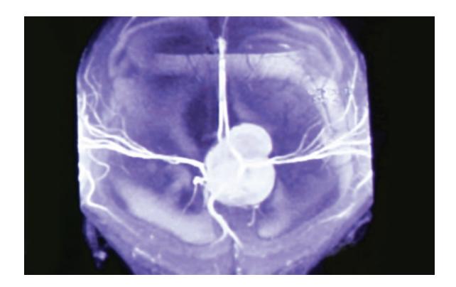
a. What is the most likely cause of the large head?
b. What clinical sign (not mentioned above) would give a clue to the diagnosis?
c. Name three signs seen on plain radiograph of the skull in hydrocephalus.
d. Name six causes of a large head (macrocephaly).
Answer
- a. Vein of Galen Aneurysmal Malformation (VGAM) causing obstructive hydrocephalus and high-output congestive heart failure.
- b. Cranial bruit auscultated over the anterior fontanelle, orbits, or temporal regions.
- c. Plain skull radiograph signs of hydrocephalus:
-
- Sutural diastasis (widening/separation of cranial sutures)
-
- "Beaten copper / beaten silver" appearance (accentuated gyral/convolutional markings)
-
- Erosion/demineralization of the dorsum sellae and posterior clinoid processes
-
- Craniofacial disproportion (large vault with small facial skeleton)
-
- d. Causes of macrocephaly:
-
- Hydrocephalus (aqueductal stenosis, Dandy-Walker malformation, Chiari malformation)
-
- Megalencephaly / Metabolic leukodystrophies (Canavan disease, Alexander disease, Tay-Sachs disease)
-
- Neurocutaneous syndromes (Neurofibromatosis type 1, Tuberous sclerosis)
-
- Sotos syndrome (cerebral gigantism)
-
- Skeletal dysplasias (Achondroplasia, Osteogenesis imperfecta)
-
- Chronic subdural hematoma / hygroma
-
- Benign familial macrocephaly
-
VVK09-011
A female neonate aged 3 days, born at home following an unbooked pregnancy to a primigravida mother, was brought to the hospital with a mass on the back. Clinically, a reddish leaking placode lesion is seen in the lumbar region. The lower limbs are hypotonic and areflexic, and the anal opening is patulous with constant dribble of urine. Response to painful stimuli cannot be elicited below the level of L1. Craniospinal MRI ruled out hydrocephalus.
a. What is the recurrence risk of this lesion in the next child? If a mother has had two such children in the past, what is the recurrence risk?
b. Name the drug and dosage schedule required to prevent the recurrence of this lesion in the next pregnancy.
c. The long-term neurological outlook for the neonate, in case of successful surgery is likely to be:
- i. Ambulant
- ii. Ambulant on crutches
- iii. Wheelchair bound
- iv. Bedridden
d. Name the associated central nervous system defect which may be present and name its components.
Answer
- a.
- Recurrence risk after 1 affected child: 3–4%.
- Recurrence risk after 2 affected children: 10%.
- b. Folic acid 4 mg/day (high dose) orally, initiated at least 1–3 months prior to planned conception and continued through the first trimester of pregnancy.
- c. iii. Wheelchair bound (lesion above L2 results in complete paraplegia and neurogenic bladder/bowel).
- d. Chiari Malformation Type II (Arnold-Chiari Malformation):
- Caudal herniation of cerebellar vermis, tonsils, pons, and medulla oblongata through the foramen magnum
- Elongation and compression of the fourth ventricle
- Kinking of the cervicomedullary junction ("Z-kink")
- Associated obstructive hydrocephalus and syringomyelia/hydromyelia
VVK09-012
A two-year-old, previously asymptomatic child is rushed to the emergency department with history of loss of consciousness and generalized tonic-clonic seizures lasting for 2 minutes. The child had coryza for one day and high-grade fever for 4 hours. Clinically, the child is drowsy, with no neck stiffness, rash, or focal neurological deficit. Other than a congested throat, rest of the clinical examination is normal.
a. What is the indication for doing a lumbar puncture in such a case?
b. For recurrent febrile seizures, which of the following have been proved to prevent recurrence of febrile seizures: (select all that apply)
- i. Antipyretics at the time of fever
- ii. Long-term phenytoin
- iii. Long-term carbamazepine
- iv. Long-term phenobarbitone
- v. Oral diazepam at the time of fever
c. What are the diagnostic criteria for complex febrile seizures?
d. Name three factors associated with increased risk of recurrence of febrile seizures.
Answer
- a. Indications for Lumbar Puncture (LP):
- Any infant
of age (especially ). - Child 12–18 months of age with complex features, prolonged postictal drowsiness, or partially treated with antibiotics.
- Presence of meningeal signs (neck stiffness, Kernig/Brudzinski sign) or clinical concern for meningitis.
- Child who is unimmunized or under-immunized against Hib and Streptococcus pneumoniae.
- Any infant
- b. iv and v (Long-term continuous Phenobarbitone / Sodium Valproate and Intermittent Oral Diazepam / Clobazam at the onset of fever).
- c. Criteria for Complex (Atypical) Febrile Seizure (any of):
-
- Focal seizure onset or focal features during seizure
-
- Prolonged duration (
)
- Prolonged duration (
-
- Recurrence / multiple episodes within 24 hours (or during the same febrile illness)
-
- Postictal neurological deficit (e.g. Todd's paresis)
-
- d. Risk Factors for Recurrence:
- Young age at onset (< 12–18 months)
- Family history of febrile seizures in first-degree relatives
- Low peak temperature / short duration of fever before seizure onset
- Complex febrile seizure at presentation
VVK09-013
A 7½-year-old girl is brought with history that she has started having episodes of inattentiveness for 2 weeks. The episodes last 10 to 15 seconds, occur several times a day, and involve sudden staring spells with immediate resumption of prior activity. There is no history of fall, convulsive movements, tongue bite, or bladder/bowel incontinence.
a. What is your diagnosis?
b. Name one biochemical test to differentiate true seizure from a pseudoseizure.
c. Name one maneuver that is likely to precipitate the episode.
d. What is the characteristic EEG finding in this disorder?
Answer
- a. Childhood Absence Epilepsy (Petit Mal Seizure).
- b. Serum Prolactin level (drawn 15–20 minutes post-event; elevated in true epileptic generalized/focal seizures, normal in pseudoseizures/PNES).
- c. Hyperventilation for 3–4 minutes (or intermittent photic stimulation).
- d. Generalized, synchronous, symmetrical 3-Hz (3 per second) spike-and-slow-wave discharges with normal background activity.
VVK09-014
A 6½-year-old boy is brought by his parents with history that over the past six months, he has undergone marked behavioral change, with inattention, irritability, poor attention span and loss of scholastic skills. In addition, the child has stopped talking and is barely able to communicate his needs with gestures (acquired auditory agnosia). He was diagnosed as having autism by a local doctor. Three months back, he started having falls with facial injuries, and in the past week, has had two generalized tonic-clonic seizures. MRI of the brain is normal.
a. What is the most likely diagnosis?
b. What is the diagnostic test for this condition?
c. What is the drug of choice for this condition?
d. What other medications can be used?
Answer
- a. Landau-Kleffner Syndrome (LKS / Acquired Epileptic Aphasia).
- b. Sleep Electroencephalogram (Overnight Sleep EEG) demonstrating continuous spike-wave discharges during slow-wave sleep (CSWS / ESES — Electrical Status Epilepticus in Sleep) involving the temporal/temporo-parietal regions.
- c. Sodium Valproate (or Clobazam / Ethosuximide).
- d. Corticosteroids (oral Prednisolone / pulse IV Methylprednisolone), IVIG, Levetiracetam, Lamotrigine, and Subpial intracortical transection (in refractory cases).
VVK09-015
A 10-year-old girl is brought with history of gradual loss of interest in studies, psychological withdrawal, irritability, and episodes of abnormal behavior starting about 8 months back. The child started having frequent falls and involuntary, periodic jerky movements of the limbs and trunk. These became frequent over the past three months and now the child is completely bedridden and uncommunicative. On examination: Afebrile, lead-pipe rigidity in all 4 limbs, generalized periodic myoclonic jerks, and poorly elicited DTRs.
a. What is the likely diagnosis?
b. Name three diagnostic tests for the condition.
c. What treatment options have been studied in this condition?
Answer
- a. Subacute Sclerosing Panencephalitis (SSPE) (slow-virus progressive neurodegenerative disorder caused by persistent mutant Measles virus infection).
- b.
-
- Elevated Measles-specific IgG antibody titers in CSF (
).
- Elevated Measles-specific IgG antibody titers in CSF (
-
- EEG: Periodic, high-amplitude, generalized, symmetrical complexes (Radermecker complexes) every 4–10 seconds on a slow background.
-
- Brain MRI showing diffuse white matter hyperintensities (periventricular/subcortical demyelination) and brain biopsy for viral antigen/RNA.
-
- c.
- Oral Isoprinosine (Inosiplex 100 mg/kg/day)
- Intrathecal / intraventricular Interferon-alpha (via Ommaya reservoir)
- Oral Ribavirin, Lamivudine, and supportive/anticonvulsant therapy (Clobazam/Valproate).
VVK09-016
A male infant aged 18 months is brought with history of regression of milestones and generalized seizures since the age of 1 year. The infant was well till one year of age but is now losing milestones in all sectors of development. Clinically, the infant is irritable, pale, with few petechial spots on the abdomen and limbs. The liver is enlarged 12 cm below the costal margin and the spleen 6 cm below the costal margin. Fundus and hearing are normal. Bone marrow examination shows the presence of PAS-positive "wrinkled tissue paper" macrophages.
a. What is the most probable diagnosis?
b. Other than bone marrow examination, what is the diagnostic modality?
c. What is the possible treatment and definitive cure?
Answer
- a. Gaucher Disease Type 2 (Acute Infantile Neuronopathic Gaucher Disease) or Type 3.
- b. Demonstration of deficient Acid
-Glucosidase (Glucocerebrosidase) enzyme activity in peripheral blood leukocytes or cultured skin fibroblasts; confirmed by GBA gene mutation analysis. - c.
- Enzyme Replacement Therapy (ERT): Recombinant
-Glucocerebrosidase (Imiglucerase / Velaglucerase alfa 60 U/kg IV every 2 weeks) improves visceral/hematologic manifestations (though does not cross blood-brain barrier). - Definitive Cure: Allogeneic Bone Marrow Transplantation (BMT) / Hematopoietic Stem Cell Transplant (HSCT).
- Enzyme Replacement Therapy (ERT): Recombinant
VVK09-017
A 4½-year-old girl from Gorakhpur (Eastern UP) is brought with high-grade fever with headache of 5 days duration and altered sensorium since 2 days. Several other cases have been reported from her neighborhood recently. Since this morning, the child has had three generalized tonic-clonic seizures. Clinically, the child is febrile, normotensive and has mild pallor. Neurological examination reveals bilateral sixth nerve palsy, dystonia, rigidity, and a coarse tremor of both upper limbs.
a. What is the most likely diagnosis?
b. Name the main transmitter of this disease to man.
c. How will you reach a diagnosis of the etiology?
d. What is the preventive measure?
Answer
- a. Japanese Encephalitis (JE) (Acute Encephalitis Syndrome with thalamic/basal ganglia involvement).
- b. Female Culex mosquito (primarily Culex tritaeniorhynchus and Culex vishnui breeding in flooded paddy fields).
- c.
-
- CSF IgM capture ELISA (MAC-ELISA) for JE-specific IgM antibodies (test of choice).
-
-fold rise in paired serum antibody titers.
-
- Viral RNA RT-PCR in CSF/brain tissue.
-
- Brain MRI showing bilateral thalamic and basal ganglia T2/FLAIR hyperintensities.
-
- d.
- Vaccination: Live attenuated SA 14-14-2 JE vaccine (2 doses at 9 and 16–24 months under Universal Immunization Programme).
- Vector control (ultra-low volume Malathion thermal fogging, insecticide-treated bed nets), keeping pig enclosures away from human dwellings.
VVK09-018
A 12-year-old boy is brought with history of progressive slurring of speech, frequent falls and clumsiness for the past 8 months. There is history of jaundice 2 years ago that lasted for 2 months. Clinically, the child is pale, with hepatomegaly (5 cm below costal margin). Neurological examination shows dystonia, intention tremor, "wing-beating" tremor, and dysarthria. Examination of his asymptomatic 6-year-old sister reveals hepatomegaly (4 cm).
a. What is the most probable diagnosis?
b. What is the pattern of inheritance?
c. What is the test to confirm the diagnosis?
d. Name two ocular findings in this disease.
e. Name the drug and dosage used in this disease.
Answer
- a. Wilson Disease (Hepatolenticular Degeneration).
- b. Autosomal recessive (ATP7B gene mutation on chromosome 13q14.3).
- c.
- Quantitative hepatic copper concentration on liver biopsy (
dry weight of liver, gold standard). - Low serum ceruloplasmin (< 20 mg/dL), elevated 24-hour urinary copper excretion (> 100 mcg/day / > 40 mcg in children), and ATP7B genetic testing.
- Quantitative hepatic copper concentration on liver biopsy (
- d.
-
- Kayser-Fleischer (KF) ring (copper deposition in Descemet's membrane on slit-lamp examination).
-
- Sunflower cataract (copper deposition in anterior lens capsule).
-
- e. D-Penicillamine (20 mg/kg/day in 2–3 divided doses given 1 hour before meals, max 1000–1500 mg/day, with oral Pyridoxine 25–50 mg/day) or Trientine (20 mg/kg/day); maintenance with oral Zinc acetate (50 mg elemental zinc tid).
VVK09-019
A mother brings her 14-month-old male infant with history of regression of all milestones since the past two months, following a diarrheal illness. The infant is exclusively milk fed (breast milk and unfortified cow's milk). Clinically, the infant is conscious, pale, plump, with sparse brownish scalp hair, hyperpigmentation of knuckles/skin, coarse tremors of upper and lower limbs that disappear during sleep, and a high-pitched "bleating" cry. The MRI shows cerebral cortical atrophy.
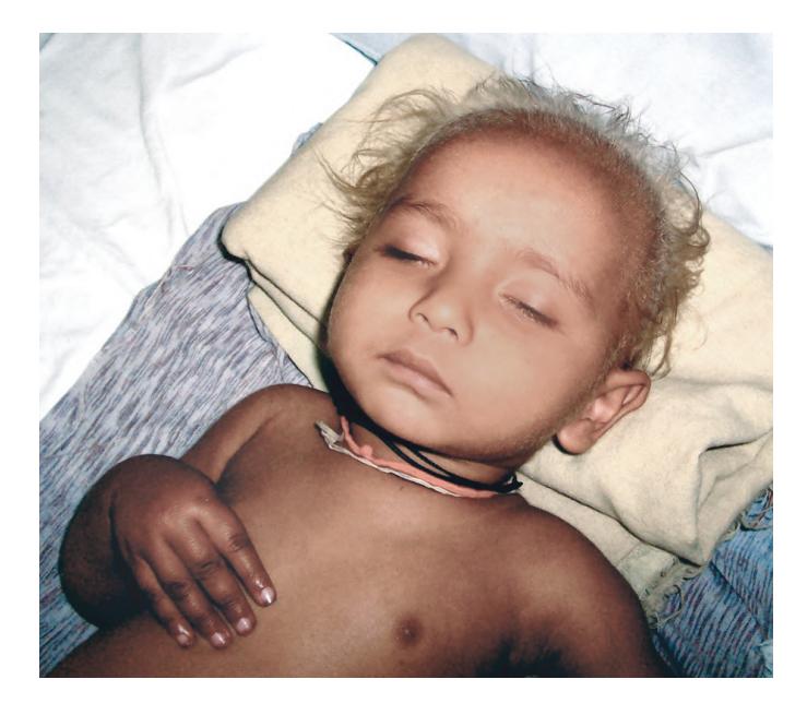
a. What is the diagnosis?
b. What is the likely peripheral smear finding?
c. What is the treatment?
Answer
- a. Infantile Tremor Syndrome (Nutritional Tremor Syndrome).
- b. Megaloblastic Anemia (macrocytes, macro-ovalocytes, and hypersegmented neutrophils with low hemoglobin and mild thrombocytopenia).
- c.
- Parenteral Vitamin B12 (Cyanocobalamin / Hydroxocobalamin) 250–1000 mcg IM daily for 1–2 weeks, then weekly, then monthly; plus oral Folic acid (1–5 mg/day) and Iron supplementation.
- Symptomatic control of tremors: Low-dose Phenobarbital, Carbamazepine, or Propranolol.
- Nutritional rehabilitation and introduction of balanced complementary weaning foods.
VVK09-020
A 13-year-old girl is brought with history of progressive weakness of both lower limbs since 3 days, making her completely bedridden. She had an episode of acute diarrhea 4 weeks back. Clinically, she has symmetrical flaccid ascending weakness of lower limbs and trunk with absent deep tendon reflexes (areflexia). There is no objective sensory loss, but calves and thighs are tender on palpation. Cranial nerves and upper limbs are currently preserved.
a. What is the most probable diagnosis?
b. What is the diagnostic test for the disorder? What are the characteristic findings?
c. What is the drug of choice and what are the indications for its use? Give the dosage schedule.
Answer
- a. Guillain-Barré Syndrome (GBS / Acute Inflammatory Demyelinating Polyradiculoneuropathy - AIDP) (post-Campylobacter jejuni or viral gastroenteritis).
- b. Lumbar Puncture (CSF Examination): Demonstrates Albuminocytological Dissociation (marked elevation of CSF protein
with normal CSF cell count ); Nerve Conduction Studies (NCS) showing slowed conduction velocities, prolonged F-wave latencies, and conduction block. - c.
- Intravenous Immunoglobulin (IVIG) (or Therapeutic Plasma Exchange).
- Indications: Rapidly progressive paralysis, inability to ambulate, respiratory compromise (falling FVC), bulbar palsy, or autonomic instability.
- Dosage: 2 g/kg total dose given as 400 mg/kg/day IV for 5 consecutive days (or 1 g/kg/day for 2 days).
VVK09-021
A 14-year-old boy has sustained an injury to the neck due to a diving accident in a swimming pool. He is breathing spontaneously but cannot move or feel his arms or legs (quadriplegia).
a. What is the recommended maneuver for opening the airway in neck injuries?
b. X-ray of the cervical spine shows no bony injury. Is it still possible for the child to have a spinal cord injury? Name the condition, its incidence, and mode of diagnosis.
c. What is the emergency drug treatment that can be offered to this child?
Answer
- a. Jaw-thrust maneuver WITHOUT head tilt (while maintaining manual in-line cervical spine stabilization).
- b. Yes.
- Condition: SCIWORA (Spinal Cord Injury Without Radiographic Abnormality).
- Incidence: Accounts for up to 15–20% of all pediatric spinal cord injuries (due to high elasticity and ligamentous laxity of the pediatric vertebral column).
- Mode of Diagnosis: Spinal MRI (reveals cord edema, contusion, transection, or ligamentous rupture).
- c. High-dose IV Methylprednisolone (NASCIS protocol: 30 mg/kg IV bolus over 15 minutes, followed 45 minutes later by continuous infusion of 5.4 mg/kg/hour for 23 hours if started within 3–8 hours of injury).
VVK09-022
A nine-month-old boy is taken to the Primary Health Center for a checkup and routine immunization. By mistake, the nurse gives the infant 5 mL (500,000 units) of vitamin A. One week later, the infant is brought back with features of vomiting and irritability. There is no history of fever or alteration in level of consciousness. Clinically, the physician finds a conscious infant with normal vitals, with a tense bulging anterior fontanelle and widening of sutures. There is no focal neurological deficit and rest of the examination is essentially normal.
a. What is the most probable diagnosis?
b. What findings do you expect the CSF to show?
c. Name three drugs that can lead to a similar clinical situation.
d. Name one complication of this disease.
Answer
- a. Pseudotumor Cerebri (Idiopathic Intracranial Hypertension / Benign Intracranial Hypertension) secondary to acute hypervitaminosis A.
- b. Normal CSF composition (normal protein, normal glucose, and no pleocytosis) with markedly elevated opening CSF pressure (
). - c.
- Tetracyclines (Tetracycline, Doxycycline, Minocycline)
- Nalidixic acid / Fluoroquinolones
- Retinoids / Isotretinoin
- Growth hormone
- Nitrofurantoin
- Systemic corticosteroid withdrawal
- d. Optic atrophy and permanent visual loss / blindness (secondary to chronic papilledema).
VVK09-023
A previously well, 3½-year-old girl comes with history of sudden onset of unsteadiness while walking for 4 days. Initially mild, the child is now unable to walk and is reluctant to sit up in bed. There is history of fever with a vesicular rash 2 weeks back. Clinically, the child is afebrile, normotensive, and has no neck stiffness or signs of raised ICP. The cranial nerves are normal. The child has marked truncal ataxia, generalized hypotonia, dysmetria with past pointing, intention tremor, horizontal nystagmus, and scanning dysarthria.
a. What is the most probable diagnosis?
b. Is there a role of CSF examination in this child? What findings do you expect?
c. What is the likely organism responsible?
Answer
- a. Acute Post-infectious Cerebellar Ataxia (Acute Cerebellitis).
- b. Yes, to rule out active CNS infections/meningitis. CSF typically shows mild lymphocytic pleocytosis (< 30 cells/cumm) and mildly elevated protein with normal glucose and sterile cultures (or may be completely normal).
- c. Varicella-Zoster Virus (VZV) (most common); also Epstein-Barr virus, enteroviruses, mumps, influenza, and Mycoplasma pneumoniae.
VVK09-024
This 7½-year-old girl was brought with history of delayed developmental milestones and generalized seizures. As an infant she had had myoclonic seizures (infantile spasms).
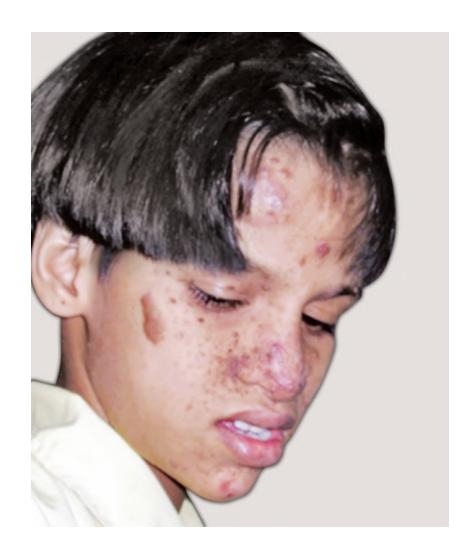
a. What is the diagnosis? What are the lesions on the face?
b. Name three other types of skin lesions seen in this condition.
c. What is the inheritance?
d. Name three organ systems other than skin and brain that may be affected.
Answer
- a. Tuberous Sclerosis Complex (Bourneville Disease); facial lesions are Facial Angiofibromas (Adenoma sebaceum) distributed in a butterfly pattern across cheeks and nose.
- b.
-
- Hypopigmented macules ("Ash-leaf spots" / confetti lesions)
-
- Shagreen patch (connective tissue nevus in lumbosacral region)
-
- Ungual/Periungual fibromas (Koenen tumors)
-
- Forehead fibrous plaque
-
- c. Autosomal dominant with variable expressivity (mutations in TSC1 hamartin or TSC2 tuberin; ~60–70% are spontaneous new mutations).
- d.
- Cardiovascular: Cardiac rhabdomyoma (often in infancy)
- Renal: Renal angiomyolipomas, multiple renal cysts, renal cell carcinoma
- Ocular: Retinal astrocytic hamartomas / phakomas
- Pulmonary: Lymphangioleiomyomatosis (LAM), multifocal micronodular pneumocyte hyperplasia
- Dental/Oral: Dental enamel pits, gingival fibromas
VVK09-025
Study the image of the 10-year-old child and answer the questions:
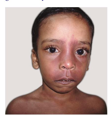
a. Name the lesion and the syndrome that it forms a part of.
b. What are the neurological morbidities of this syndrome? (Name three)
c. What is the ocular complication that can accompany?
d. Name the radiological sign associated with cranial involvement.
Answer
- a. Port-Wine Stain (Nevus Flammeus) along the ophthalmic (
) / maxillary ( ) divisions of the trigeminal nerve, forming part of Sturge-Weber Syndrome (Encephalotrigeminal Angiomatosis). - b.
-
- Refractory focal or generalized epileptic seizures
-
- Contralateral hemiparesis / hemiplegia and stroke-like episodes
-
- Intellectual disability / developmental delay
-
- c. Ipsilateral Glaucoma (congenital buphthalmos or juvenile open-angle glaucoma) and choroidal hemangioma.
- d. "Tram-track" (Railroad-track) gyral calcifications in the parieto-occipital cortex on plain skull X-ray / CT head (leptomeningeal angiomatosis).
VVK09-026
A 2½-year-old child is brought by the mother with history of poor development of speech. The child plays with his toys but not with other children. He also does not respond to his name being called. On examination, the motor system, sensory system, hearing and vision are normal. The child is able to say a few words. He does not interact with the examiner.
a. What is the most probable diagnosis?
b. What are the three characteristic features of this condition?
c. Which parts of the brain are affected in this condition?
d. What is the scale used to assess this condition?
Answer
- a. Autism Spectrum Disorder (ASD).
- b. Core Domains (DSM-5 / DSM-IV):
-
- Persistent deficits in social communication and social interaction across multiple contexts (poor eye contact, lack of joint attention, delayed speech).
-
- Restricted, repetitive patterns of behavior, interests, or activities (stereotypies, insistence on sameness, echolalia).
-
- Atypical sensory reactivity (hyper- or hypo-reactivity to sensory input).
-
- c. Cerebellum (Purkinje cell reduction), amygdala, hippocampus, frontal and temporal lobes, and corpus callosum.
- d. Childhood Autism Rating Scale (CARS), Autism Diagnostic Observation Schedule (ADOS-2), or M-CHAT (Modified Checklist for Autism in Toddlers).
VVK09-027
Match the abnormal involuntary movement with its clinical description:
| Movement | Clinical Description |
|---|---|
| 1. Chorea | A. Rhythmic, oscillatory movement caused by alternating/simultaneous contractions of antagonistic muscles |
| 2. Athetosis | B. Brief, shock-like, involuntary contraction of a muscle or muscle group resulting in a sudden jerk |
| 3. Dystonia | C. Rapid, unsustained, irregular, purposeless, jerky, non-patterned movements |
| 4. Tremor | D. Slow, continuous, sinuous, writhing movements that are more pronounced in distal extremities |
| 5. Myoclonus | E. Sustained or intermittent muscle contractions causing twisting and repetitive movements or abnormal fixed postures |
Answer
- 1. Chorea — C. Rapid, unsustained, irregular, purposeless, jerky, non-patterned movements
- 2. Athetosis — D. Slow, continuous, sinuous, writhing movements that are more pronounced in distal extremities
- 3. Dystonia — E. Sustained or intermittent muscle contractions causing twisting and repetitive movements or abnormal fixed postures
- 4. Tremor — A. Rhythmic, oscillatory movement caused by alternating/simultaneous contractions of antagonistic muscles
- 5. Myoclonus — B. Brief, shock-like, involuntary contraction of a muscle or muscle group resulting in a sudden jerk
VVK09-028
Study the plain CT scan film of a breastfed 3-month-old infant who was brought to the hospital with history of left-sided focal seizures:
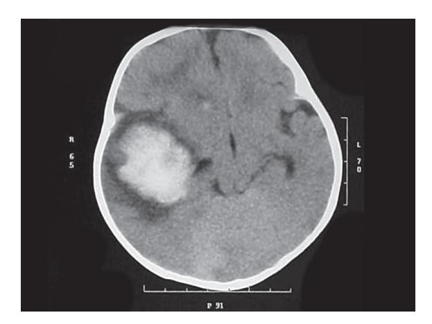
a. What is the CT scan finding suggestive of and what is the most probable diagnosis?
b. How would you confirm the etiological diagnosis? Name three tests.
c. What is the preventive measure for this condition?
Answer
- a.
- CT Finding: Hyperdense acute Intracranial Hemorrhage (ICH) in the right temporoparietal / subdural region with significant mass effect and midline shift.
- Probable Diagnosis: Late-onset Vitamin K Deficiency Bleeding (VKDB) (Late-onset Hemorrhagic Disease of the Newborn) in an exclusively breastfed infant who missed birth vitamin K prophylaxis.
- b. Confirmation of Etiological Diagnosis:
-
- Markedly prolonged Prothrombin Time (PT / INR) and prolonged aPTT, with normal platelet count and fibrinogen.
-
- Elevated PIVKA-II (Protein Induced by Vitamin K Absence / Incomplete prothrombin).
-
- Rapid normalization of PT/INR within 2–6 hours after IV/SC Vitamin K administration.
-
- c. Prevention: Routine administration of Injection Vitamin K1 (Phytonadione 1 mg IM) to all newborns at birth.