MNJ19-001
Match the following lysosomal storage disorders with their enzyme defects:
| Disease | Enzyme Defect |
|---|---|
| 1. Metachromatic leukodystrophy | a. |
| 2. Krabbe disease | b. |
| 3. Fabry disease | c. Ceramidase |
| 4. Farber disease | d. Acid lipase |
| 5. Wolman disease | e. Arylsulfatase A |
Answer
- 1 - e: Metachromatic leukodystrophy
Arylsulfatase A - 2 - a: Krabbe disease
-galactocerebrosidase - 3 - b: Fabry disease
-galactosidase A - 4 - c: Farber disease
Ceramidase - 5 - d: Wolman disease
Acid lipase (lysosomal acid lipase)
MNJ19-002
Observe the clinical and radiological images below:
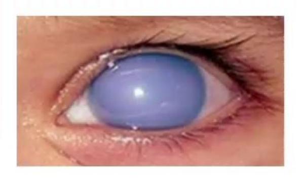
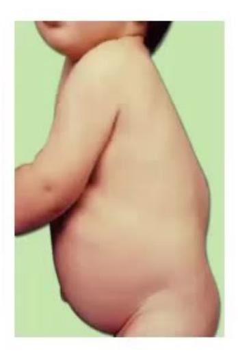

- Diagnose this eye problem and the disease.
- Name five types of this condition.
- What is the urinary test for diagnosis?
- Name three metabolites seen in the urine.
- Name the main organs involved.
- What are the X-ray findings called?
- For what types is enzyme replacement available?
- Write down causes of corneal clouding in infancy/childhood.
Answer
- Diagnosis: Corneal clouding in Mucopolysaccharidosis (MPS).
- Five types of MPS:
- Type I: Hurler / Scheie / Hurler-Scheie (
-L-iduronidase deficiency) - Type II: Hunter (Iduronate-2-sulfatase deficiency)
- Type III: Sanfilippo (Heparan N-sulfatase / NAGLU / HGSNAT / GNS deficiency)
- Type IV: Morquio (Galactose-6-sulfate sulfatase /
-galactosidase deficiency) - Type VI: Maroteaux-Lamy (Arylsulfatase B deficiency)
- Type VII: Sly syndrome (
-glucuronidase deficiency)
- Type I: Hurler / Scheie / Hurler-Scheie (
- Urinary test: Urine glycosaminoglycan (GAG) screening (toluidine blue / DMB test) followed by urine GAG electrophoresis/chromatography.
- Metabolites in urine: Dermatan sulfate, Heparan sulfate, Keratan sulfate, Chondroitin sulfate.
- Main organs involved: Liver, spleen (hepatosplenomegaly), skeleton/bones, heart (valvular disease, cardiomyopathy), eyes (corneal clouding, retinal degeneration), ears (sensorineural/conductive hearing loss), brain/CNS.
- X-ray findings: Dysostosis multiplex (oar-shaped/spatulate ribs, anterior beaking/ovoid vertebral bodies, hook-like hypoplasia of lumbar vertebrae, bullet-shaped metacarpals/phalanges).
- Types with Enzyme Replacement Therapy (ERT) available:
- Type I (Hurler/Scheie): Laronidase (Aldurazyme)
- Type II (Hunter): Idursulfase (Elaprase)
- Type IVA (Morquio A): Elosulfase alfa (Vimizim)
- Type VI (Maroteaux-Lamy): Galsulfase (Naglazyme)
- Type VII (Sly): Vestronidase alfa (Mepsevii)
- Causes of corneal clouding:
- Mucopolysaccharidosis (Hurler, Scheie, Morquio, Maroteaux-Lamy, Sly — absent in Hunter and Sanfilippo)
- Mucolipidoses (GM1 gangliosidosis)
- Zellweger syndrome (peroxisomal disorder)
- Congenital glaucoma (buphthalmos)
- Congenital rubella / intrauterine infections
- Birth trauma (forceps injury with rupture of Descemet membrane)
- Cystinosis.
MNJ19-003
True statements regarding classic galactosemia include all of the following except:
a. It is autosomal recessive
b. It is caused by a deficiency in the enzyme galactose-1-phosphate uridyltransferase
c. Treatment is by eliminating galactose from diet
d. The majority of the hyperbilirubinemia is indirect
e. Hypoglycemia is frequent
f. E. coli sepsis is specific for this condition
Answer
Answer: d (The majority of the hyperbilirubinemia is indirect)
- Explanation: In classic galactosemia, neonatal cholestasis develops with predominantly conjugated (direct) hyperbilirubinemia (hepatocellular injury), although mixed hyperbilirubinemia can occur.
- Statements a, b, c, e, and f are true:
- Mode of inheritance is autosomal recessive (GALT gene mutation).
- Enzyme deficient is galactose-1-phosphate uridyltransferase (GALT).
- Treatment is lifelong elimination of dietary lactose and galactose.
- Hypoglycemia occurs due to accumulation of galactose-1-phosphate inhibiting glycogenolysis and gluconeogenesis.
- Fulminant E. coli sepsis/neonatal septicemia is a classic early complication.
MNJ19-004
Observe the clinical and radiological image below:
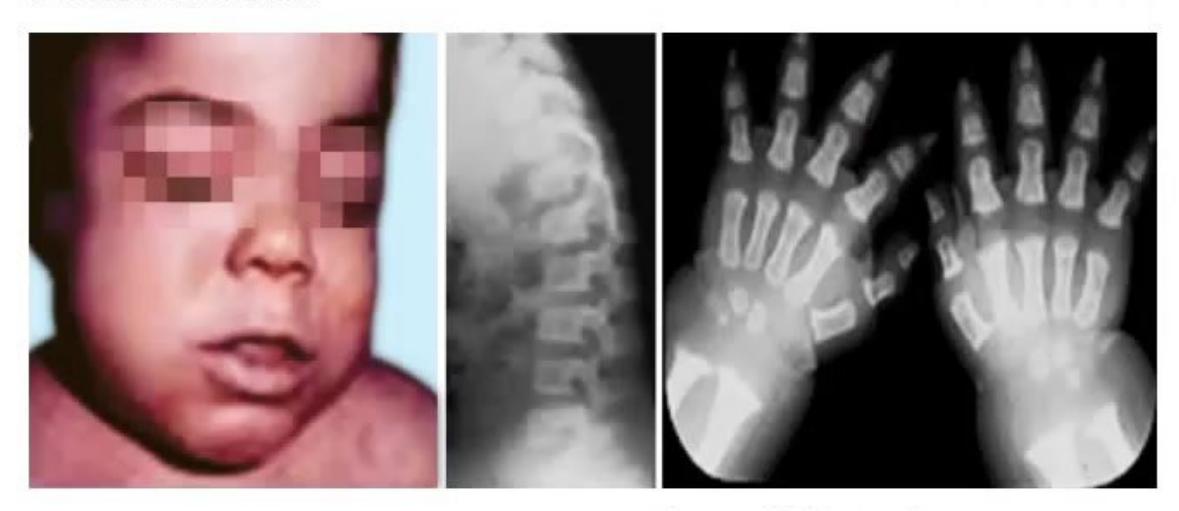
- Spot the diagnosis based on facial characteristics, X-ray findings, and identify the enzyme deficiency.
- What do tissues and urine contain?
- What is the mode of inheritance?
Answer
- Diagnosis & Enzyme:
- Diagnosis: Mucopolysaccharidosis Type II (Hunter syndrome).
- Facial characteristics: Hunter facies (coarse facial features, macrocephaly, prominent forehead, flat nasal bridge, thick lips, macroglossia).
- X-ray findings: Dysostosis multiplex (anterior beaking/ovoid vertebral bodies, bullet-shaped phalanges).
- Enzyme deficiency: Iduronate-2-sulfatase (IDS) deficiency.
- Tissues and urine content: Dermatan sulfate and Heparan sulfate.
- Mode of inheritance: X-linked recessive (Hunter syndrome is the only X-linked MPS; all other MPS types are autosomal recessive).
MNJ19-005
A 6-month-old child was noted to be normal at birth, but over the ensuing months you have been somewhat concerned about his slow weight gain and his mild delay in achieving developmental milestones. The family calls you urgently at 7:00 AM noting that their child seems unable to move the right side of his body:
- Which of the following conditions might explain this child's condition?
- a. Phenylketonuria
- b. Homocystinuria
- c. Cystathioninuria
- d. Maple syrup urine disease
- Which one other systemic examination would you like to do?
- What investigation would confirm the etiology?
- What treatment would you institute as a long-term measure?
Answer
- Condition: b. Homocystinuria (presents with developmental delay, failure to thrive, and acute thromboembolic episodes such as hemiparesis/stroke due to hypercoagulability).
- Systemic examination: Ophthalmologic examination / slit-lamp examination to look for ectopia lentis (characteristically downward and inward subluxation of the lens, in contrast to upward-outward in Marfan syndrome).
- Confirmatory investigation: Elevated plasma total homocysteine and methionine levels, positive urinary cyanide-nitroprusside test, and confirmation by enzymatic assay (cystathionine
-synthase [CBS] deficiency) or genetic testing (CBS gene). - Long-term treatment:
- High-dose Pyridoxine (Vitamin
) (200–1000 mg/day) in -responsive patients - Folic acid (1–5 mg/day) and Vitamin
(1–2 mg/day) - Betaine (trimethylglycine) to remethylate homocysteine to methionine
- Low-methionine diet with cysteine supplementation in
-non-responsive patients.
- High-dose Pyridoxine (Vitamin
MNJ19-006
At birth, an infant is noted to have an abnormal neurologic examination. Over the next 1–2 weeks he develops severe progressive central nervous system degeneration, an enlarged liver and spleen, macroglossia, coarse facial features, and a cherry-red spot in the eye:
- The laboratory finding likely to explain this child's problem is:
- a. Reduced serum hexosaminidase A activity
- b. Deficient activity of acid
-galactosidase - c. A defective gene on the X-chromosome
- d. Complete lack of acid
-galactosidase activity - e. Deficient activity of galactosyl-3-sulfate-ceramide sulfatase
- What is the diagnosis?
- What is a specific skin finding seen in related disorders?
- If this same child has hyperacusis and macrocephaly but NO organomegaly, what is the diagnosis?
Answer
- Laboratory finding: b. Deficient activity of acid
-galactosidase. - Diagnosis: Type 1 GM1 Gangliosidosis (Infantile form) (characterized by cherry-red spot, severe CNS degeneration, coarse facies, skeletal dysplasia, and hepatosplenomegaly).
- Specific skin finding: Mongolian spots (extensive bluish-gray macules) in GM1 gangliosidosis; or Angiokeratoma (angiokeratoma corporis diffusum) seen in Fabry disease / Fucosidosis.
- Diagnosis if hyperacusis, macrocephaly, cherry-red spot, and ABSENT organomegaly: Tay-Sachs Disease (GM2 gangliosidosis / Hexosaminidase A deficiency).
MNJ19-007
An 8-day-old child presented with yellowish sclera, whitish stool, turmeric color urine, and seizures on the 3rd day of septicemia who was exclusively breastfed:
- What could be the diagnosis?
- What is the enzyme deficient?
- What is the commonest organism causing infection?
- What are the ophthalmic manifestations?
- What is the treatment?
Answer
- Diagnosis: Classic Galactosemia.
- Enzyme deficient: Galactose-1-phosphate uridyltransferase (GALT).
- Commonest organism: Escherichia coli (E. coli septicemia).
- Ophthalmic manifestations: "Oil droplet" cataracts (due to accumulation of galactitol in the lens), vitreous hemorrhage, retinal detachment.
- Treatment: Immediate and lifelong elimination of galactose and lactose from the diet (switch from breast milk/cow milk formula to soy-based or elemental formula).
MNJ19-008
A six-week-old infant is referred for poor weight gain and seizures. On examination, he looks cachectic and is dysmorphic with low-set ears and a very small lower jaw (micrognathia). He also has severe thrush affecting the oral cavity and perineum. Cardiac apex is displaced into the left sixth intercostal space in anterior axillary line and there is a palpable thrill. A loud pansystolic murmur and an apical mid-diastolic murmur can be heard. He is tachypneic but the lung fields are clear on auscultation. The liver is 3 cm below the costal margin in the midclavicular line.
Investigations: Na: 136, K: 4.9, Ca: 1.7 mmol/L, Albumin: 4.2 g/dL, Urea: 7.8 mmol/L, Hb: 10.2 g/dL, WBC: 3800/
- What is the diagnosis?
- What is the commonest presentation during the newborn period?
- Most likely explanation for the low plasma calcium?
- What is the cause of immunodeficiency?
- What is the etiopathogenesis of all these features?
Answer
- Diagnosis: DiGeorge Syndrome (22q11.2 deletion syndrome / CATCH-22).
- Commonest newborn presentation: Hypocalcemic seizures / tetany and cyanosis/heart failure due to congenital conotruncal cardiac defect.
- Explanation for hypocalcemia: Primary hypoparathyroidism due to parathyroid gland hypoplasia or aplasia.
- Cause of immunodeficiency: Thymic aplasia or hypoplasia leading to defective cell-mediated (T-cell) immunity and susceptibility to opportunistic infections (e.g., Candida, Pneumocystis).
- Etiopathogenesis: Dysmorphogenesis of the 3rd and 4th pharyngeal pouches and arches secondary to microdeletion on chromosome 22q11.2.
MNJ19-009
Baby of Neelu, a term newborn baby with birth weight of 2.6 kg, was admitted in NICU on day 3 of life with poor feeding, lethargy, and nonbilious forceful vomiting. He was the 2nd baby of 2nd-degree consanguineous parents with normal antenatal, natal, and immediate postnatal period. Elder sibling who expired on day 10 of life had similar complaints, but was not investigated.
Condition of baby worsened despite supportive care and he developed convulsions; on next day he went into a comatose state. Baby was flaccid, dehydrated.
Investigations showed: Hb: 12 g/dL, TC: 3000/

- Can you suggest a diagnosis?
- What abnormal odor may be seen in these cases?
- How will you tide over the acute crisis?
Answer
- Diagnosis: Organic Acidemia (e.g., Isovaleric Acidemia, Methylmalonic Acidemia, or Propionic Acidemia) presenting with high anion gap metabolic acidosis, mild-moderate hyperammonemia, pancytopenia/leukopenia, and encephalopathy.
(Note: In urea cycle defects, there is severe hyperammonemia presenting with respiratory alkalosis without metabolic acidosis). - Abnormal odor:
- Sweaty feet / cheesy odor: Isovaleric acidemia / Glutaric acidemia type II
- Fruity / sweet odor: Methylmalonic acidemia / Propionic acidemia
- Maple syrup / burnt sugar odor: Maple syrup urine disease (MSUD)
- Mousy / musty odor: Phenylketonuria (PKU).
- Acute management:
- Stop all protein intake immediately (NPO).
- Provide high intravenous caloric support with 10% dextrose (glucose infusion rate 8–10 mg/kg/min) and lipids to suppress catabolism.
- Correct metabolic acidosis with cautious IV sodium bicarbonate.
- L-Carnitine (100–300 mg/kg/day IV/oral) to detoxify acyl-CoA intermediates and excrete acylcarnitine esters.
- L-Glycine (250–300 mg/kg/day) for isovaleric acidemia (forms non-toxic isovalerylglycine).
- Cofactor trial: Vitamin
(1–2 mg IM), Biotin (10 mg/day), Thiamine (300 mg/day). - Prompt hemodialysis, peritoneal dialysis, or exchange transfusion if ammonia/acidosis is severe and unresponsive to medical therapy.
MNJ19-010
A 7-year-old boy is admitted with dehydration. He is the 3rd child whose 2 elder brothers died of similar illness. He gives history of worsening school performance and progressive defective vision. Recently he is having some difficulty in walking.
Exam: Wasted child, hyperpigmentation of lips and hands.
Inv: Serum sodium: 112 mEq/L, potassium: 7.0 mEq/L, RBS: 60 mg/dL:
- Identify this condition.
- What is the mode of inheritance?
- Which test is confirmatory?
- Discuss the management.
- Any dietary oil/drug tried in managing this disease?
Answer
- Diagnosis: X-Linked Adrenoleukodystrophy (X-ALD) (presenting with childhood cerebral demyelination and primary adrenal insufficiency / Addison disease).
- Mode of inheritance: X-linked recessive (ABCD1 gene mutation on Xq28).
- Confirmatory test: Elevated plasma levels of Very Long Chain Fatty Acids (VLCFA), specifically hexacosanoic acid (C26:0) and elevated C26:0/C22:0 and C24:0/C22:0 ratios.
- Management:
- Replacement therapy for adrenal insufficiency: Glucocorticoids (hydrocortisone) and mineralocorticoids (fludrocortisone).
- Early allogeneic Hematopoietic Stem Cell Transplantation (HSCT) or ex-vivo lentiviral gene therapy in early-stage cerebral ALD.
- Symptomatic and supportive care for spasticity, seizures, and visual/hearing loss.
- Dietary therapy: Lorenzo's oil (a 4:1 mixture of glyceryl trioleate and glyceryl trierucate) to lower saturated VLCFA levels.
MNJ19-011
Identify the fundus pathology shown below. Give four conditions in which this could be a finding:
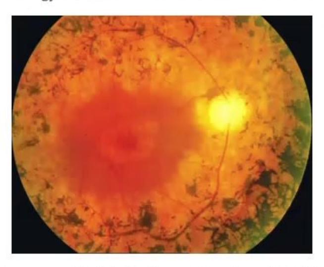
Answer
- Pathology: Retinitis Pigmentosa (Bone-spicule pigmentation of the retina with attenuated retinal vessels and waxy disc pallor).
- Four conditions with Retinitis Pigmentosa / Retinal Dystrophy:
- Mucopolysaccharidosis (MPS I Hurler/Scheie, MPS II Hunter, MPS III Sanfilippo)
- Laurence-Moon-Biedl / Bardet-Biedl syndrome
- Kearns-Sayre syndrome (mitochondrial cytopathy)
- Refsum disease (phytanic acid storage disease)
- Neuronal ceroid lipofuscinosis (Batten disease)
- Abetalipoproteinemia (Bassen-Kornzweig syndrome)
- Usher syndrome.
MNJ19-012
A 6-year-old boy presented with obesity, polydactyly, and diminished night vision:
- What is the likely diagnosis?
- What are the other abnormalities likely to be present in this child?
- What ophthalmologic finding will you get in this child?
Answer
- Likely diagnosis: Laurence-Moon-Biedl / Bardet-Biedl Syndrome (BBS).
- Other abnormalities:
- Intellectual disability / developmental delay
- Hypogonadism (microgenitalia in males, genital anomalies in females)
- Postaxial polydactyly / syndactyly / brachydactyly
- Renal structural anomalies and chronic renal failure
- Hepatic fibrosis and diabetes mellitus.
- Ophthalmologic finding: Atypical Retinitis pigmentosa / tapetoretinal degeneration leading to early night blindness (nyctalopia) and progressive visual loss.
MNJ19-013
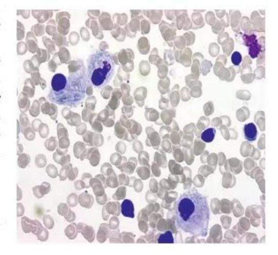
A 3-year-old child Monu with developmental delay is admitted for evaluation. On examination, he had anemia with marked organomegaly. Lab evaluation showed anemia with thrombocytopenia. Bone marrow examination was performed:
- What is the diagnosis?
- What are the marrow findings?
- What is the etiology for such findings?
- What are the typical radiological findings?
- What is the definitive treatment?
Answer
- Diagnosis: Gaucher Disease (Type 1 non-neuronopathic or Type 3 chronic neuronopathic).
- Marrow findings: Presence of Gaucher cells (large lipid-laden histiocytes/macrophages with characteristic "wrinkled tissue paper" or "crumpled tissue paper" cytoplasm, eccentric small nuclei, strongly PAS-positive).
- Etiology: Autosomal recessive deficiency of the lysosomal enzyme Acid
-glucosidase (Glucocerebrosidase) leading to accumulation of glucocerebroside (glucosylceramide) in reticuloendothelial macrophages. - Radiological findings:
- Erlenmeyer flask deformity of distal femora
- Generalized osteopenia, cortical thinning, osteosclerosis
- Avascular necrosis of femoral heads
- Bone marrow infiltration and pathologic fractures.
- Definitive treatment:
- Enzyme Replacement Therapy (ERT): Recombinant human acid
-glucosidase (e.g., Imiglucerase [Cerezyme], Velaglucerase alfa, Taliglucerase alfa) 60 U/kg IV every alternate week. - Substrate Reduction Therapy (SRT) (e.g., Miglustat, Eliglustat).
- Hematopoietic Stem Cell Transplantation (HSCT) for selected severe cases.
- Enzyme Replacement Therapy (ERT): Recombinant human acid