THG33-001
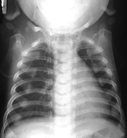
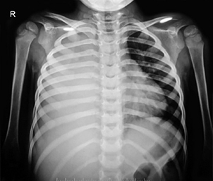
This 10-month-old boy presented with cough. He is feeding well. Respiratory rate is 36 breaths/minute and heart rate (HR) is 96 beats/minute. Work of breathing is normal. SpO2 is 98% in room air. He was treated with antibiotics and inhaled bronchodilators, but there was no response.
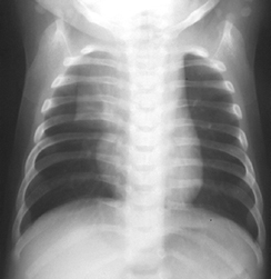
a. What is the radiological finding in above image?
b. Substantiate your observation.
c. What is the next step of preferred investigation to confirm the observation?
d. What will be the appearance in this preferred imaging?
Answer
- a. Thymic sail sign: Seen as a triangular extension of the normal thymus laterally. It may also be seen as thymus wave sign (scalloped border from rib compression). It causes no mass effect on vascular structures or airway. The inferior margin of the thymus merges with the margin of the cardiac silhouette, producing the "notch sign".
- b. Normal thymus: Upper mediastinal shadow mostly on the right side in any asymptomatic child < 2 years is normal thymus.
- c. Ultrasonography (USG) of the chest or CT chest. USG is noninvasive and preferred.
- d. On USG, it appears homogenous with echotexture similar to the liver (less than muscle) and shows multiple echogenic foci or strands giving rise to a "starry sky" appearance.
THG33-002
An 8-year-old child fell down from the cycle and cycle spoke valve punctured her heel and emptied the air in the tube into the foot. She complained of vague chest pain. No breathlessness was found. Respiratory rate was 28 breaths/minute, HR 100 beats/minute, and SpO2 98%. Computed tomography (CT) chest did not reveal any bony injury.
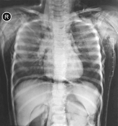
a. List out any four radiological findings in below image.
b. Is it life-threatening?
c. What is the next step of intervention?
Answer
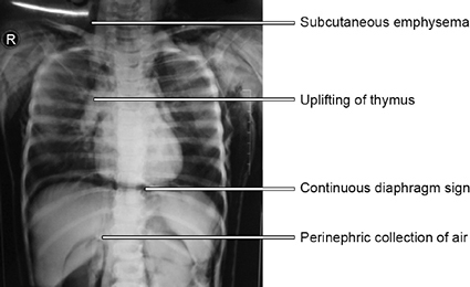
- a. Radiological findings:
- Pneumomediastinum (continuous diaphragm sign)
- Perinephric air collection on both sides
- Subcutaneous emphysema in neck and chest wall
- Uplifted thymic lobes ("spinnaker sail" sign)
- b. No, because this is isolated pneumomediastinum with stable vitals (no desaturation or tachypnea, and no tension pneumothorax or pneumopericardium).
- c. Monitor closely for respiratory distress and desaturation. Repeat CXR if symptoms develop. High-flow oxygen supplementation promotes faster nitrogen washout and absorption of mediastinal air.
THG33-003
A 3-month-old boy developed fever with blood-stained stools, followed by respiratory distress, grunting, and shock.
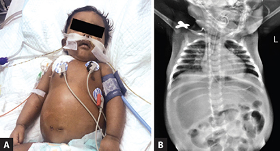
a. What do you observe in the child (image A)?
b. List out the X-ray findings, in both diseases aspect as well as interventions aspect (image B).
c. What is the probable diagnosis?
d. What are the next steps in intervention?
Answer
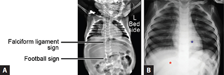
- a. Pneumoperitoneum: Football sign (large oval lucency across abdomen) and falciform ligament sign (falciform ligament outlined by free air). Normally liver and heart densities are equal; here the liver density is markedly reduced due to overlying free gas.
- b.
- Disease-related findings: Massive pneumoperitoneum with abdominal distension and elevation of diaphragms.
- Intervention-related findings: External jugular vein (EJV) line, nasogastric (NG) tube, and endotracheal tube (ETT).
- c. Pneumoperitoneum secondary to gastrointestinal perforation (e.g., necrotizing enterocolitis or ruptured viscous).
- d. Emergency resuscitation: Shock management with IV fluid boluses, inotropes, broad-spectrum IV antibiotics, correction of metabolic acidosis, stomach decompression via NG tube, and emergent exploratory laparotomy.
THG33-004
This 2-year-old girl presented with abdominal pain, upper abdominal distension, shock, and nonbilious vomiting.
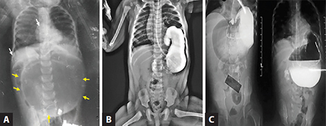
a. What are the radiological findings (images A to C)?
b. What are the clinical impressions and one close differential diagnosis?
c. What are the further investigations after seeing the initial X-ray (image A)?
d. Name the further steps of management.
Answer
- a. Radiological findings:
- Image A (Plain X-ray abdomen): Distended gas-filled stomach occupying the upper abdomen (yellow arrow); heart and liver density look identical (not pneumoperitoneum).
- Image B (NG tube X-ray): NG tube seen arrested or curled; pyloric end of stomach is upturned due to torsion/malrotation.
- Image C (Barium meal study): Inversion of stomach with obstruction at pyloro-duodenal junction confirming gastric volvulus.
- b. Diagnosis: Gastric volvulus. Close differential diagnosis: Pneumoperitoneum.
- c. Upper GI water-soluble contrast/barium study or CT scan of abdomen.
- d. Immediate resuscitation (shock and acidosis management), nasogastric decompression, and emergent surgical detorsion and gastropexy (with repair of diaphragmatic hernia if present).
THG33-005
This is a preterm neonate with respiratory distress, intubated, and ventilated. Suddenly he developed cardiopulmonary failure and cardiac arrest.
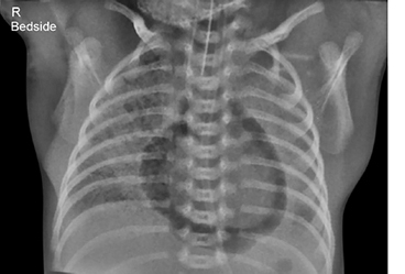
a. What are the radiological findings (as shown in above image), disease related and intervention related?
b. What is the diagnosis?
c. What is the immediate intervention?
Answer
- a.
- Disease-related: Continuous halo/rim of gas sharply outlining the pericardial sac around the heart, not extending beyond the pericardial reflection into the superior mediastinum or pleural space; bilateral lung haziness (RDS).
- Intervention-related: Endotracheal tube in situ.
- b. Diagnosis: Tension pneumopericardium.
- c. Immediate emergency subxiphoid pericardiocentesis / needle aspiration to relieve cardiac tamponade.
THG33-006
A 4-year-old child initially presented with multiple abscesses and pneumonia. He was admitted in pediatric intensive care unit (PICU), then developed empyema drained by intercostal tube drainage (ICD) and mechanically ventilated. But remained sick, and this chest X-ray (CXR) showed the missing complications. Previously well child did not have any cardiac disease. This child was treated in a district hospital and the CXR was taken early morning. Further advanced investigatory modalities are not immediately available.
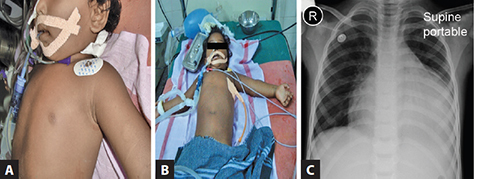
a. What are the radiological findings (as shown in image C)?
b. What is the close differential diagnosis?
c. What is the next lifesaving intervention?
d. What is the procedure to perform the investigation?
e. If the child is treated in a tertiary care center with availability of point of care investigations, how the diagnosis and management would have differed?
Answer
- a. Marked globular cardiomegaly ("water-bottle" appearance), altered cardiac contour, and obtuse cardiophrenic angles with left ICD in place.
- b. Pyopericardium / purulent pericardial effusion (with cardiac tamponade).
- c. Emergency pericardiocentesis.
- d. Procedure: Under continuous ECG and pulse oximetry monitoring, insert a long lumbar puncture needle at a 45° angle to the skin on the left side of the xiphoid process, directing the tip at 45° toward the left shoulder while continuously aspirating until purulent pericardial fluid is obtained.
- e. In a tertiary care center: Point-of-care ultrasound (POCUS/echocardiography) would rapidly confirm fluid in the pericardial space with tamponade physiology (diastolic RV collapse), followed by ultrasound-guided pericardial pigtail catheter placement for continuous closed drainage for 24–48 hours.
THG33-007
These are the CXR of two children, who did not maintain SpO2, endotracheal tube (ETT) position confirmed by capnography.
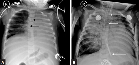
a. What are the radiological findings in images A and B?
b. Name the standard method to confirm the right placement of ETT?
c. What intervention to be done immediately in both the clinical situations?
Answer
- a.
- Image A: ETT tip inserted too deeply into the right main bronchus resulting in left lung collapse/atelectasis.
- Image B: ETT tip positioned too high (above T1), risking accidental extubation.
- b. Five-point auscultation, continuous end-tidal capnography (
), and bedside CXR / POCUS confirming the tip between T2 and T3 vertebrae (mid-trachea, midway between clavicles and carina). - c.
- For Case A: Withdraw/pull back the ETT by 1–2 cm until breath sounds are equal bilaterally.
- For Case B: Advance/push in the ETT by 1–2 cm to secure position.
THG33-008
An 8-year-old underwent a surgical procedure by a team of neurosurgeon and orthopedic surgeon. Just look at the CXR (image given below) for any peculiar things.
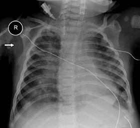
a. Do you observe any difference in ETT?
b. What are the indications of the usage of this tube?
c. Is it inserted at right depth?
d. Does it need reposition?
Answer
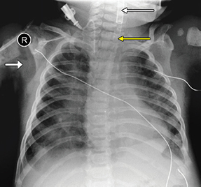
- a. Armored / reinforced endotracheal tube: Contains an embedded metallic wire spiral along its wall to prevent kinking and occlusion.
- b. Indications: Head, neck, craniofacial, and spinal/neurosurgical procedures where the patient's head/neck may be flexed, extended, or rotated, or prone positioning is required. (These tubes are MRI-conditional).
- c. No, the nonmetal tip is placed too high up (above the clavicular level) rather than between T2 and T3.
- d. Yes, it needs to be carefully advanced into the mid-trachea.
THG33-009
A 2-year-old child under follow up with pediatrician, now admitted for breathlessness for 5 days followed by convulsions. This CXR gives clue for both the complaints.
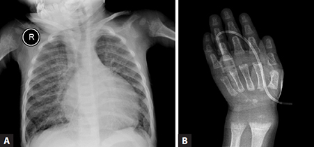
a. List the radiological findings?
b. What are the next investigations?
c. What are the next interventions?
d. What are the complications of this problem?
Answer
- a. Radiological findings: Bilateral beading at costochondral junctions (rachitic rosary), metaphyseal widening, cupping and fraying of the proximal humerus, cardiomegaly, and pulmonary plethora.
- b. Serum calcium, ionic calcium, serum phosphorus, alkaline phosphatase, 25-hydroxy vitamin D, and serum creatinine.
- c.
- Acute management of hypocalcemia: IV 10% calcium gluconate (1–2 mL/kg diluted 1:1 with 5% Dextrose over 10–20 minutes under ECG monitoring).
- Treatment of heart failure (diuretics, inotropes).
- Vitamin D therapy: Stoss therapy / cholecalciferol 60,000 IU orally once every fortnight for 5 doses, along with oral elemental calcium (50–75 mg/kg/day) for 3 months.
- d. Hypocalcemic seizures, hypocalcemic dilated cardiomyopathy (heart failure), laryngospasm, and pathological fractures.
THG33-010
A 4-year-old child with pica presented with afebrile convulsions. His father is working in battery workshop and their house is located above the work place. CXR gives a clue.
a. What are the radiological findings (as shown in below image)?
b. What is the probable diagnosis?
c. What is the gold standard test?
d. What is the management?
e. What is the associated problem?
Answer
- a. Dense radiopaque metaphyseal bands ("lead lines") at the upper end of the humeri and costochondral junctions; unusually prominent medial border of the scapula.
- b. Chronic lead poisoning (plumbism) with lead encephalopathy.
- c. Blood lead level (BLL) measurement (
is toxic; requires chelation). - d. Airway protection, seizure control (benzodiazepines), management of cerebral edema (mannitol/hypertonic saline), and chelation therapy with Dimercaprol (BAL) followed by Calcium EDTA or oral Succimer (DMSA).
- e. Microcytic hypochromic anemia with basophilic stippling on peripheral blood smear, renal Fanconi syndrome (glycosuria, aminoaciduria, proteinuria), and cognitive/behavioral deficits.
THG33-011
These are the serial CXRs of a 5-month-old child brought with features of bronchiolitis. The child is yet to attain head control, but had social smile. Child is floppy and knee jerks not elicitable. Interpret the X-rays in this background which are taken on day 1, 3, and 5 of admission.
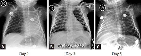
a. What are the radiological findings of all three X-rays in images A to C?
b. What is the impression after reading these X-rays, mention one term used to describe this situation?
c. What are the possible causes, any three?
d. What is the more likely diagnosis in this child?
Answer
- a.
- Image A (Day 1): Complete left lung collapse (homogenous dense opacity of left hemithorax with ipsilateral mediastinal shift), NG tube, and ETT in place.
- Image B (Day 3): Re-expansion and aeration of the left lung.
- Image C (Day 5): Collapse of the right lung with rib crowding on the right and mediastinal shift to the right.
- b. Shifting atelectasis / shifting lung collapse.
- c. Causes: Mucus plugging due to weak cough/hypotonia, migrating aspirated foreign body, and neuromuscular diseases (e.g., Spinal Muscular Atrophy, congenital myopathy, Guillain-Barré syndrome).
- d. Spinal Muscular Atrophy (SMA Type 1 / Werdnig-Hoffmann disease) in view of hypotonia, absent deep tendon reflexes, preserved cognition/social smile, and severe intercostal weakness with diaphragmatic breathing.
THG33-012
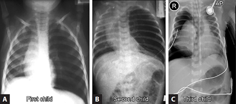
These are the X-rays of three different children presenting with breathlessness. Identify the diagnosis of these three.
- First child (image A): A toddler, playing with elder sibling. He was well after but an hour after his breakfast, he developed coughing and became breathless. Mother is confused about this sudden development.
- Second child (image B): Ten months old child admitted for breathlessness on four occasions in a district hospital. Now he admitted in PICU with the same complaints.
- Third child (image C): Child admitted for bronchopneumonia, developed respiratory failure, hence intubated and mechanically ventilated. Suddenly he developed bradycardia, hypotension and desaturation.
Answer
- First child (Image A): Foreign body aspiration with check-valve air trapping causing obstructive emphysema of the affected lung.
- Second child (Image B): Congenital lobar emphysema (CLE) of the left upper lobe with herniation across the anterior mediastinum and compressive atelectasis of the left lower lobe.
- Third child (Image C): Tension pneumothorax (hyperlucent right hemithorax devoid of vascular markings, marked mediastinal shift to opposite side, and flattened right hemidiaphragm).
THG33-013
A 5-year-old boy previously healthy child brought to emergency room (ER) at 2 AM with distress, sweating, and cold limbs. He was well child sleeping in his village home and got up in the night suddenly screaming. On arrival, he had cardiopulmonary failure, gasping, proximal pulse absent. ECG revealed ventricular tachycardia (VT) followed by ventricular fibrillation (VF).
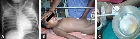
a. What are the radiological findings (image A)?
b. What are the possible causes and diagnosis?
c. What is the mechanism of action?
d. What are the clinical effects?
e. Name the steps in the management, any four.
Answer
- a. Bilateral perihilar fluffy opacities with bat-wing appearance (acute noncardiogenic/cardiogenic pulmonary edema).
- b. Diagnosis: Scorpion sting envenomation (Mesobuthus tamulus / Indian red scorpion).
- c. Mechanism: Scorpion venom contains neurotoxins (
-toxin) that open neuronal sodium channels, triggering an autonomic storm with massive endogenous release of catecholamines (epinephrine, norepinephrine, endothelin) leading to severe vasoconstriction, increased systemic afterload, and myocardial stunning. - d. Myocardial dysfunction, acute pulmonary edema, tachyarrhythmias (VT/VF), hypertension transitioning into shock, cold clammy extremities, diaphoresis, and priapism.
- e. Management:
- Prazosin (selective
-blocker) orally at 0, 3, and 6 hours. - High-flow oxygen; noninvasive or invasive mechanical ventilation with PEEP if refractory.
- Inotrope support with dobutamine infusion for cardiogenic failure.
- Judicious fluid restriction and vasodilators (e.g., nitroglycerin / sodium nitroprusside) if systemic vascular resistance is markedly elevated.
- Prazosin (selective
THG33-014
X-ray of 4-year-old child, who presented with fever, grunt, and respiratory distress, is given in image below.
a. What are the radiological findings (image below)? Mention one close differential diagnosis.
b. What could be the causes of grunting in this child?
c. What is the radiological diagnosis here?
d. What investigations will confirm the diagnosis?
e. What are the causative organisms? List the antibiotics.
f. Describe the steps in the management?
Answer
- a. Radiological findings: Dense homogenous opacity occupying the entire left lower hemithorax with obliteration of the left costophrenic and cardiophrenic angles and meniscus sign. Close differential diagnosis: Massive lobar consolidation / diaphragmatic hernia.
- b. Causes of grunting: Physiologic expiration against a partially closed glottis to generate auto-PEEP, maintain functional residual capacity, and splint painful pleuritic inflammation.
- c. Left-sided pleural empyema / massive parapneumonic effusion.
- d. Bedside chest ultrasonography (POCUS) and diagnostic thoracentesis with pleural fluid analysis (cell count, Gram stain, culture, pH, LDH, glucose).
- e. Causative organisms & Antibiotics:
- Streptococcus pneumoniae: IV Ceftriaxone / Ampicillin.
- Staphylococcus aureus (MSSA/MRSA): IV Cloxacillin / Cefazolin (MSSA) or IV Vancomycin / Linezolid / Clindamycin (MRSA).
- Haemophilus influenzae: IV Ceftriaxone.
- f. Management steps:
- Needle aspiration / thoracentesis for diagnostic confirmation.
- Intercostal drainage (ICD) tube insertion with underwater seal.
- Intrapleural fibrinolytic therapy (urokinase/alteplase) for multiloculated empyema.
- Video-assisted thoracoscopic surgery (VATS) or decortication if medical/ICD drainage fails.
THG33-015
A 5-year-old boy presented with increasing breathlessness of 3 weeks duration. There was no history of fever or any trauma.
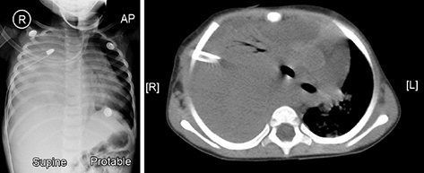
a. Identify the radiological findings in the X-ray chest, both diseases related and treatment related.
b. What is the possible differential diagnosis?
c. What are the next level investigations needed after seeing the initial X-ray chest?
d. If it is a space occupying lesion, list out any three causes for it.
Answer
- a.
- Disease-related: Complete opacification ("whiteout") of the right hemithorax with mediastinal shift away from the lesion.
- Treatment-related: Right-sided intercostal chest drainage tube and ECG monitoring leads in situ.
- b. Differential diagnosis: Massive pleural effusion, intrathoracic tumor / mediastinal mass, severe consolidated pneumonia, or total lung collapse (though collapse shifts mediastinum towards the lesion).
- c. Contrast-enhanced CT scan (CECT) of the chest and chest ultrasonography.
- d. Causes of intrathoracic mass: Mediastinal lymphoma, teratoma, neuroblastoma / ganglioneuroma, and pleuropulmonary blastoma.
THG33-016
A 4-week-old infant presented with respiratory distress noted in the early days of life. The first image (image A) is CXR and the second image (image B) is chest CT.
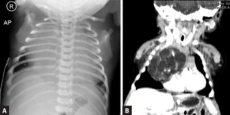
a. What are the radiological findings in X-ray (images A and B)?
b. What is the differential diagnosis with X-ray findings?
c. What are the next level investigations after the initial X-ray chest?
d. What is the diagnosis after seeing the CT?
e. What are the management strategies?
Answer
- a.
- Image A (CXR): Right-sided thoracic whiteout with massive cardiomegaly and mediastinal shift.
- Image B (Chest CT): Large heterogenous, multicystic anterior mediastinal mass with internal calcifications compressing the tracheobronchial tree.
- b. Congenital cystic adenomatoid malformation (CPAM), congenital diaphragmatic hernia, mediastinal mass (teratoma/lymphangioma), or pericardial cyst.
- c. Contrast-enhanced CT chest / MRI chest and echocardiography.
- d. Diagnosis: Mediastinal teratoma.
- e. Preoperative cardiorespiratory stabilization followed by complete surgical excision and histopathological examination (HPE).
THG33-017
A 3-year-old child presented with noisy breathing, no fever or breathlessness. But mother suspects that he might have swallowed some objects. There are two X-rays. First one (image A) is taken when the child arrived in ER. He was relatively stable at that time. This foreign body was removed after about 12 hours and the second X-ray (image B) was taken after the removal of foreign body, which was a smooth procedure under anesthesia. At the time of second X-ray, he developed breathlessness and fall in SpO2.
a. What are the radiological findings in the second X-ray (image B) after the removal of foreign body?
b. What is the possible round shadow?
c. What is the mechanism of injury?
d. If the nature of foreign body is known earlier, what intervention would have prevented this?
e. In what situation these objects should be removed immediately?
f. What is the prehospital first aid that can be done to reduce the complications?
Answer
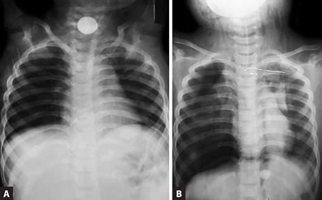
- a. Right-sided tension pneumothorax (hyperlucent right hemithorax, flattened right hemidiaphragm, continuous diaphragm sign, mediastinal shift to left) and subcutaneous emphysema in the neck and chest wall following esophageal/tracheal perforation.
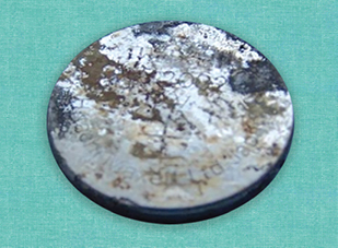
- b. Button battery (disc battery): Demonstrating the characteristic "double-ring" or "halo" sign on AP view and "step-off" on lateral view.
- c. Mechanism of tissue destruction:
- Hydroxide generation due to external electrical current discharge.
- Alkaline chemical caustic injury from electrolyte leakage.
- Physical pressure necrosis.
- d. Emergency endoscopic removal within 2–4 hours of ingestion.
- e. Button batteries lodged in the esophagus, pharynx, cricopharyngeus, nasal cavity, or external auditory canal must be removed emergently.
- f. Prehospital first aid: Administration of oral honey (10 mL every 10 minutes, up to 6 doses) or sucralfate suspension to coat the battery and neutralize alkaline injury before hospital arrival.
THG33-018
This is a 3-year-old boy presented with fever of 5 days duration and respiratory distress. The first CXR (image A) was taken at the time of admission to PICU, second CXR (image B) after 10 hours and third CXR (image C) after 24 hours.
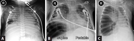
a. What are the radiological findings in the first CXR (image A), both disease related and treatment related?
b. What are the changes observed in the second (image B) and third CXR (image C) and the interventions likely to be helpful?
Answer
- a.
- Disease-related: Right-sided round consolidation with early thin-walled air cavity (pneumatocele) characteristic of staphylococcal pneumonia.
- Treatment-related: Endotracheal tube in trachea, ECG leads, and NG tube.
- b.
- Image B: Rapid enlargement and expansion of the pneumatocele.
- Image C: Rupture of the tension pneumatocele into the pleural space resulting in a large pneumothorax with collapse of the right lung.
- Intervention: Emergent placement of an intercostal drainage tube (ICD) with underwater seal, optimization of ventilator settings (avoiding high peak inspiratory pressures), and targeted antistaphylococcal antibiotic therapy.
THG33-019
This 8-year-old girl presented with defective vision, squint, and hard of hearing. She also has anemia and abdominal distension. This CXR (image A) was taken for cough without breathlessness. Second X-ray (image B) is a normal one placed adjacently to compare and identify the abnormalities.
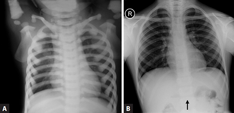
a. What are the radiological findings in the first X-ray (image A)?
b. Give two differential diagnoses.
c. Explain the clinical findings based on the final diagnosis.
d. What is the underlying pathophysiology?
e. What is the definitive treatment here?
Answer
- a. Diffuse, symmetric, generalized bone sclerosis ("marble bone" appearance) with loss of corticomedullary differentiation involving the ribs, clavicles, and vertebrae.
- b. Differential diagnosis: Osteopetrosis and Pyknodysostosis.
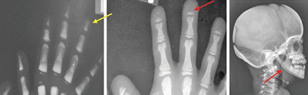
| Feature | Osteopetrosis | Pyknodysostosis |
|---|---|---|
| Bone density | Diffusely increased (sclerosis) | Diffusely increased (sclerosis) |
| Terminal phalanges | Normal terminal phalanges | Acro-osteolysis (resorption of distal phalanges) |
| Mandible angle | Normal mandibular angle | Obtuse mandibular angle |
| Cranial sutures | Sclerotic base of skull | Delayed fontanelle closure, wormian bones |
- c.
- Anemia & Abdominal distension: Obliteration of medullary cavity (myelophthisis) causing bone marrow failure and extramedullary hematopoiesis (hepatosplenomegaly).
- Vision defect, Squint, Deafness: Narrowing and bony encroachment of cranial nerve foramina (optic, oculomotor, vestibulocochlear nerves) leading to compressive cranial neuropathies.
- d. Defective osteoclast-mediated bone resorption due to mutations in the TCIRG1 or CLCN7 genes.
- e. Allogeneic Hematopoietic Stem Cell Transplantation (HSCT).
THG33-020
This 2.5-year-old child has delayed motor milestones. Yet to walk, but his comprehension and language milestones are normal.
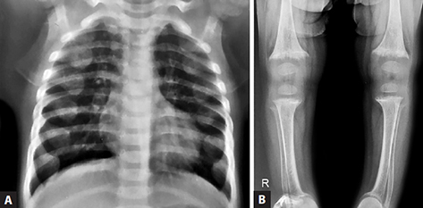
a. What are the radiological findings (images A and B)?
b. What is the diagnosis?
c. What is the investigation you would do next?
Answer
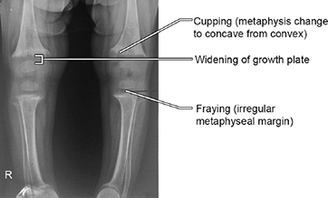
- a.
- Image A (Chest X-ray): Nodular expansion/beading of costochondral junctions (rachitic rosary).
- Image B (Lower limb X-ray): Widening of growth plates, cupping, fraying, and splaying of the distal femoral and proximal tibial metaphyses with cortical osteopenia.
- b. Diagnosis: Active Rickets (nutritional or metabolic).
- c. Serum calcium, ionic calcium, serum phosphorus, alkaline phosphatase, 25-hydroxy vitamin D, parathyroid hormone (PTH), and renal function tests.
THG33-021
A 4-year-old child with neuromuscular weakness admitted in PICU for breathlessness. These two X-rays (images A and B) are taken 24 hours apart.
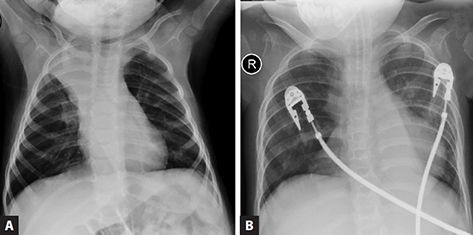
a. What are the radiological findings?
b. What are the assist devices which are helpful in chest physiotherapy?
Answer
- a.
- Image A: Triangular wedge-shaped opacity in the right upper zone with inferior concavity, indicating right upper lobe collapse; ETT, NG tube, and ECG leads visible.
- Image B (24 hours later): Resolution of right upper lobe collapse, with new bilateral lower lobe atelectasis.
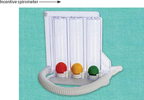
- b. Mechanical insufflator-exsufflator (CoughAssist), high-frequency chest wall oscillation (airway clearance vest), mechanical percussor/vibrator, and incentive spirometry.
THG33-022
This 2-year-old boy admitted for left-sided consolidation, treated with IV amoxicillin clavulanic acid. High-grade fever is persisting even on fourth day. Repeat CXR is shown in image here.
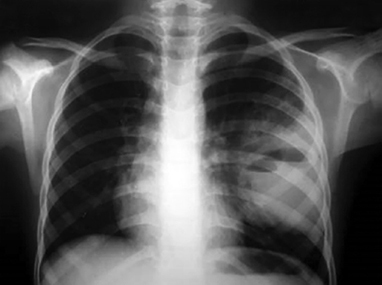
a. What are the radiological findings (as shown in above image) and possible diagnosis?
b. What are the probable causative organisms and empirical antibiotics?
c. What further investigations may be needed?
Answer
- a. Dense left mid- and lower-zone consolidation containing a well-defined cavity with an internal air–fluid level, consistent with necrotizing pneumonia / lung abscess.
- b.
- Causative organisms: Streptococcus pneumoniae, Staphylococcus aureus (MSSA/MRSA), Klebsiella pneumoniae, and oral anaerobes.
- Empirical antibiotics: IV Vancomycin / Linezolid plus IV Ceftriaxone or Meropenem plus Clindamycin.
- c. Contrast-enhanced CT scan of the chest (CECT), blood culture, sputum/tracheal aspirate culture, and pleural fluid examination if effusion is present.
THG33-023
A 3-month-old child presented with fever for 5 days and increasing respiratory distress, hospitalized, and started on amoxicillin and clavulanic acid. Following CXRs were taken on day 1, day 3, day 5, and day 8. Blood culture grew methicillin-sensitive Staphylococcus aureus (MSSA).
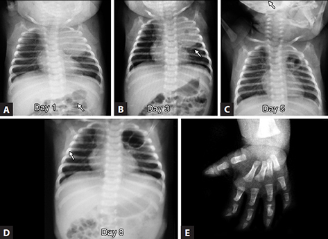
a. What are the radiological findings in the first four CXRs?
b. What are the radiological findings in the last hand X-ray (image E)?
c. What is the possible diagnosis?
d. What are the other causative organisms which can manifest like this?
e. What is the management based on the types of bacteria?
Answer
- a.
- Day 1 (Image A): Round pneumonia in the left upper lobe.
- Day 3 (Image B): Internal radiolucencies/air pockets developing within the consolidated area (necrotizing pneumonia).
- Day 5 (Image C): Enlargement of cavitation and pneumatocele formation.
- Day 8 (Image D): Evacuation of abscess contents leaving a large thin-walled cavity.
- b. Osteolytic destruction, periosteal reaction, and cortical disruption of the 4th middle phalanx (osteomyelitis / dactylitis).
- c. Staphylococcal pneumonia with lung abscess complicated by hematogenous osteomyelitis.
- d. Streptococcus pneumoniae, Methicillin-Resistant Staphylococcus aureus (MRSA), Klebsiella pneumoniae, and Pseudomonas aeruginosa.
- e.
- MSSA: IV Cloxacillin / Cefazolin for 3–4 weeks.
- MRSA: IV Vancomycin / Linezolid or Clindamycin.
- Drainage of associated empyema or subperiosteal abscess if present.
THG33-024
A 4-year-old child fell down from chair at home and sustained injury and brought with pain in forearm.
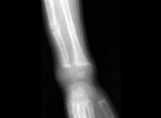
a. What are the radiological findings (as shown in above image)?
b. What is the possible diagnosis?
c. Are any further investigations needed?
d. Describe the steps in management.
Answer
- a. Metaphyseal cupping, fraying, and splaying of the distal radius and ulna with complete transverse pathological fractures through the weakened metaphyseal bone of both radius and ulna.
- b. Pathological fracture of distal radius and ulna secondary to active rickets.
- c. Serum calcium, ionic calcium, serum phosphorus, alkaline phosphatase, 25-hydroxy vitamin D, and renal function test.
- d.
- Orthopedic immobilization (forearm slab/cast).
- High-dose vitamin D therapy: Cholecalciferol 60,000 IU orally once every fortnight for 5 doses.
- Elemental calcium supplementation (50–75 mg/kg/day) for 3 months followed by maintenance vitamin D (400 IU/day).
THG33-025
A 2-year-old boy presented with fever, neck pain, difficulty in swallowing, and neck retraction. No breathlessness or seizures or altered consciousness was found. This child’s X-ray neck lateral view is given in image A below. The second image is a normal X-ray (image B) given for comparison.
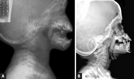
a. What are the radiological findings and probable diagnosis?
b. What are the differential diagnoses for a child presenting fever, swallowing difficulty, and retractions?
c. What further investigations will confirm?
d. What are the causative organisms, the choice of antibiotics, and further management?
Answer
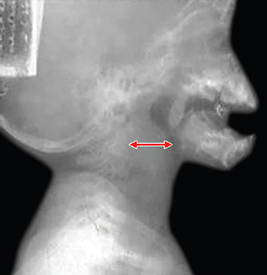
- a. Marked widening of the prevertebral / retropharyngeal soft tissue space (width > adjacent vertebral body width) with anterior displacement of the airway column, confirming a retropharyngeal abscess.
- b. Differential diagnosis: Acute epiglottitis, parapharyngeal abscess, peritonsillar abscess (quinsy), foreign body in upper esophagus, and bacterial tracheitis.
- c. Contrast-enhanced CT scan (CECT) of the neck.
- d.
- Causative organisms: Streptococcus pyogenes (Group A Strep), Staphylococcus aureus, Haemophilus influenzae, and oral anaerobes.
- Antibiotics: IV Ampicillin-Sulbactam or IV Ceftriaxone plus Clindamycin / Vancomycin.
- Management: Airway protection, transoral or transcervical surgical incision and drainage under general anesthesia.