MNJ14-001
A 13-year-old girl Reema comes to your hospital with complaints of acute onset painful swelling below her knee joint down to the ankle. She recently visited a school camp for 3 days where she shared a room with her batchmates. On examination, you find painful erythematous nodular lesions over the shins, and her pharynx is erythematous:
- What is the most likely diagnosis of these lesions?
- What are the infectious causes of these lesions (list 4)?
- Which drugs can be responsible for these?
Answer
- Diagnosis: Erythema nodosum (septal panniculitis).
- Infectious causes:
- Streptococcal pharyngitis (Group A Streptococcus - most common)
- Tuberculosis (Mycobacterium tuberculosis)
- Mycoplasma pneumoniae
- Salmonella / Yersinia enterocolitica
- Epstein-Barr virus (EBV) / Coccidioidomycosis / Histoplasmosis
- Drugs: Sulfonamides, penicillins, oral contraceptive pills (OCPs), NSAIDs, bromides/iodides.
MNJ14-002
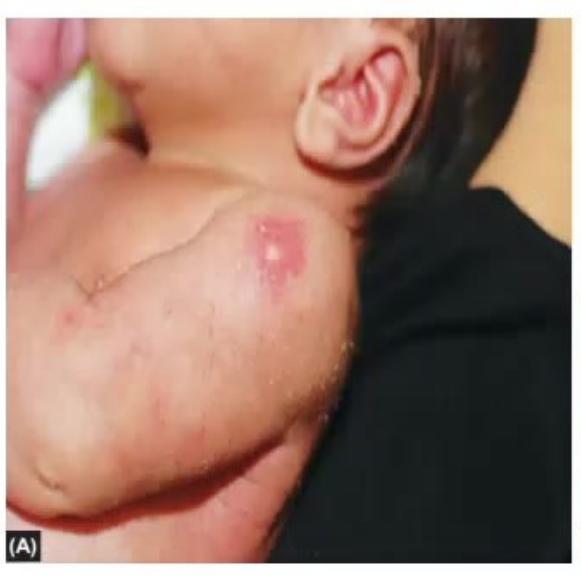
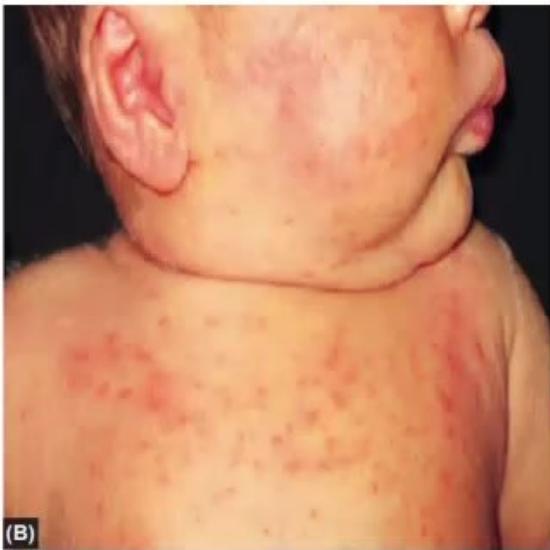
See the images of 2 neonates (Image A and Image B) and answer the following questions:
- What is the diagnosis of Image A and Image B?
- How long do these persist in a normal neonate?
- In which subgroup of gestational age are these two more commonly seen?
- What are the treatment options for Image A and Image B?
Answer
- Diagnosis:
- Image A: Erythema toxicum neonatorum (ETN).
- Image B: Neonatal cephalic pustulosis / Acne neonatorum.
- Duration / Course:
- Image A (Erythema toxicum): Appears at 24–48 hours of life and resolves spontaneously within 7–10 days.
- Image B (Acne neonatorum): Appears around 2–3 weeks of life and may persist for several weeks up to 6–8 months.
- Gestation: Both are significantly more common in full-term, healthy newborns (rare in premature infants).
- Treatment:
- Image A: Reassurance and gentle skin care; no treatment required.
- Image B: Usually self-limiting; gentle cleansing with water. If severe/persistent, topical 2% ketoconazole cream or mild topical hydrocortisone.
MNJ14-003
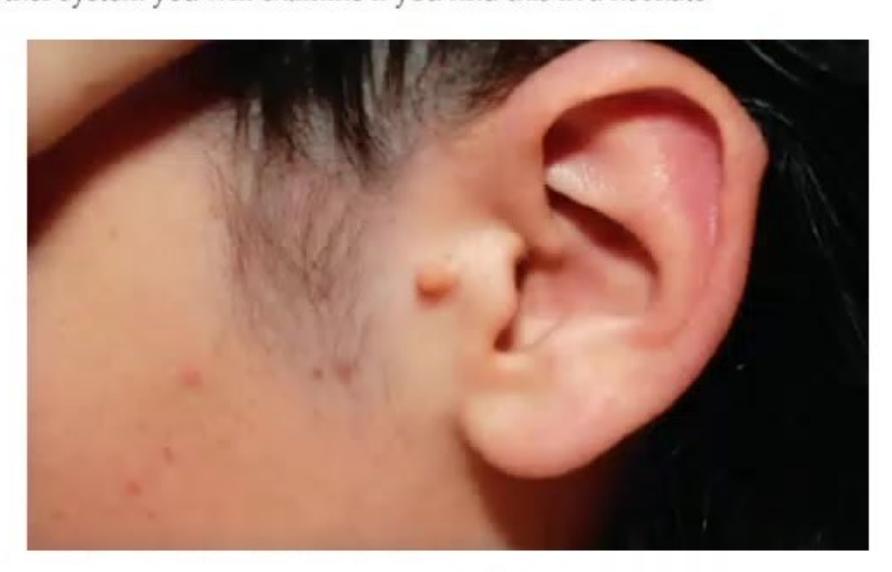
See the image and answer the following questions:
- What is the clinical finding in this picture?
- What is the embryological pathophysiology for this?
- What are the differential diagnoses (list 2)?
- What is its natural course in a child?
- What other organ system should you screen/examine if you find this in a neonate?
Answer
- Finding: Preauricular skin tag (accessory tragus).
- Pathophysiology: Defective or abnormal fusion of the first and second branchial arches (specifically the hillocks of His) during embryogenesis.
- Differential diagnosis: Epidermal inclusion cyst, preauricular fibroma, adnexal tumor, lipoma, neurofibroma.
- Course: Benign and asymptomatic; persists lifelong unless surgically excised.
- Other system: Renal system (routine screening / USG KUB if associated with other dysmorphic features or hearing impairment; screen for Goldenhar syndrome, BOR syndrome, Treacher Collins syndrome).
MNJ14-004
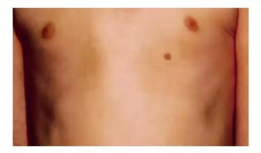
A 6-year-old child comes to you with fever and cough. During chest examination, you notice an unusual finding along the chest wall:
- What is the clinical finding in this picture?
- What is the milk line?
- What is its male-to-female ratio and recommended management?
Answer
- Finding: Polythelia / Accessory (supernumerary) nipple.
- Milk line (mammary ridge): An imaginary embryological line extending bilaterally from the anterior axillary fold down to the inguinal region along which mammary glands develop in mammals.
- Demographics & Management:
- Equal frequency in males and females (
). - Management: Reassurance; benign anomaly requiring no treatment. Surgical excision may be performed for cosmetic reasons or recurrent irritation.
- Equal frequency in males and females (
MNJ14-005
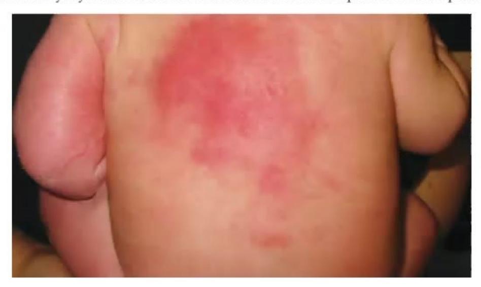
- What is the diagnosis shown in this newborn?
- What are the common causes/triggers in a neonate?
- What is the pathophysiology for this finding?
- What is the clinical course?
- What is the treatment?
- What electrolyte should you monitor in this case and what complication can develop?
Answer
- Diagnosis: Subcutaneous fat necrosis of the newborn (SCFN).
- Causes / Predisposing factors: Perinatal asphyxia, hypothermia/therapeutic hypothermia, birth trauma, maternal gestational diabetes, pre-eclampsia, meconium aspiration.
- Pathophysiology: Neonatal subcutaneous fat contains a high concentration of saturated fatty acids (stearic and palmitic acids) with a high melting point. Cold injury or tissue hypoperfusion induces fat crystallization within adipocytes, leading to necrosis, giant cell reaction, and granulomatous panniculitis.
- Course: Lesions evolve over weeks to months from firm, red-purple indurated plaques/nodules to soft, fluctuant areas with spontaneous resolution.
- Treatment: Supportive care and watchful monitoring; lesions resolve spontaneously without active treatment.
- Electrolyte monitoring & Complication:
- Serum Calcium: Monitor bi-weekly for up to 6 months.
- Complication: Severe hypercalcemia (due to extra-renal production of 1,25-dihydroxyvitamin D by activated macrophages in granulomas), which can lead to nephrocalcinosis, cardiac arrhythmias, and failure to thrive.
MNJ14-006
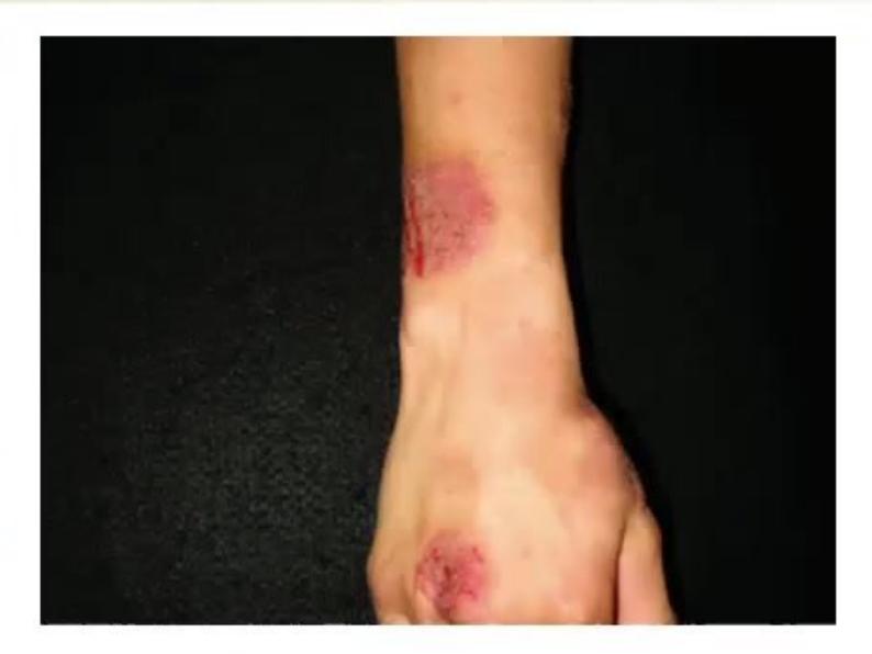
A 13-year-old child Rajeev with known atopic dermatitis comes to your clinic with these new itchy lesions that began in December and increased in size over a month. He is on regular emollients and has not traveled recently:
- What is the diagnosis of this condition?
- What are the differential diagnoses?
- How will you manage these lesions?
Answer
- Diagnosis: Nummular eczema (discoid eczema).
- Differential diagnosis:
- Tinea corporis (fungal infection)
- Contact dermatitis
- Plaque psoriasis
- Pityriasis rosea (herald patch)
- Management:
- Gentle, soap-free cleansers and warm (not hot) baths.
- Frequent application of thick bland emollients/moisturizers (petrolatum/ceramide creams).
- Mid- to high-potency topical corticosteroids applied to active lesions.
- Topical calcineurin inhibitors (tacrolimus/pimecrolimus) for maintenance.
- Oral antihistamines for pruritus control.
MNJ14-007
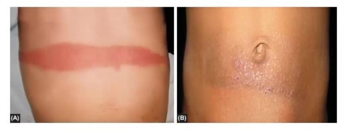
These 2 patients presented with cutaneous lesions: Patient A is hospitalized and had adhesive tape applied across his abdomen; Patient B wears a tight metallic belt buckle regularly:
- What is the diagnosis in both images? Do they represent the same underlying problem?
- Which type of allergic hypersensitivity reaction is this?
- What treatment can you offer for these patients?
Answer
- Diagnosis: Allergic contact dermatitis (Patient A due to adhesive acrylate/tape; Patient B due to nickel/metal buckle). Yes, both represent allergic contact dermatitis.
- Immunologic reaction: Type IV (delayed-type / cell-mediated) hypersensitivity reaction.
- Treatment:
- Strict avoidance/removal of the offending allergen (hypoallergenic paper tape; nickel-free belt buckle or barrier cloth).
- Application of topical corticosteroids (hydrocortisone or betamethasone).
- Calamine lotion / cool compresses and oral antihistamines for symptomatic itch relief.
MNJ14-008
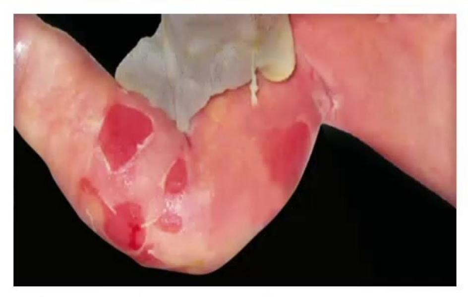
A 7-day-old neonate comes with complaints of recurrent blister and skin peel formation following minor friction, rubbing, or clothing contact:
- What is the most likely diagnosis?
- What is the recurrence risk in future pregnancies?
- What is the pathophysiology and genetic basis?
- What is the usual course and prognosis?
Answer
- Diagnosis: Epidermolysis bullosa (EB - simplex / junctional / dystrophic).
- Recurrence risk:
- Autosomal Dominant (e.g., EB Simplex): 50% risk for each pregnancy.
- Autosomal Recessive (e.g., Junctional or recessive Dystrophic EB): 25% risk per pregnancy.
- Pathophysiology & Genetics: Mutations in structural proteins that maintain epidermal-dermal integrity:
- KRT5 / KRT14 on chromosomes 12q and 17q (EB simplex)
- LAMA3, LAMB3, LAMC2, COL17A1 (Junctional EB)
- COL7A1 on chromosome 3p (Dystrophic EB)
- Course: Fragility often persists throughout life; managed with non-adherent silicone dressings, skin protection, prevention of secondary infections, and nutritional support.
MNJ14-009
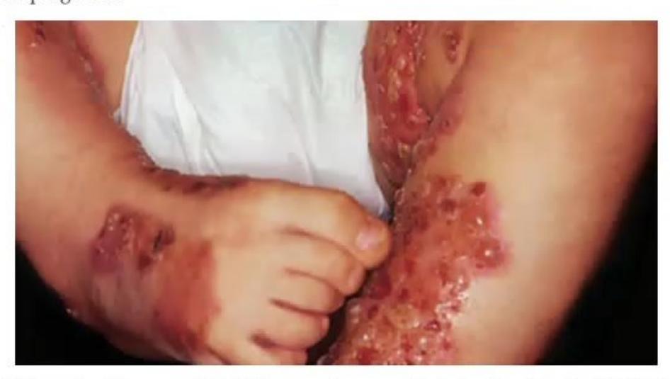
A 4-month-old boy Rahul presents with itchy, grouped tense blisters over the extremities:
- What is the likely diagnosis?
- What is the characteristic pattern of these lesions called?
- What is found on direct immunofluorescence (DIF) / electron microscopy?
- What is the prognosis?
Answer
- Diagnosis: Linear IgA bullous dermatosis of childhood (Chronic bullous disease of childhood [CBDC]).
- Pattern: "String of pearls" or "Cluster of jewels" pattern (rosette-like arrangement of new tense vesicles around resolving crusted plaques).
- Immunofluorescence / EM finding: Continuous linear deposition of IgA along the basement membrane zone (subepidermal split with cleavage through lamina lucida).
- Prognosis: Excellent; usually a benign, self-limiting disorder that responds dramatically to oral Dapsone and resolves spontaneously within 2–4 years.
MNJ14-010
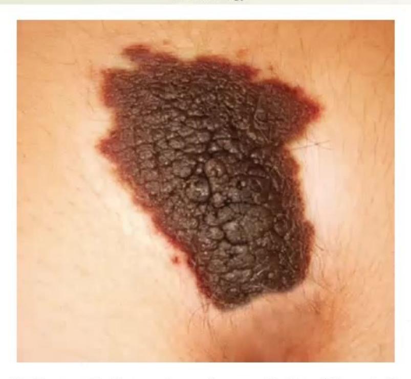
A mother of a 13-year-old child presents for URI and shows an abnormal hyperpigmented lesion on her child's neck that has darkened and developed irregular surface changes:
- What is the likely diagnosis?
- What are 4 possible differential diagnoses?
- What advice will you give the mother regarding future observation?
Answer
- Diagnosis: Congenital melanocytic nevus (CMN) / Giant pigmented nevus.
- Differential diagnosis:
- Mongolian spot (congenital dermal melanocytosis)
- Nevus of Ota / Nevus of Ito
- Blue nevus
- Becker's nevus
- Plexiform neurofibroma
- Advice / Follow-up:
- Monitor for signs of malignant melanoma transformation using the ABCDE criteria:
- A: Asymmetry
- B: Border irregularity
- C: Color variegation / darkening
- D: Diameter
or rapid expansion - E: Evolution / Elevation / Ulceration / Bleeding
- Regular clinical examination and prompt biopsy if any rapid changes occur.
- Monitor for signs of malignant melanoma transformation using the ABCDE criteria:
MNJ14-011
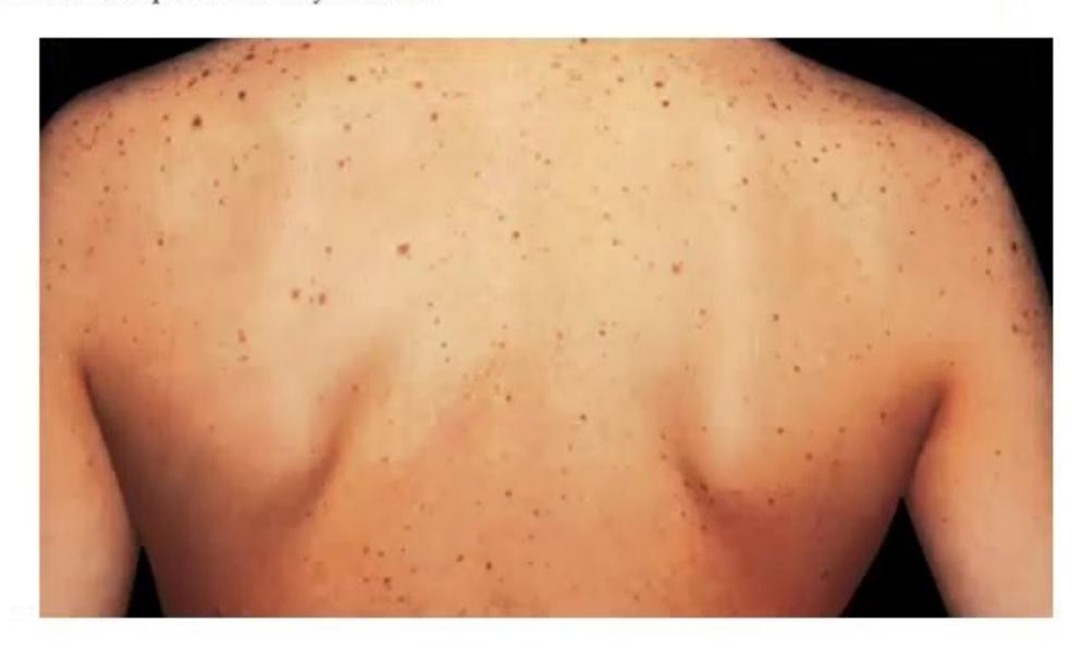
Mother of a 5-year-old girl comes for consultation regarding multiple brown macules on her back present since birth. The child also has poor growth, sensorineural hearing loss, and a congenital heart lesion on echocardiography:
- What are these skin lesions called?
- What is the associated genetic syndrome?
- What are the clinical components of this syndrome?
Answer
- Lesions: Multiple lentigines (scattered hyperpigmented macules).
- Syndrome: LEOPARD syndrome (Noonan syndrome with multiple lentigines; PTPN11 mutation).
- Mnemonic Components (LEOPARD):
- L: Lentigines (multiple dark-brown macules)
- E: ECG conduction abnormalities
- O: Ocular hypertelorism
- P: Pulmonary stenosis
- A: Abnormalities of genitalia (hypogonadism, cryptorchidism)
- R: Retardation of growth
- D: Deafness (sensorineural hearing loss)
MNJ14-012
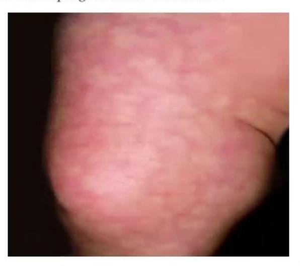
A 24-hour-old neonate of a diabetic mother is in the NICU. On changing the diaper in a cool room, a lace-like reticular reddish-blue mottling is observed over the lower extremities:
- What is the likely diagnosis of this condition?
- What is the cause/pathophysiology?
- What is the prognosis?
Answer
- Diagnosis: Cutis marmorata (physiologic mottling).
- Cause / Pathophysiology: Normal physiological vasomotor response to cold ambient temperature, resulting from immature autonomic vascular regulation leading to alternating venular dilation and arteriolar constriction.
- Prognosis: Completely benign and transient; disappears upon re-warming and gradually resolves as the infant's autonomic vasomotor control matures over early infancy.
MNJ14-013
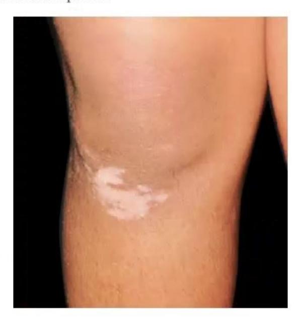
See the image of well-demarcated depigmented patches on the skin:
- What is the diagnosis of this lesion?
- What endocrine screening test will you order for this child?
- What are the primary treatment options?
Answer
- Diagnosis: Vitiligo (autoimmune melanocyte destruction).
- Endocrine test: Thyroid profile (Free
, , anti-TPO antibodies), as autoimmune thyroid disease (Hashimoto thyroiditis/Grave's) occurs in up to 20–30% of vitiligo patients. - Treatment options:
- Topical corticosteroids (mid-to-high potency) or topical calcineurin inhibitors (tacrolimus 0.03%–0.1%, pimecrolimus).
- Phototherapy: Narrowband UVB (NB-UVB) or targeted excimer laser.
- Systemic steroids (oral mini-pulse) for rapidly progressive disease.
- Strict photoprotection with broad-spectrum sunscreens.
MNJ14-014
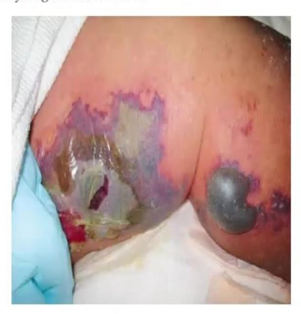
A child presents with acute onset fever, respiratory distress, septic shock, and rapidly spreading ecchymoses and necrotic purpura. Blood tests show a deranged coagulation profile:
- What is the diagnosis of this condition?
- What are 4 possible etiologies?
- What are the expected results for D-dimer, fibrin degradation products (FDPs), and plasma fibrinogen?
- What is the emergency treatment for this child?
Answer
- Diagnosis: Purpura fulminans (Disseminated Intravascular Coagulation with microvascular skin necrosis).
- Etiologies:
- Severe bacterial sepsis (Neisseria meningitidis / meningococcemia, Streptococcus pneumoniae, Staphylococcus aureus, Gram-negative rods)
- Severe viral infections (varicella, measles)
- Congenital or acquired Protein C / Protein S / Antithrombin III deficiency
- Catastrophic antiphospholipid syndrome
- Lab results:
- D-dimer & FDPs: Markedly elevated
- Fibrinogen level: Significantly decreased (consumed)
- (Prolonged PT/INR and aPTT, severe thrombocytopenia)
- Treatment:
- Immediate aggressive resuscitation (airway, high-flow
, IV fluid boluses, vasopressors). - Prompt broad-spectrum parenteral antibiotics (e.g., 3rd gen cephalosporin + vancomycin).
- Blood product replacement: Fresh Frozen Plasma (FFP), cryoprecipitate (to replete fibrinogen), and platelet concentrates.
- Protein C concentrate replacement if indicated.
- Immediate aggressive resuscitation (airway, high-flow
MNJ14-015
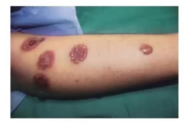
A 5-year-old boy Pankaj Gulati presents with a 1-week history of widespread pruritic and painful vesicular and bullous lesions arranged in an annular configuration with crusting:
- What is the diagnosis?
- What is the characteristic description of this lesion pattern?
- Which immunoglobulin deposition is classically identified on direct immunofluorescence (DIF)?
Answer
- Diagnosis: Linear IgA bullous dermatosis of childhood (Chronic bullous disease of childhood).
- Pattern: "String of pearls" or "Cluster of jewels" appearance (new tense vesicles ringing the periphery of resolving central lesions).
- Immunopathology: Continuous linear deposition of IgA along the basement membrane zone (BMZ) on direct immunofluorescence.