VVK05-001
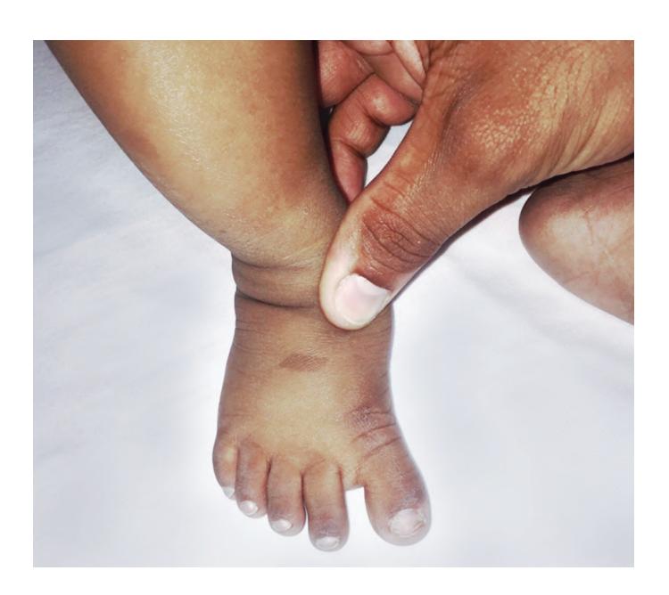
- a. What is the clinical finding in the foot?
- b. What is the commonest genetic condition associated with the above finding?
- c. Can this be a normal variant in healthy individuals? (Yes/No)
- d. Enlist 1 antenatal/environmental cause it can be associated with.
Answer
- a. Sandal gap (wide space / increased gap between 1st and 2nd toes).
- b. Trisomy 21 (Down syndrome).
- c. Yes (can be present as an isolated benign normal variant in healthy individuals).
- d. Amniotic band constriction / disruption sequence.
VVK05-002
Match the clinical findings with the corresponding pediatric genetic / neuromuscular conditions:
Findings:
- a. Tongue fasciculation
- b. Gower sign
- c. Heliotrope rash
- d. Non-thrombocytopenic purpura
- e. Thrombocytopenic purpura with eczema and immunodeficiency
Conditions:
- Spinal Muscular Atrophy (SMA)
- Juvenile Dermatomyositis
- Duchenne Muscular Dystrophy (DMD)
- Henoch-Schönlein Purpura (IgA Vasculitis)
- Wiskott-Aldrich Syndrome
Answer
- a. Tongue fasciculation
1. Spinal Muscular Atrophy (SMA) - b. Gower sign
3. Duchenne Muscular Dystrophy (DMD) - c. Heliotrope rash
2. Juvenile Dermatomyositis - d. Non-thrombocytopenic purpura
4. Henoch-Schönlein Purpura - e. Thrombocytopenic purpura with eczema
5. Wiskott-Aldrich Syndrome
VVK05-003
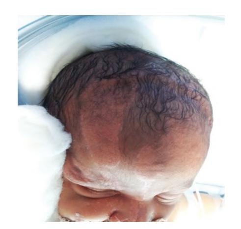
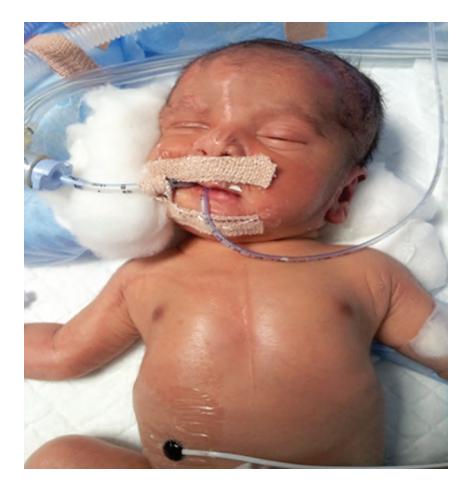
- a. What is the diagnosis?
- b. What can be the associated systemic abnormalities?
Answer
- a. Linear Nevus Sebaceous Syndrome (Schimmelpenning-Feuerstein-Mims syndrome) / Epidermal Nevus Syndrome.
- b. Associated multisystem abnormalities:
- CNS: Hemimegalencephaly, cortical dysplasia, intractable infantile seizures, intellectual disability, lateral ventriculomegaly.
- Ophthalmologic: Colobomas (iris/choroid), strabismus, corneal vascularization, scleral choristomas, microphthalmia.
- Skeletal: Craniofacial asymmetry, localized fibrous dysplasia, hypophosphatemic vitamin D-resistant rickets, kyphoscoliosis.
- Cardiovascular: Coarctation of aorta, ventricular septal defect.
- Renal: Horseshoe kidney, renal dysplasia.
VVK05-004
Given below is a karyotype of a 15-day-old child:
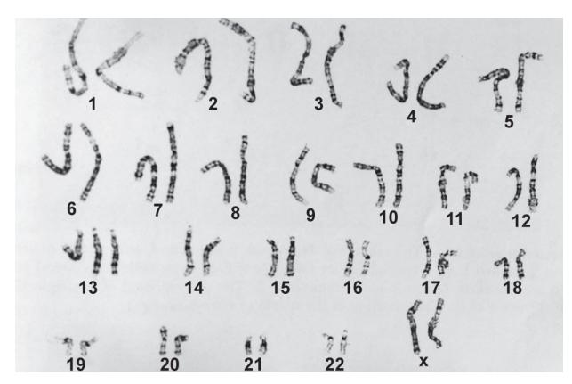
- a. Identify the condition and sex of the child.
- b. Enlist 3 characteristic features across organ systems.
Answer
- a. Trisomy 13 (Patau Syndrome), Female (
). - b. Characteristic clinical features:
- Craniofacial / CNS: Holoprosencephaly, microcephaly, sloping forehead, microphthalmia/anophthalmia, coloboma, midline cleft lip and palate, low-set malformed ears.
- Dermatologic / Extremities: Aplasia cutis congenita (scalp defects over parieto-occipital area), postaxial polydactyly, prominent calcaneus (rocker-bottom feet).
- Cardiovascular (80%): VSD, PDA, ASD, dextrocardia.
- Genitourinary: Bicornuate uterus (females), cryptorchidism (males), polycystic kidneys.
(Note: >80% mortality within the first month of life).
VVK05-005
Enlist the chromosomal location and associated cancer/tumor in relation to each gene/oncogene:
| Gene / Oncogene | Chromosomal Location | Associated Cancer / Tumor |
|---|---|---|
| RB1 gene | ||
| APC gene | ||
| WT1 gene | ||
| BRCA1 gene | ||
| NF1 gene | ||
| NF2 gene | ||
| N-MYC (MYCN) | ||
| ABL1 (BCR-ABL) | ||
| PML-RARA (ERBA) | ||
| RET proto-oncogene | ||
| SIS (PDGFB) |
Answer
| Gene / Oncogene | Chromosomal Location | Associated Cancer / Tumor |
|---|---|---|
| RB1 | 13q14 | Retinoblastoma, Osteosarcoma |
| APC | 5q21 | Familial Adenomatous Polyposis (FAP), Colorectal carcinoma |
| WT1 | 11p13 | Wilms tumor, WAGR syndrome |
| BRCA1 | 17q21 | Hereditary Breast and Ovarian Cancer |
| NF1 | 17q11.2 | Neurofibromatosis type 1, Optic pathway glioma |
| NF2 | 22q12.2 | Neurofibromatosis type 2, Bilateral vestibular schwannomas |
| N-MYC (MYCN) | 2p24 | Neuroblastoma |
| ABL1 (BCR-ABL) | 9q34 (t(9;22)) | Chronic Myeloid Leukemia (CML), B-ALL (Philadelphia chromosome) |
| PML-RARA | 17q21 (t(15;17)) | Acute Promyelocytic Leukemia (APL, AML M3) |
| RET | 10q11.2 | MEN 2A, MEN 2B, Medullary Thyroid Carcinoma |
| SIS (PDGFB) | 22q12.3 | Dermatofibrosarcoma protuberans, Glioblastoma / Glioma |
VVK05-006
Study the pedigree chart and answer the questions:
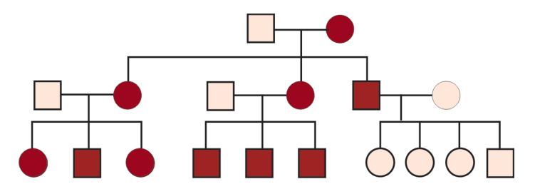
- a. Identify the pattern of inheritance in the given pedigree with explanation.
- b. Explain the mechanism of this inheritance.
- c. Give 2 examples of this pattern of inheritance.
Answer
- a. Mitochondrial (Maternal) Inheritance.
- Explanation: All offspring of an affected female inherit the trait/disease, regardless of sex. Affected males never transmit the disorder to any of their children.
- b. Mechanism: The human ovum contains abundant cytoplasm and mitochondria (along with circular mitochondrial DNA), whereas sperm mitochondria located in the tail/midpiece are destroyed upon fertilization. Hence, all mitochondrial DNA is exclusively maternally inherited.
- c. Examples:
-
- Leber's Hereditary Optic Neuropathy (LHON)
-
- Mitochondrial Encephalomyopathy with Lactic Acidosis and Stroke-like episodes (MELAS)
-
- Myoclonic Epilepsy with Ragged Red Fibers (MERRF)
-
- Leigh syndrome (subacute necrotizing encephalopathy)
-
VVK05-007
Study the pedigree chart given below and answer the questions:
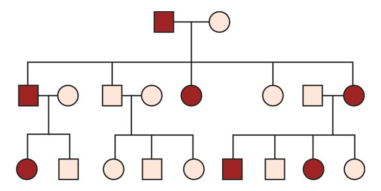
- a. What is the pattern of inheritance?
- b. Name three conditions with a similar pattern of inheritance.
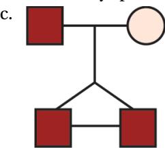
- c. Draw a pedigree chart showing parents with a pair of identical (monozygotic) twins.
Answer
- a. Autosomal Dominant Inheritance (Vertical transmission across successive generations without skipping, equal male and female involvement, transmission from affected parent to ~50% of offspring).
- b. Examples:
-
- Neurofibromatosis type 1 (NF-1)
-
- Tuberous Sclerosis
-
- Marfan syndrome
-
- Achondroplasia
-
- Hereditary Spherocytosis
-
- c. Pedigree representation of monozygotic twins:
VVK05-008
Given below is a pedigree chart of a male child with mental retardation:
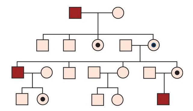
- a. Identify the pattern of inheritance.
- b. Name three conditions with similar inheritance.
- c. What is the risk of getting affected in each pregnancy for a carrier female?
Answer
- a. X-Linked Recessive Inheritance (Diagonal transmission: unaffected carrier females transmit the disease to 50% of sons; affected males do not transmit to sons but all daughters become obligate carriers).
- b. Examples:
-
- Duchenne Muscular Dystrophy (DMD) / Becker Muscular Dystrophy
-
- Hemophilia A and B
-
- Glucose-6-Phosphate Dehydrogenase (G6PD) deficiency
-
- Fragile X syndrome (X-linked dominant with reduced penetrance) / Menkes disease
-
- X-linked Adrenoleukodystrophy
-
- c. For a carrier mother (
): - 50% risk for each male child to be affected.
- 50% chance for each female child to be an asymptomatic carrier (Overall 25% risk per pregnancy of having an affected child).
VVK05-009
Identify the degree of consanguinity in the relationships shown with dotted lines in the following pedigree charts:
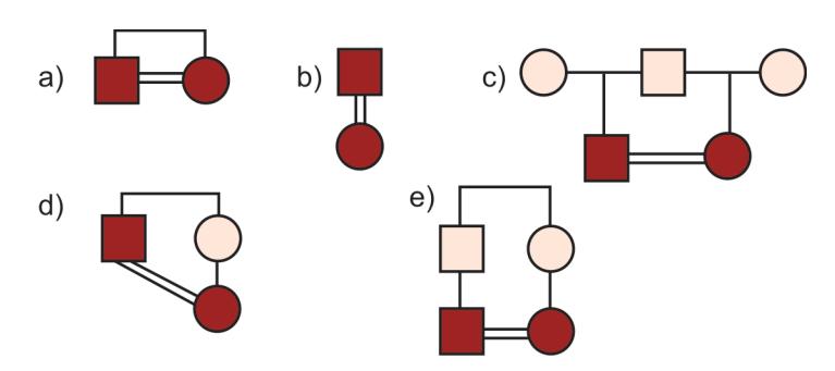
Answer
- a. First-degree relationship / incest (Brother–sister mating; coefficient of relationship
). - b. First-degree relationship / incest (Parent–offspring mating;
). - c. Second-degree consanguinity (Uncle–niece or Aunt–nephew mating;
). - d. Second-degree relationship (Half-siblings mating;
). - e. Third-degree consanguinity (First cousins mating: children of brother and sister;
).
VVK05-010
Study the pedigree chart shown below and answer the questions:
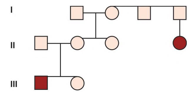
- a. Identify the pattern of inheritance.
- b. Give two examples of this type of inheritance.
- c. If one child of parents with this disorder is already affected, what would be the recurrence risk in the next offspring?
- d. If two earlier pregnancies are affected by this disease, what is the recurrence risk in the third offspring?
Answer
- a. Multifactorial / Polygenic Inheritance (Interaction of multiple minor additive genes with environmental factors).
- b. Examples:
-
- Neural Tube Defects (Anencephaly, Spina bifida)
-
- Cleft lip with or without cleft palate
-
- Congenital hypertrophic pyloric stenosis
-
- Congenital heart defects (isolated VSD, ASD)
-
- Hirschsprung disease / Clubfoot (talipes equinovarus)
-
- c. Recurrence risk after 1 affected child: 3% to 5%.
- d. Recurrence risk after 2 affected children: 10% to 12%.
VVK05-011
A 1-year-old female infant, born of a consanguineous marriage is brought to you with history of developmental delay. Clinically, there is dysmorphism consisting of coarse facies, short stature and corneal clouding. Liver is palpable, 4 cm below costal margin. There is no other family history of a similarly affected child. You make a clinical diagnosis of Hurler syndrome (MPS I).
- a. What features suggest that the diagnosis is Hurler syndrome and not Hunter syndrome (MPS II)?
- b. How will you evaluate this child to support the diagnosis?
- c. Name two modalities of treatment for this child.
Answer
- a. Distinguishing features:
-
- Sex and inheritance: The infant is female (Hurler is Autosomal Recessive; Hunter syndrome is X-Linked Recessive and affects almost exclusively males).
-
- Corneal clouding: Corneal clouding is characteristically PRESENT in Hurler syndrome (MPS I) and ABSENT in Hunter syndrome (MPS II).
-
- b. Diagnostic evaluation:
-
- Urine screening for glycosaminoglycans (GAGs): elevated dermatan sulfate and heparan sulfate.
-
- Skeletal survey: features of Dysostosis Multiplex (hooked anterior lumbar vertebrae / anterior beaking, spatulate ribs, claw hand deformity).
-
- Specific enzyme assay in leukocytes/plasma: severe deficiency of
-L-iduronidase (confirmatory).
- Specific enzyme assay in leukocytes/plasma: severe deficiency of
-
- Molecular genetic testing for IDUA gene mutations.
-
- c. Treatment modalities:
-
- Enzyme Replacement Therapy (ERT): Recombinant human
-L-iduronidase (Laronidase / Aldurazyme).
- Enzyme Replacement Therapy (ERT): Recombinant human
-
- Hematopoietic Stem Cell Transplantation (HSCT / Bone Marrow Transplant): Curative if performed early (<2 years of age before significant neurocognitive decline).
-
VVK05-012
Study the pedigree chart and answer the questions:
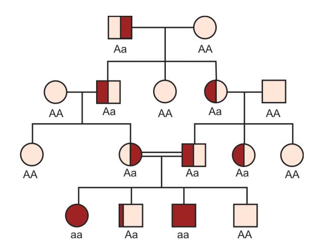
- a. What type of inheritance is shown?
- b. What is the risk of recurrence in the next pregnancy?
- c. Of the following, which is consistent with this type of inheritance?
- i. Glucose-6-phosphate dehydrogenase deficiency
- ii. Niemann-Pick disease
- iii. Meningomyelocele
- iv. Tuberous sclerosis
Answer
- a. Autosomal Recessive Inheritance (Horizontal transmission: affected individuals usually in a single sibship of healthy carrier parents, high association with parental consanguinity, equal male and female ratio).
- b. 25% (1 in 4) recurrence risk in each pregnancy.
- c. ii. Niemann-Pick disease (G6PD is X-linked recessive; Meningomyelocele is multifactorial; Tuberous sclerosis is autosomal dominant).
VVK05-013
State the pattern of inheritance for each of the following genetic disorders:
- a. Achondroplasia
- b. Hemophilia B (Christmas disease)
- c. Congenital adrenal hyperplasia
- d. Glucose-6-phosphate dehydrogenase deficiency
- e. Sickle cell disease
- f. Hemophilia A
- g. Phenylketonuria (PKU)
- h. Duchenne muscular dystrophy (DMD)
- i. Marfan syndrome
- j. Huntington's chorea
- k. Neurofibromatosis type 1
Answer
- a. Achondroplasia
Autosomal Dominant - b. Hemophilia B
X-Linked Recessive - c. Congenital adrenal hyperplasia
Autosomal Recessive - d. G6PD deficiency
X-Linked Recessive - e. Sickle cell disease
Autosomal Recessive - f. Hemophilia A
X-Linked Recessive - g. Phenylketonuria
Autosomal Recessive - h. Duchenne muscular dystrophy
X-Linked Recessive - i. Marfan syndrome
Autosomal Dominant - j. Huntington's disease
Autosomal Dominant - k. Neurofibromatosis type 1
Autosomal Dominant
VVK05-014
What do the following standard symbols represent in a pedigree chart?
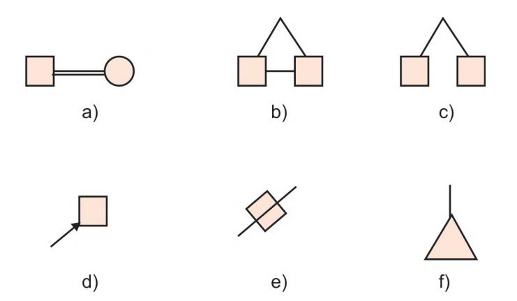
Answer
- a. Consanguineous marriage / relationship (double horizontal bar).
- b. Monozygotic (identical) twins (diagonal lines joined by a horizontal bar).
- c. Dizygotic (fraternal) twins (diagonal lines originating from a common point without horizontal bar).
- d. Proband / Index case (indicated by an arrow pointing to the affected individual).
- e. Stillbirth (symbol with a horizontal strike / diagonal line through shape).
- f. Spontaneous abortion / Miscarriage (small filled or unfilled triangle).
VVK05-015
Study the pedigree chart given below and answer the questions:
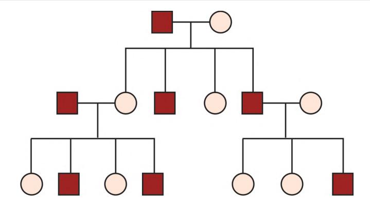
- a. What is the mode of inheritance?
- b. What is the recurrence risk of the disease to the offspring of an affected individual?
- c. Is it possible for phenotypically normal members of the family to transmit this disease?
- d. Which of the following is transmitted in this fashion?
- i. Noonan syndrome
- ii. Galactosemia
- iii. Diabetes mellitus
- iv. Hypophosphatemic rickets
Answer
- a. Autosomal Dominant Inheritance.
- b. 50% (1 in 2) risk of transmission to each child.
- c. No (in fully penetrant autosomal dominant traits, unaffected individuals do not carry the gene and cannot transmit it; exceptions include non-penetrance or germline mosaicism).
- d. i. Noonan syndrome (Galactosemia is autosomal recessive; Diabetes is multifactorial; Hypophosphatemic rickets is X-linked dominant).
VVK05-016
A full-term male neonate was born by normal vaginal delivery to a 23-year-old primigravida mother and was noted to have the following dysmorphic features:
-
Prominent occiput
-
Microcephaly (Head circumference 29 cm)
-
Clenched fists with overlapping fingers (2nd over 3rd, 5th over 4th)
-
Rocker-bottom feet with prominent calcaneus
-
a. What is the most probable diagnosis?
-
b. What is the likely chromosomal configuration of this neonate?
-
c. Name two congenital heart diseases commonly associated with this syndrome.
Answer
- a. Edwards Syndrome (Trisomy 18).
- b.
. - c. Associated congenital heart diseases (>90% incidence):
-
- Ventricular Septal Defect (VSD)
-
- Patent Ductus Arteriosus (PDA)
-
- Polyvalvular disease / Atrial Septal Defect (ASD) / Coarctation of aorta.
-
VVK05-017
This female child was brought for evaluation of dysmorphic features:
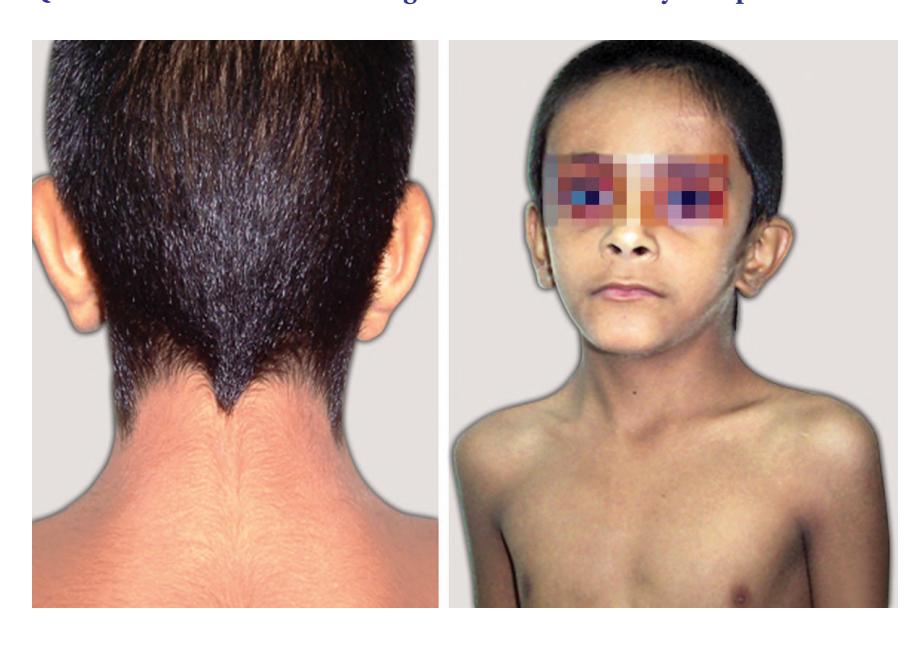
- a. What is the most probable diagnosis?
- b. Name one cardiac and one renal malformation associated most commonly with this condition.
- c. What drug therapy can be provided to these patients?
- d. Name one easy OPD procedure to reach a provisional diagnosis.
- e. What is the male counterpart / phenotypic mimic of this disease?
Answer
- a. Turner Syndrome (
). - b.
- Cardiac: Bicuspid aortic valve (most common), Coarctation of the aorta, Aortic root dilation.
- Renal: Horseshoe kidney (most common), double collecting system, pelvic kidney.
- c. Medical therapy:
-
- Recombinant Human Growth Hormone (rhGH): started early to optimize final adult height.
-
- Estrogen and Progesterone replacement therapy: to induce secondary sexual characteristics and maintain bone mineral density.
-
- Levothyroxine (for autoimmune hypothyroidism).
-
- d. Buccal smear for Barr body (shows absent Barr body, i.e., chromatin-negative; definitive test is chromosomal karyotype).
- e. Noonan Syndrome (Autosomal dominant condition with normal karyotype affecting both males and females).
VVK05-018
A 32-year-old pregnant lady is at 20 weeks of gestation. She is referred with a positive triple test result.
- a. What are the components of the triple test, what pattern constitutes a screen-positive result for Down syndrome, and which conditions does it screen for?
- b. Name three radiological findings on ultrasound that suggest the presence of the disease.
- c. How can a definitive diagnosis be established at this gestational age?
Answer
- a.
- Triple Test Components:
-
- Maternal Serum Alpha-Fetoprotein (MSAFP)
Low
- Maternal Serum Alpha-Fetoprotein (MSAFP)
-
- Unconjugated Estriol (
) Low
- Unconjugated Estriol (
-
- Human Chorionic Gonadotropin (
) High
- Human Chorionic Gonadotropin (
-
- Screening Targets: Screens for Down syndrome (Trisomy 21), Edwards syndrome (Trisomy 18 - all 3 markers low), and Neural Tube Defects (elevated MSAFP).
- Triple Test Components:
- b. Second-trimester ultrasound soft markers for Down syndrome:
-
- Increased nuchal fold thickness (
).
- Increased nuchal fold thickness (
-
- Absent / hypoplastic nasal bone.
-
- Echogenic intracardiac focus (EICF) / Endocardial cushion defect (AV canal defect).
-
- Echogenic bowel, "double bubble" sign (duodenal atresia), short femur/humerus length, mild pyelectasis.
-
- c. Amniocentesis with fetal karyotyping / Chromosomal Microarray / QF-PCR (performed at 16–20 weeks).
VVK05-019
Study the karyotype of a male neonate born to a 26-year-old primigravida mother with an uneventful antenatal history:
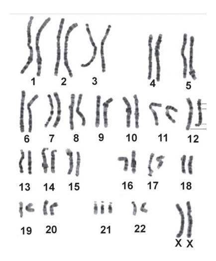
- a. The recurrence risk of the condition in the next pregnancy is _____%.
- b. If the father is a Robertsonian translocation carrier
, the recurrence risk in the next pregnancy is _____%. - c. _____% of these cases occur due to primary non-disjunction.
Answer
- a. 1% (or maternal age-specific risk, whichever is higher, for free trisomy 21).
- b. 1% to 2% (or 2.5% when father is carrier; if mother is carrier of
, recurrence risk is 10–15%; if either parent carries , recurrence risk is 100%). - c. 95% of Down syndrome cases result from maternal meiotic non-disjunction (
Robertsonian translocation, mosaicism).
VVK05-020
A 14-year-old boy is brought with history of poor scholastic performance, poor social interaction with family and peers, and hyperactive behavior. Clinically, he has large prominent ears, a long face with prominent mandible, high-arched palate, and testicular volume of 36 cc bilaterally (macroorchidism). IQ testing shows his IQ to be 75.
- a. What is the most probable diagnosis?
- b. What is the diagnostic test for the condition?
- c. Name the chromosomal site and gene defect in this individual.
Answer
- a. Fragile X Syndrome (Martin-Bell Syndrome).
- b. Molecular genetic testing: PCR and Southern blot analysis for FMR1 CGG trinucleotide repeat expansion (>200 repeats = full mutation).
- c. Chromosomal locus: Xq27.3 (FMR1 gene on the long arm of the X chromosome, causing transcriptional silencing and loss of FMRP protein).
VVK05-021
Match the mechanisms of abnormal morphogenesis with their definitions:
- a. Malformation
- b. Deformation
- c. Disruption
- d. Dysplasia
Definitions:
- Mechanical / extrinsic physical forces that distort an intrinsically normal tissue structure
- Abnormal organization of cells into tissues and morphologic result (histogenesis defect)
- Primary intrinsic structural defect of an organ or body part resulting from intrinsically abnormal developmental process
- Extrinsic breakdown or interference with an originally normal developmental process (in utero tissue destruction)
Answer
- a. Malformation
3. Primary intrinsic developmental arrest/defect (e.g., ventricular septal defect, cleft lip). - b. Deformation
1. Extrinsic mechanical force distorting normal tissue (e.g., oligohydramnios causing Potter sequence, uterine constraint causing clubfoot). - c. Disruption
4. Destruction/breakdown of previously normal tissue (e.g., amniotic band syndrome causing digital amputations). - d. Dysplasia
2. Abnormal cellular architecture/histogenesis throughout a tissue (e.g., skeletal dysplasia, polycystic kidney disease).
VVK05-022
Match the clinical terms with their prototype examples:
Terms:
- a. Syndrome
- b. Sequence
- c. Association
Prototypes:
- Trisomy 21
- Pierre Robin
- VACTERL
Answer
- a. Syndrome
Trisomy 21 (Down syndrome) / Fetal alcohol syndrome - b. Sequence
Pierre Robin sequence / Potter sequence - c. Association
VACTERL association / CHARGE association
Definitions:
- Syndrome: A recognizable pattern of multiple congenital anomalies causally related by a single defined etiology.
- Sequence: A cascade of secondary and tertiary structural anomalies all arising from a single primary localized embryonic defect (e.g., Micrognathia
Glossoptosis Cleft palate in Pierre Robin). - Association: A non-random statistical co-occurrence of multiple congenital anomalies not explainable by a single sequence or syndrome (Vertebral, Anal, Cardiac, Tracheoesophageal, Renal, Limb).
VVK05-023
A 15-year-old male child was brought to pediatric OPD for evaluation of small testes and underdeveloped secondary sex characters. On detailed physical examination, he was found to have prominent breasts bilaterally (gynecomastia). He was suspected to have a chromosomal anomaly and a karyotype was done, as shown below:
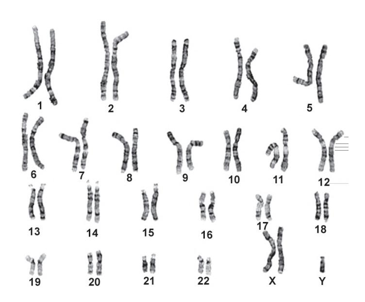
- a. What is your diagnosis?
- b. Give three salient features of this syndrome.
- c. What is the effect on stature in this condition?
Answer
- a. Klinefelter Syndrome (
). - b. Salient features:
-
- Small, firm testes (
) with hypergonadotropic hypogonadism (infertility/azoospermia).
- Small, firm testes (
-
- Bilateral gynecomastia (increased risk of male breast carcinoma).
-
- Sparse facial/body hair and female escutcheon.
-
- Learning disabilities, speech delay, psychosocial and behavioral challenges.
-
- c. Tall stature with eunuchoid proportions (disproportionately long legs, decreased Upper Segment to Lower Segment ratio, and arm span exceeding height).
VVK05-024
A lady has come to the OPD. Her husband has hemophilia A. There is no history of hemophilia in her family. She is now pregnant and wants genetic counseling. Answer the following questions:
- a. Is her son at risk for hemophilia?
- b. Her daughter marries a normal man. What are the chances of hemophilia occurring in their children?
- c. How will you offer prenatal diagnosis for hemophilia in the first trimester?
Answer
- a. No (An affected father passes his Y chromosome to his sons; all sons are completely normal and cannot inherit hemophilia from him).
- b. The daughter is an obligate carrier (
). When she marries a normal man: - Each son has a 50% risk of having hemophilia.
- Each daughter has a 50% chance of being a carrier (none will be affected).
- Overall risk of an affected child per pregnancy is 25%.
- c. First-trimester prenatal diagnosis: Chorionic Villus Sampling (CVS) at 10–13 weeks of gestation followed by fetal DNA mutation analysis (e.g., Factor VIII gene inversion/mutation testing via PCR).
VVK05-025
Answer the following questions regarding Turner syndrome:
- a. What is the characteristic physical external neonatal manifestation?
- b. Name 2 characteristic cardiac anomalies in Turner syndrome.
- c. Name 2 endocrine problems in Turner syndrome.
- d. Which GI problems can occur?
- e. Name 3 hormones used in treatment of Turner syndrome.
- f. What are the most common skeletal abnormalities?
Answer
- a. Lymphedema of the dorsum of hands and feet, redundant loose nuchal skin folds (pterygium colli / webbed neck), and low posterior hairline.
- b. Cardiac: Bicuspid aortic valve and Coarctation of the aorta.
- c. Endocrine: Primary ovarian insufficiency (hypergonadotropic hypogonadism), Autoimmune Hashimoto's hypothyroidism, and Type 2 diabetes mellitus.
- d. GI: Celiac disease, inflammatory bowel disease, and gastrointestinal telangiectasias/bleeding.
- e. Hormones:
-
- Recombinant human Growth Hormone (rhGH)
-
- Estrogen (low-dose 17
-estradiol)
- Estrogen (low-dose 17
-
- Progesterone (cyclical after 2 years of estrogen)
-
- Levothyroxine (if hypothyroid)
-
- f. Skeletal: Short 4th metacarpal/metatarsal bones, cubitus valgus, Madelung deformity of wrist, high-arched palate, and scoliosis.
VVK05-026
Answer the following questions with regard to a 5-year-old male child with Down syndrome and mental retardation:
- a. Which single joint will you prefer to examine the most and why?
- b. What would you like to test as a treatable cause for worsening developmental delay?
- c. Name two blood tests you would like to do on periodic follow-up and why?
- d. Elaborate the cytogenetic nomenclature:
.
Answer
- a. Atlantoaxial joint (C1–C2): Screen for Atlantoaxial Instability (AAI) (anterior atlanto-dens interval
on cervical flexion-extension radiographs) because hyperlaxity risks subluxation, spinal cord compression, quadriplegia, and death. - b. Thyroid function tests (Free T4 and TSH) to screen for treatable acquired autoimmune hypothyroidism.
- c.
-
- Complete Blood Count with peripheral smear: surveillance for transient myeloproliferative disorder (TMD), acute megakaryoblastic leukemia (AML-M7), and B-ALL.
-
- TSH and anti-tTG IgA serology: annual surveillance for hypothyroidism and celiac disease.
-
- d. Cytogenetic elaboration:
- 46: Total number of chromosomes.
- XY: Sex chromosomes (genetically male).
- t: Reciprocal translocation.
- (4;8): Chromosomes involved in the translocation (chromosome 4 and chromosome 8).
- (p21;q22): Specific breakpoints on the chromosomes: band 21 on short arm (p) of chromosome 4, and band 22 on long arm (q) of chromosome 8.
VVK05-027
A 2-year-old girl is brought to the clinic with headache, vomiting, and pallor. Her blood pressure is 130/80 mm Hg. On physical examination, she is noted to have aniridia and a large abdominal mass. Abdominal scanning reveals a poorly vascularized tumor in the upper pole of the right kidney.
- a. What is your most likely diagnosis?
- b. What is the chromosomal defect seen in this condition?
- c. What other abnormalities are associated with this condition?
- d. Name 2 other syndromes associated with Wilms tumor.
Answer
- a. WAGR Syndrome (Wilms tumor, Aniridia, Genitourinary anomalies, and Range of developmental delays).
- b. Contiguous gene deletion / microdeletion at 11p13 (involving contiguous loss of PAX6 and WT1 genes).
- c. Components of WAGR:
- W: Wilms tumor (nephroblastoma).
- A: Aniridia (congenital absence of iris).
- G: Genitourinary anomalies (cryptorchidism, ambiguous genitalia, streak ovaries).
- R: Retardation (intellectual disability / developmental delay).
- d. Other Wilms tumor syndromes:
-
- Beckwith-Wiedemann Syndrome (BWS) (11p15.5)
-
- Denys-Drash Syndrome (DDS) (WT1 point mutation: Wilms tumor, diffuse mesangial sclerosis, male pseudohermaphroditism)
-
- Simpson-Golabi-Behmel syndrome / Perlman syndrome.
-
VVK05-028
Study the photograph of this child whose X-ray of one hand is shown:
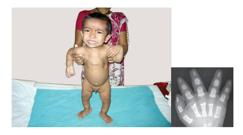
- a. What is your likely diagnosis?
- b. What inheritance pattern does this disease follow?
- c. What is the average IQ in these children?
- d. What complications of the vertebral column are likely to occur in this condition?
- e. What is this characteristic shortening of limbs called?
- f. Where is the abnormal gene in this condition located?
Answer
- a. Achondroplasia.
- b. Autosomal Dominant (approx. 80% arise as de novo mutations associated with advanced paternal age).
- c. Normal IQ / normal intelligence.
- d. Vertebral complications:
- Infancy: Foramen magnum stenosis leading to cervicomedullary compression, central sleep apnea, hypotonia, and sudden infant death.
- Childhood/Adulthood: Lumbar spinal canal stenosis (due to narrowing interpedicular distances) causing neurogenic claudication, root compression, paraparesis, and thoracolumbar kyphosis.
- e. Rhizomelic micromelia (proximal limb segment shortening: humerus and femur shorter than forearms and shins).
- f. Chromosome 4p16.3 (FGFR3 gene: Fibroblast Growth Factor Receptor 3; >98% due to G380R point mutation).
VVK05-029
Match the genetic concepts with their descriptions:
Concepts:
- a. Germline mosaicism
- b. Reduced penetrance
- c. Variable expressivity
- d. Anticipation
- e. Non-paternity
Descriptions:
- Some individuals who inherit the pathogenic genotype do not manifest the phenotypic features of the disease
- Development of progressively earlier onset and more severe disease expression through successive generations
- Mutation that affects a clone of germ cells in one parent, causing recurrence in multiple offspring despite unaffected parents
- Genetic discrepancy where the biological father is not the presumed parent
- Individuals carrying the same gene mutation exhibit varying severity and clinical manifestations of the phenotype
Answer
- a. Germline mosaicism
3. Mutation in germline progenitor cells of an unaffected parent (causes disease recurrence in siblings, e.g., Osteogenesis imperfecta, DMD). - b. Reduced penetrance
1. Proportion of individuals with a genotype who express the phenotype (e.g., Retinoblastoma with 90% penetrance). - c. Variable expressivity
5. Degree of clinical severity differs among individuals with the same genotype (e.g., Neurofibromatosis type 1, Tuberous sclerosis). - d. Anticipation
2. Earlier onset and increased severity in successive generations due to unstable trinucleotide repeat expansion (e.g., Huntington's, Myotonic dystrophy). - e. Non-paternity
4. Apparent genetic conflict arising because the assumed father is not the biological parent.
VVK05-030
Match the trinucleotide repeat expansion disorder with its abnormal repeat motif:
| Disease | Repeat Sequence |
|---|---|
| a. Myotonic Dystrophy (DM1) | GAA |
| b. Friedreich's Ataxia | CGG |
| c. Fragile X Syndrome | CTG |
| d. Huntington's Disease | CAG |
Answer
| Disease | Gene / Location | Repeat Sequence |
|---|---|---|
| a. Myotonic Dystrophy (DM1) | DMPK (3' UTR) | CTG |
| b. Friedreich's Ataxia | FXN (Intron 1) | GAA |
| c. Fragile X Syndrome | FMR1 (5' UTR) | CGG |
| d. Huntington's Disease | HTT (Exon 1 / Polyglutamine) | CAG |
VVK05-031
Given below is a karyotype of a 1-month-old child who presented with a characteristic high-pitched sound produced during crying:
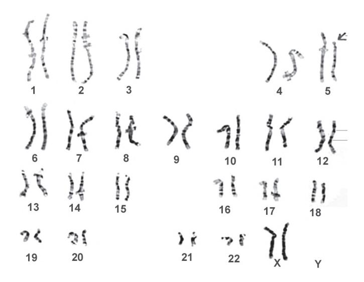
- a. Identify the condition and the sex of the child.
- b. What is the characteristic presentation in a neonate?
- c. What is the life expectancy in this condition?
- d. What is the chromosomal defect in this condition?
- e. What is the parental origin of the abnormal chromosome in the majority of cases?
Answer
- a. Cri-du-chat Syndrome (5p- syndrome), Female (
). - b. High-pitched, kitten-like / cat-like cry (due to laryngeal hypoplasia and abnormal vocal cord development), severe microcephaly, round "moon-like" face, hypertelorism, epicanthic folds, low-set ears, micrognathia, hypotonia, and severe intellectual disability.
- c. Most patients survive into adulthood with supportive medical care (normal or near-normal life expectancy).
- d. Terminal or interstitial deletion of the short arm of chromosome 5 (5p15).
- e. In approximately 80% to 85% of cases, the deleted chromosome is of paternal origin (de novo during spermatogenesis).
VVK05-032
This male child aged 10 months has been brought with a large tongue. He was born macrosomic weighing 4.2 kg and had no perinatal or subsequent complication:
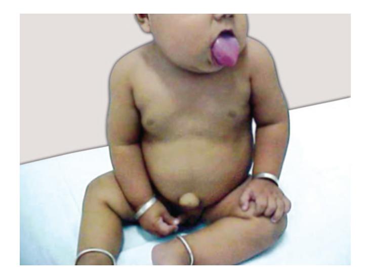
- a. What is the most probable diagnosis?
- b. What are the classical clinical triad/components of this syndrome?
- c. Which chromosome region carries the genes responsible for this disorder?
- d. What embryonic tumors is this child predisposed to?
Answer
- a. Beckwith-Wiedemann Syndrome (BWS) (Overgrowth syndrome).
- b. Classical components:
-
- Macroglossia (large protruding tongue).
-
- Macrosomia / Somatic overgrowth and Hemihypertrophy (asymmetric growth of limbs/body).
-
- Abdominal wall defects (Omphalocele, umbilical hernia, diastasis recti).
-
- Neonatal hypoglycemia (due to pancreatic islet cell hyperplasia / hyperinsulinism).
-
- Anterior ear lobe creases and posterior helical indentations/pits.
-
- c. Chromosome 11p15.5 (imprinting center defects involving IGF2, H19, and CDKN1C genes).
- d. Embryonal pediatric tumors:
-
- Wilms tumor (Nephroblastoma) (most common)
-
- Hepatoblastoma
-
- Adrenocortical carcinoma, Neuroblastoma, Rhabdomyosarcoma.
(Requires abdominal USG every 3 months until age 8 and serum alpha-fetoprotein every 3 months until age 4).
- Adrenocortical carcinoma, Neuroblastoma, Rhabdomyosarcoma.
-