MNJ05-001
Identify the anatomical structures labeled A to K in the neuroanatomy and brain imaging sections below:
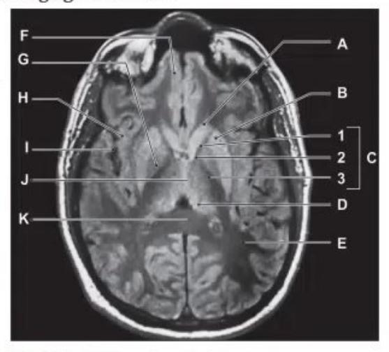
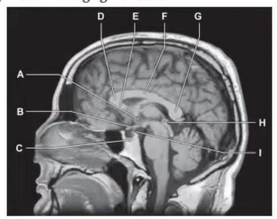
- Identify the anatomical landmarks and structures labeled on the CT/MRI brain sections.
Answer
- Anterior Horn of Lateral Ventricle
- Caudate Nucleus (Head)
- Internal Capsule (Anterior limb, Genu, Posterior limb)
- Lentiform Nucleus (Putamen and Globus Pallidus)
- Thalamus
- Third Ventricle
- Corpus Callosum (Genu and Splenium)
- Sylvian Fissure / Insular Cortex
- Occipital Horn / Trigone of Lateral Ventricle
- Fourth Ventricle and Cerebellar Hemispheres
- Pons / Brainstem
MNJ05-002
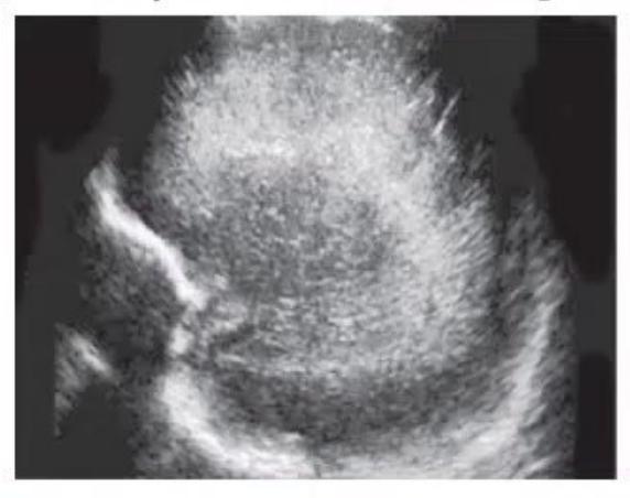
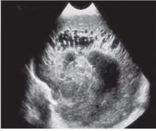
Review the neonatal cranial ultrasound images showing Periventricular Leukomalacia (PVL):
- Identify the imaging planes and grade of PVL shown in the sagittal views.
- State the de Vries ultrasound grading system for PVL.
Answer
- Parasagittal and coronal cranial ultrasound views showing Periventricular Leukomalacia (PVL): Grade 1 (prolonged periventricular flares) and Grade 3 (extensive cystic changes in periventricular white matter).
- de Vries Ultrasound Classification of PVL:
- Grade I: Prolonged periventricular echogenicity / flaring persisting for ≥ 7 days.
- Grade II: Localized periventricular cysts developing in the frontoparietal white matter.
- Grade III: Extensive periventricular cystic lesions extending to frontoparietal and occipital white matter.
- Grade IV: Extensive subcortical white matter cystic lesions involving deep and peripheral parenchyma.
MNJ05-003
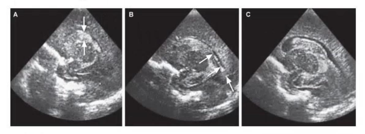
Review the cranial ultrasound images of a preterm newborn below:
- What are the major risk factors for intraventricular / germinal matrix hemorrhage (IVH/GMH)?
- State the Papile (CT) and Volpe (Ultrasound) grading criteria.
- What are the key preventive strategies?
- What are the possible long-term neurological complications in a preterm infant?
- Grade the IVH seen in these ultrasound images.
Answer
- Risk factors:
- Extreme prematurity (<32 weeks) and very low birth weight (<1500 g)
- Fluctuating cerebral blood flow (hypotension, rapid volume expansion/boluses, rapid sodium bicarbonate infusion)
- Severe respiratory distress syndrome, pneumothorax, hypoxia, hypercapnia
- Sepsis, acidosis, coagulopathy
- Grading Systems:
- Papile Grading (CT):
- Grade I: Subependymal / Germinal matrix hemorrhage alone.
- Grade II: Intraventricular hemorrhage without ventricular dilatation (fills <50% of lateral ventricle).
- Grade III: Intraventricular hemorrhage with ventricular dilatation (fills ≥50% of lateral ventricle).
- Grade IV: Intraventricular hemorrhage with parenchymal hemorrhage / hemorrhagic infarction.
- Volpe Grading (Cranial USG):
- Grade 1: Germinal matrix hemorrhage with minimal/no IVH (<10% ventricular area).
- Grade 2: IVH occupying 10–50% of ventricular area.
- Grade 3: IVH occupying >50% of ventricular area with acute ventricular distension.
- Periventricular Hemorrhagic Infarction (PVHI / IPE): Concomitant venous intraparenchymal echodensity.
- Papile Grading (CT):
- Preventive strategies:
- Antenatal corticosteroids in threatened preterm labor
- Gentle resuscitation and avoid rapid volume/bicarbonate boluses
- Synchronized gentle ventilation to avoid fluctuating cerebral venous pressures
- Minimal handling and neutral head positioning in the first 72 hours of life
- Complications: Post-hemorrhagic ventricular dilatation (PHVD), obstructive hydrocephalus, cerebral palsy (spastic diplegia/quadriplegia), developmental delay, epilepsy.
- Grading of Images:
- Grade I: Echogenic clot localized to the caudothalamic groove.
- Grade II: Clot in the dependent portion of the lateral ventricle without dilatation.
- Grade III: Large distending echogenic cast filling and ballooning the entire lateral ventricle.
MNJ05-004
Diagnose the two conditions shown on brain MRI (Image A and Image B) and outline their specific medical management:
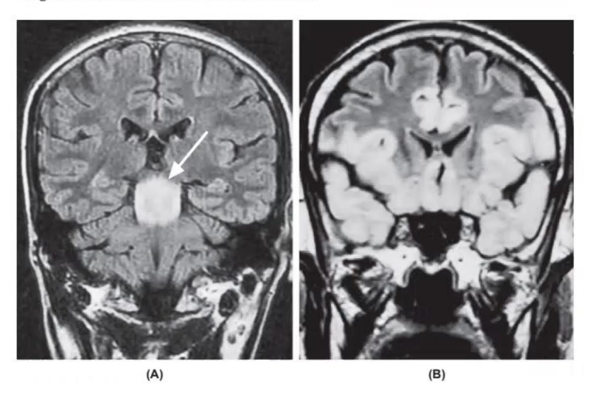
- Identify the condition in Image A and state the treatment.
- Identify the condition in Image B and state the treatment.
Answer
- Image A: Japanese Encephalitis (JE)
- Imaging: Characteristic bilateral symmetrical hyperintensity in the thalamus, basal ganglia, and midbrain/brainstem on T2/FLAIR.
- Treatment: Supportive care (airway protection, control of raised intracranial pressure with mannitol/hypertonic saline, seizure management, fluid and electrolyte maintenance; no specific antiviral therapy).
- Image B: Herpes Simplex Encephalitis (HSE)
- Imaging: Asymmetric hyperintensity and edema involving the temporal lobes, inferior frontal lobes, and insular cortex on T2/FLAIR with gyral enhancement.
- Treatment: Intravenous Acyclovir (10–15 mg/kg/dose or 500 mg/m²/dose IV every 8 hours) for 14–21 days.
MNJ05-005
An 8-year-old child presents with acute onset of seizures, multiple cranial nerve palsies, dysarthria, and dysphagia following a febrile viral illness 2 weeks prior:
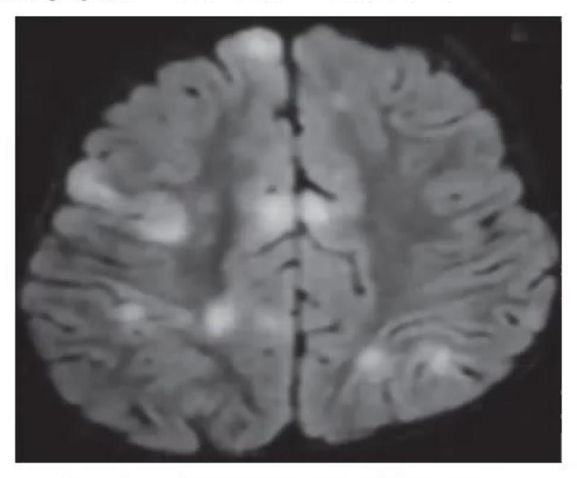
- Identify the condition from the clinical presentation and brain MRI.
- What is the treatment and expected prognosis?
Answer
- Diagnosis: Acute Disseminated Encephalomyelitis (ADEM) (showing widespread, asymmetric, poorly demarcated T2/FLAIR hyperintense demyelinating lesions involving the deep white matter, subcortical white matter, brainstem, and basal ganglia).
- Treatment & Prognosis:
- Treatment: High-dose intravenous Methylprednisolone (30 mg/kg/day, max 1000 mg/day) for 3–5 days, followed by oral prednisolone taper over 2–4 weeks. Refractory cases: IVIG (2 g/kg over 2 days) or plasmapheresis (plasma exchange).
- Prognosis: Generally good; complete neurological recovery in 70–80% of children following a monophasic course.
MNJ05-006
Review the cerebral angiogram / MRI below:
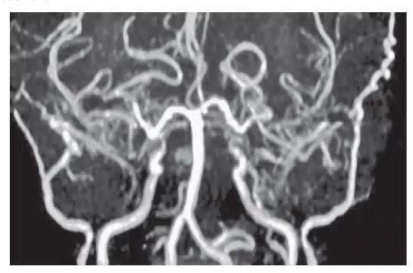
- Diagnose the condition shown in the image.
- List 4 predisposing / associated risk factors.
- Describe the typical clinical presentation.
- What is the definitive surgical treatment?
Answer
- Diagnosis: Moyamoya Disease / Moyamoya Syndrome (progressive stenosis of the terminal internal carotid arteries and circle of Willis with characteristic "puff of smoke" collateral vessel network).
- Associated conditions:
- Down syndrome (Trisomy 21)
- Neurofibromatosis type 1 (NF-1)
- Sickle cell disease
- Post-radiation vasculopathy
- Asian descent (familial RNF213 gene mutation)
- Clinical presentation: Recurrent transient ischemic attacks (TIAs) or ischemic strokes triggered by crying, hyperventilation, dehydration, or coughing; alternating hemiparesis, headaches, seizures, chorea, cognitive decline.
- Surgical management: Cerebral revascularization procedures:
- Direct bypass: Superficial Temporal Artery to Middle Cerebral Artery (STA-MCA) anastomosis.
- Indirect bypass: Encephaloduroarteriosynangiosis (EDAS) or Encephalomyosynangiosis (EMS).
MNJ05-007
CT scan of an 8-year-old child:
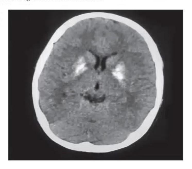
- Diagnose the abnormality seen on this non-contrast CT scan.
- Give 4 differential diagnoses / etiologies of this condition in children.
Answer
- Diagnosis: Bilateral symmetrical Basal Ganglia Calcification (hyperdense calcification involving globus pallidus, putamen, and caudate nuclei).
- Differential diagnoses / Causes:
- Idiopathic basal ganglia calcification (Fahr disease)
- Hypoparathyroidism and Pseudohypoparathyroidism
- Congenital TORCH infections (Toxoplasmosis, CMV, Congenital HIV)
- Mitochondrial disorders (MELAS, Leigh syndrome, Kearns-Sayre syndrome)
- Post-infectious / post-inflammatory (Tuberculous meningitis, Neurocysticercosis)
- Carbon monoxide or lead poisoning
MNJ05-008
Review the two brain MRI scans below:
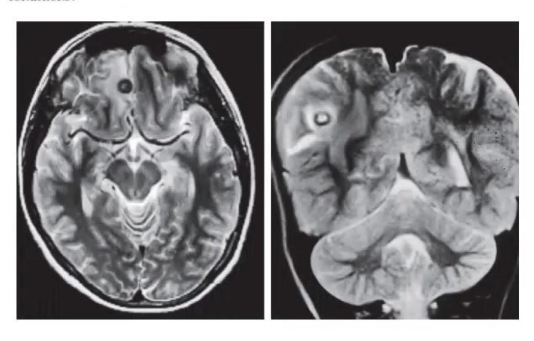
- What is this imaging modality / sequence?
- Identify the lesions in Image A and Image B, and explain how to differentiate them on MRI.
Answer
- Modality: T2-weighted / FLAIR and Contrast-Enhanced T1-weighted Magnetic Resonance Imaging (MRI).
- Differentiation:
- Image A (Tuberculoma): Solid granulatous lesions with central hypointensity on T2-weighted MRI, thick nodular or ring enhancement on T1+contrast, and marked perilesional edema.
- Image B (Neurocysticercosis - NCC): Cystic lesions with thin smooth ring enhancement; T2W hyperintense cyst fluid with an eccentric hypointense mural nodule representing the scolex ("hole-with-dot" sign).
MNJ05-009
Diagnose the four leukodystrophies shown in A, B, C, and D based on their characteristic white matter involvement:
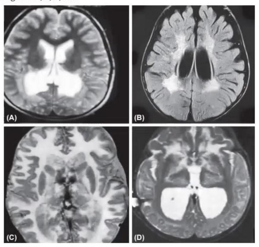
- Identify leukodystrophy A.
- Identify leukodystrophy B.
- Identify leukodystrophy C.
- Identify leukodystrophy D.
Answer
- A: X-linked Adrenoleukodystrophy (X-ALD): Confluent, symmetric demyelination in the parieto-occipital white matter and splenium of the corpus callosum with leading-edge intermediate rim enhancement.
- B: Metachromatic Leukodystrophy (MLD): Symmetrical confluent periventricular white matter demyelination with sparing of subcortical U-fibers, showing a characteristic tigroid / leopard-skin pattern.
- C: Canavan Disease: Diffuse, bilateral, symmetrical white matter involvement with prominent early involvement of subcortical U-fibers and macrocephaly (markedly elevated NAA peak on MR spectroscopy).
- D: Alexander Disease: Symmetrical white matter demyelination with frontal lobe predominance, frontal cystic degeneration, basal ganglia involvement, and macrocephaly (GFAP mutation).
MNJ05-010
Review the contrast-enhanced CT brain scans of a 6-year-old child with cyanotic congenital heart disease:
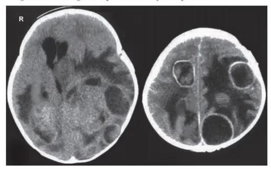

- Interpret the CT brain findings.
- What are the predisposing factors for this condition in pediatric patients (mention at least 5)?
- What long-term neurological sequelae can occur in this child?
Answer
- CT findings: Multiple well-defined, thin ring-enhancing lesions with surrounding vasogenic perilesional edema and mass effect, consistent with Multiple Brain Abscesses.
- Predisposing factors:
- Cyanotic congenital heart disease with right-to-left shunt (e.g., Tetralogy of Fallot - loss of pulmonary capillary filtering)
- Contiguous focus of infection: Chronic suppurative otitis media (CSOM), mastoiditis, paranasal sinusitis, orbital cellulitis, dental infection
- Penetrating head trauma or neurosurgical procedures (VP shunt infection)
- Immunodeficiency (HIV, severe combined immunodeficiency, post-chemotherapy neutropenia)
- Infective endocarditis with septic emboli
- Long-term sequelae: Focal neurological deficits (hemiparesis, cranial nerve palsies), secondary epilepsy / seizure disorder, hydrocephalus, cognitive impairment, learning disabilities.
MNJ05-011
Review the T2-weighted brain MRI scan below:
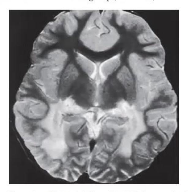
- Identify the imaging sequence and anatomical level.
- Give the diagnosis based on the distribution of white matter hyperintensity.
- Mention two other peroxisomal / metabolic disorders from the same group.
Answer
- Sequence & Level: Axial T2-weighted MRI at the level of the basal ganglia, internal capsules, and lateral ventricles.
- Diagnosis: X-linked Adrenoleukodystrophy (X-ALD) (showing confluent, symmetric T2 hyperintensity predominantly in the parieto-occipital periventricular white matter and splenium of the corpus callosum).
- Other peroxisomal disorders:
- Zellweger syndrome spectrum (Zellweger syndrome, Neonatal Adrenoleukodystrophy, Infantile Refsum disease)
- Refsum disease (phytanic acid storage disease)
- Rhizomelic chondrodysplasia punctata (RCDP)
MNJ05-012
A 10-month-old infant is brought with complaints of rapidly increasing head size, developmental delay, and a prominent occiput:
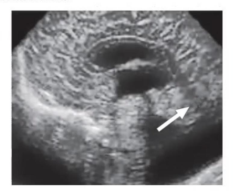
- Mention the abnormalities noted on these neuroimaging studies.
- Give your diagnosis.
- Mention associated anomalies.
Answer
- Abnormalities:
- Massive cystic dilatation of the fourth ventricle ballooning into the posterior fossa.
- Complete or partial agenesis/hypoplasia of the cerebellar vermis.
- Upward displacement of the tentorium cerebelli and torcula (high torcular Herophili).
- Enlarged posterior fossa with secondary non-communicating hydrocephalus (dilated third and lateral ventricles).
- Diagnosis: Dandy-Walker Malformation.
- Associated anomalies: Agenesis of corpus callosum, polymicrogyria, schizencephaly, holoprosencephaly, congenital heart disease, cleft palate, polycystic kidneys.
MNJ05-013
Identify the cortical and developmental brain malformations shown in Image A and Image B:
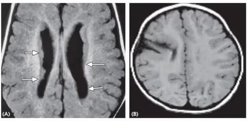
- Identify malformation in Image A.
- Identify malformation in Image B.
Answer
- Image A: Grey Matter Heterotopia (nodular or band-like collections of normal neurons arrested along the periventricular ependymal lining or in subcortical white matter, isointense to cortex on all MRI sequences).
- Image B: Schizencephaly (full-thickness CSF-filled cleft extending from the ependymal wall of the lateral ventricle to the pial surface of the cerebral cortex, lined by dysplastic/polymicrogyric grey matter; open-lip or closed-lip type).
MNJ05-014
A 10-year-old male child presents with recurrent falls during walking, gait ataxia, and worsening early morning headache with vomiting for 3 months:
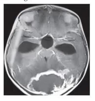
- Diagnose the condition given in the MRI.
- Outline the treatment plan.
Answer
- Diagnosis: Medulloblastoma (high-grade embryonal tumor presenting as a contrast-enhancing mass arising from the cerebellar vermis in the roof of the 4th ventricle, causing obstructive hydrocephalus).
- Treatment:
- Emergency management of raised intracranial pressure / hydrocephalus (corticosteroids, temporary EVD or VP shunt).
- Maximal safe surgical resection / craniotomy.
- Craniospinal radiation therapy (in children >3 years of age) plus adjuvant combination chemotherapy (e.g., cisplatin, vincristine, cyclophosphamide).
MNJ05-015
Review the neuroimaging studies showing brain malformations below:
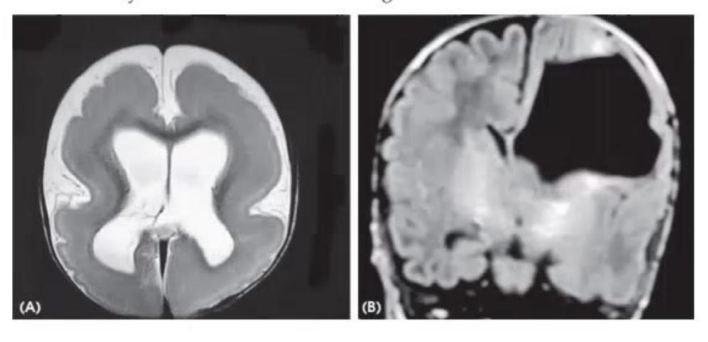

- Identify the brain abnormalities in Image A and Image B.
- Name one genetic syndrome associated with Image A and state its key clinical features.
Answer
- Identifications:
- Image A: Lissencephaly (Smooth brain / Agyria-Pachygyria): Loss of normal cerebral sulcation and gyration with thickened cortex and a "figure-of-8" / hourglass appearance of the brain.
- Image B: Porencephalic Cyst: A focal, fluid-filled cavity within the cerebral parenchyma that communicates with the ventricular system, typically secondary to perinatal vascular occlusion, hemorrhage, or trauma.
- Syndrome for Image A: Miller-Dieker Syndrome (17p13.3 microdeletion involving LIS1 gene).
- Features: Severe lissencephaly (Grade 1), distinctive facial dysmorphism (prominent forehead, bitemporal hollowing, upturned nose with anteverted nares, micrognathia, thin vermilion of upper lip), severe infantile spasms/epilepsy, profound developmental delay, and early mortality.
MNJ05-016
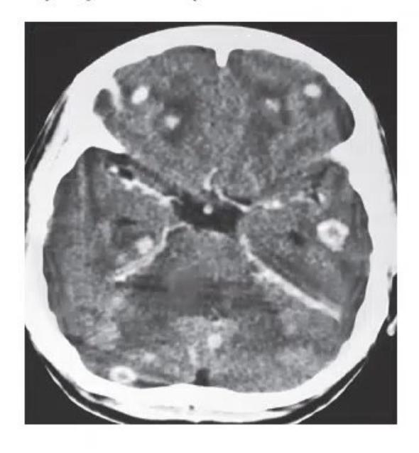
A 9-year-old child presents with new-onset focal seizures with secondary generalization. Brain CT/MRI is shown below:
- What is the diagnosis?
- Describe the stepwise management protocol and mandatory preliminary investigations.
- How long should antiepileptic drug therapy be continued?
- What preventive advice should you provide to the family?
Answer
- Diagnosis: Neurocysticercosis (NCC) (multiple parenchymal cysticerci in various stages: colloidal vesicular and granular nodular stages with perilesional edema).
- Stepwise Management:
- Assess and manage acute seizures and raised intracranial pressure.
- Initiate antiepileptic drugs (e.g., Levetiracetam, Carbamazepine, or Valproate).
- Mandatory Preliminary Investigation: Fundoscopic / ophthalmologic examination to rule out ocular/intraretinal cysticercosis (cysticidal therapy can cause severe intraocular inflammation and blindness) and CT/MRI spine if indicated.
- Steroid Coverage: Start systemic corticosteroids (Prednisolone 1–2 mg/kg/day or Dexamethasone) for 2 days prior to cysticidal therapy and continue throughout the course to suppress the host inflammatory response to dying parasites.
- Cysticidal Therapy: Start Albendazole (15 mg/kg/day in two divided doses for 10–14 days) ± Praziquantel (50 mg/kg/day for dual therapy in multiple viable lesions).
- Duration of AED Therapy: Continue antiepileptic therapy until complete CT/MRI resolution of active cystic lesions and edema, plus at least 6–12 months of seizure-free period (or up to 2 years if calcified residues remain).
- Preventive Advice:
- Strict personal hygiene and handwashing with soap before eating and after using the toilet.
- Thorough washing of all raw vegetables, salads, and fruits.
- Safe drinking water (boiled/filtered).
- Proper cooking of pork and meat to ensure larvae are destroyed.
- Screen household contacts for intestinal Taenia solium carriage (tapeworm carrier).