Grading System for Hydronephrosis (SFU Classification)
| Grade | Central Renal Complex | Renal Parenchymal Thickness |
|---|---|---|
| Grade 0 | Intact / No pelvicalyceal dilatation | Normal |
| Grade 1 | Slight pelvicalyceal splitting / renal pelvis dilatation only | Normal |
| Grade 2 | Evident splitting; pelvis dilated with few calyces (within renal border) | Normal |
| Grade 3 | Wide splitting; pelvis dilated outside renal border, calyces uniformly dilated | Normal |
| Grade 4 | Further severe dilatation of pelvis and calyces (ballooning) | Parenchymal thinning / loss |
MNJ31-001
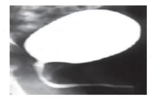
- Name the radiological investigation.
- What is the diagnosis?
- What further nuclear imaging investigations does this child need?
Answer
- Micturating Cystourethrogram (MCU / VCUG).
- Posterior Urethral Valves (PUV) (showing dilated, elongated posterior urethra with abrupt caliber change at valve level, hypertrophied bladder neck, trabeculated bladder, and secondary vesicoureteral reflux).
- Further nuclear renograms:
-DTPA or -MAG3 scan (Dynamic renogram to evaluate GFR, differential renal function, and drainage/obstruction). -DMSA scan (Static cortical scan to detect renal parenchymal scarring and dysplasia).
MNJ31-002
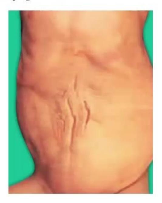
- Give the diagnosis of this condition.
- Give four important clinical components.
- State the most important prognostic factors.
Answer
- Prune-Belly Syndrome (Eagle-Barrett Syndrome / Triad Syndrome).
- Clinical components:
- Partial or complete absence/deficiency of abdominal wall musculature (wrinkled "prune-like" abdominal wall)
- Bilateral intra-abdominal cryptorchidism (undescended testes)
- Complex urinary tract anomalies (massive bilateral hydroureteronephrosis, megacystis, dilated prostatic urethra, VUR)
- Secondary oligohydramnios-related pulmonary hypoplasia.
- Prognostic factors:
- Severity of pulmonary hypoplasia at birth (leading cause of neonatal mortality)
- Degree of underlying renal dysplasia and progression to End-Stage Renal Disease (ESRD).
MNJ31-003
- How do you determine GFR using serum creatinine by the modified Schwartz formula?
- What defines Stage 2 Chronic Kidney Disease (CKD) in terms of GFR?
- State True or False:
i. Recombinant Growth Hormone (rGH) is an approved therapy for growth failure / short stature due to CKD.
ii. Anemia in CKD is primarily caused by urinary loss of ferritin.
iii. The most common form of renal osteodystrophy in pediatric CKD is high-turnover bone disease (osteitis fibrosa).
Answer
-
Modified Schwartz Formula:
Wherevalues are: for low-birth-weight preterm infants year for full-term infants year for children and adolescent females for adolescent males (or standard bedside constant with enzymatic IDMS-traceable creatinine assay).
-
Stage 2 CKD:
with evidence of kidney damage.
| CKD Stage | Description | GFR ( |
|---|---|---|
| Stage 1 | Kidney damage with normal or |
|
| Stage 2 | Kidney damage with mild |
|
| Stage 3 | Moderate |
|
| Stage 4 | Severe |
|
| Stage 5 | Kidney Failure / ESRD |
- True/False:
i. True (rGH improves linear growth velocity in children with CKD).
ii. False (Anemia of CKD is primarily due to Erythropoietin deficiency by peritubular interstitial cells).
iii. True (Secondary hyperparathyroidism leads to high-turnover osteitis fibrosa cystica).
(Answers: i-True, ii-False, iii-True)
MNJ31-004
A 6-year-old boy Rajeev presented with periorbital puffiness and cola-colored urine. He had a history of pharyngitis 12 days back. On examination he has tachycardia, tachypnea, and BP 142/80 mmHg:
- What is the most likely clinical diagnosis?
- What investigations will you do to confirm the diagnosis?
- What is the drug class of choice for acute fluid overload and hypertension?
Answer
- Acute Post-Streptococcal Glomerulonephritis (APSGN).
- Confirmatory investigations:
- Evidence of streptococcal infection: Elevated ASO titer (post-pharyngitis) or Anti-DNase B titer (post-impetigo).
- Serum Complement: Markedly decreased Serum C3 (with normal C4); normalizes within 6–8 weeks.
- Urinalysis: Dysmorphic RBCs, microscopic/gross hematuria, RBC casts, and subnephrotic proteinuria.
- Renal function tests (elevated BUN and serum creatinine).
- First-line antihypertensive: Loop diuretics (IV Furosemide
) to manage hypervolemia, hypertension, and pulmonary edema; add Calcium Channel Blockers (Amlodipine/Nifedipine) if BP remains elevated.
MNJ31-005
A 12-year-old boy Vivek presents with 2 episodes of gross hematuria and sensorineural hearing impairment. Other systemic examination is normal:
- What is the likely diagnosis?
- Which other extrarenal manifestations are present in this condition?
- What are the patterns of genetic inheritance?
Answer
- Alport Syndrome (Hereditary Nephritis, defect in Type IV collagen
chains). - Extrarenal manifestations:
- Bilateral sensorineural hearing loss (SNHL) (high-frequency; progressive)
- Ocular abnormalities: Anterior lenticonus (pathognomonic), perimacular retinal flecks, corneal endothelial vesicles / recurrent corneal erosions
- Diffuse Leiomyomatosis of the esophagus, tracheobronchial tree, and female genitalia (in deletion involving COL4A5 and COL4A6).
- Inheritance patterns:
- X-linked dominant (85%): COL4A5 gene mutation on chromosome Xq22 (severe in males).
- Autosomal recessive (10–15%): COL4A3 or COL4A4 on chromosome 2q36.
- Autosomal dominant (~5%): Heterozygous COL4A3 or COL4A4.
MNJ31-006
A 14-month-old male child Bhanu presents with severe failure to thrive, recurrent vomiting, and polyuria. On examination he is moderately dehydrated:

- What is the most likely diagnosis?
- What are the various clinical types of this disorder?
- Give three secondary causes of this condition.
- How will you manage this child?
Answer
- Distal Renal Tubular Acidosis (Type 1 RTA / Classic dRTA) with bilateral Nephrocalcinosis (due to failure of distal
-intercalated cells to secrete ions, causing urine despite systemic metabolic acidosis). - Classification of RTA:
- Type 1 (Distal RTA): Impaired distal
excretion ( , hypokalemia, nephrocalcinosis). - Type 2 (Proximal RTA): Impaired proximal
reabsorption (hypokalemia, associated Fanconi syndrome). - Type 3 (Mixed RTA): Rare variant with combined proximal and distal defects (Carbonic anhydrase II deficiency with osteopetrosis).
- Type 4 (Hyperkalemic RTA): Aldosterone deficiency or distal tubular resistance (Hyperkalemia, urine
).
- Type 1 (Distal RTA): Impaired distal
- Secondary causes of Distal RTA:
- Autoimmune: Systemic Lupus Erythematosus (SLE), Sjögren syndrome
- Hereditary: Sickle cell nephropathy, Ehlers-Danlos syndrome, Wilson disease
- Obstructive uropathy / chronic pyelonephritis / medullary sponge kidney
- Drugs/toxins: Amphotericin B, Lithium, Toluene inhalation.
- Management:
- Oral alkali therapy: Sodium bicarbonate or Potassium citrate (Bicitra/Polycitra) at
in 3–4 divided doses to maintain normal serum bicarbonate. - Potassium supplementation (Potassium citrate prevents hypokalemia and alkalinizes urine to dissolve/prevent nephrocalcinosis).
- Adequate hydration and nutritional rehabilitation for catch-up growth.
- Oral alkali therapy: Sodium bicarbonate or Potassium citrate (Bicitra/Polycitra) at
MNJ31-007
A 6-year-old girl Megha is referred with an abdominal mass extending from the right renal angle medially to the midline. Her BP is 160/110 mmHg. Renal functions are normal, urine microscopy shows microscopic hematuria:
- What are the 2 most likely differential diagnoses?
- Explain the mechanism of hypertension in these 2 conditions.
- For each diagnosis, give one common site where metastatic lesions are found.
Answer
- Differential diagnoses: Wilms' Tumor (Nephroblastoma) and Neuroblastoma.
- Mechanism of hypertension:
- In Wilms' Tumor: Renal ischemia induced by tumoral compression of the adjacent renal parenchyma and renal artery, leading to activation of the Renin-Angiotensin-Aldosterone System (RAAS) and hyperreninism.
- In Neuroblastoma: Hypersecretion of Catecholamines (Epinephrine, Norepinephrine, and Dopamine) by tumor chromaffin neuroblasts causing systemic vasoconstriction.
- Common metastatic sites:
- For Wilms' Tumor: Lungs (most common, 85%) and liver.
- For Neuroblastoma: Cortical bones, Bone Marrow, orbit ("raccoon eyes"), and liver.
MNJ31-008
A 4-day-old newborn male baby born to Anita with a history of antenatal oligohydramnios undergoes MCU:
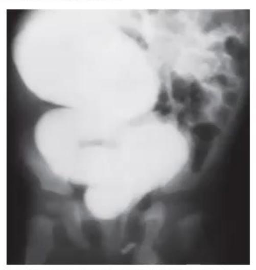
- Describe the findings in this MCU image.
- What is the absolute indication for MCU in a newborn male?
- What antenatal intervention could have been offered?
- What is the lifetime risk of developing CKD in these children?
- What are the surgical procedures available?
Answer
- MCU findings: Marked dilatation and elongation of the posterior urethra, narrow thread-like anterior urethral stream with caliber transition, hypertrophied bladder neck, heavily trabeculated "Christmas tree" bladder with multiple pseudodiverticula, and secondary high-grade Vesicoureteral Reflux (VUR).
- Indication: Suspicion of Posterior Urethral Valves (PUV) (antenatal bilateral hydronephrosis with distended bladder, poor urinary stream, or palpable bladder/renal masses).
- Antenatal intervention: Ultrasound-guided Fetal Vesicoamniotic Shunting (Harrison catheter) or fetal cystoscopy/valve ablation in select cases with oligohydramnios and preserved fetal renal cortical function.
- Risk of CKD:
of affected children progress to ESRD by adolescence/young adulthood despite successful valve ablation (due to congenital renal dysplasia and "valve bladder" syndrome). - Surgical procedures:
- Primary endoscopic transurethral Valve Ablation / Fulguration (treatment of choice)
- Cutaneous Vesicostomy (in small preterms
where scope cannot be negotiated) - High diverting ureterostomy / pyelostomy (for persistent severe upper tract obstruction).
MNJ31-009
A 2-year-old male Rahul with acute diarrheal illness develops anuria. Hb: 8.5 g/dL, platelet count: 80,000/µL, PT: 24/20 sec, aPTT: 28/30 sec:
- Describe the peripheral blood smear findings of this child.
- Mention two primary differential diagnoses.
- Which single imaging test helps differentiate these conditions?
- The baby's renal functions and hemolysis acutely deteriorated after 2 units of FFP. What specific etiology does this suggest?
- What are indications for systemic corticosteroids in this scenario?
- Which other conditions share similar microangiopathic hemolytic anemia findings?
Answer
- Microangiopathic Hemolytic Anemia (MAHA): Fragmented red blood cells (Schistocytes, helmet cells, burr cells, microspherocytes), polychromasia, and thrombocytopenia with reduced large platelets.
- Differential diagnoses: Hemolytic Uremic Syndrome (HUS) and Renal Vein Thrombosis (RVT).
- Diagnostic modality: Renal Doppler Ultrasonography (demonstrates absent flow in renal vein in RVT; intact flow in HUS).
- Streptococcus pneumoniae-associated HUS (SP-HUS / Neuraminidase-induced HUS): Bacterial neuraminidase cleaves sialic acid from RBC/platelet/glomerular membranes, exposing the hidden Thomsen-Friedenreich (T) antigen. Transfusion of plasma (FFP) containing normal donor anti-T IgM antibodies triggers massive complement-mediated hemolysis and worsening renal failure.
- Indications for corticosteroids: Not indicated in typical STEC-D+ HUS; used only in atypical HUS associated with autoimmune diseases (SLE, secondary vasculitis).
- Other MAHA conditions: Thrombotic Thrombocytopenic Purpura (TTP), Disseminated Intravascular Coagulation (DIC), Malignant hypertension, Systemic sclerosis (renal crisis).
MNJ31-010
An 8-year-old boy Bhushan presents with fever, epistaxis, red eyes, recurrent hemoptysis, breathlessness, hoarseness of voice (subglottic stenosis), and hematuria:
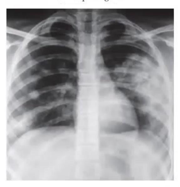
- What is the clinical diagnosis?
- Name the confirmatory serological laboratory test for this disease.
- Write 2 major immunosuppressive treatment options.
- Name 2 other small-vessel vasculitides with similar pathogenesis (ANCA-associated vasculitides).
Answer
- Granulomatosis with Polyangiitis (GPA / Wegener's Granulomatosis).
- Serological marker: c-ANCA (Cytoplasmic antineutrophil cytoplasmic antibody) / Anti-Proteinase 3 (Anti-PR3) antibodies (positive in
). - Induction treatment options:
- IV Pulse Methylprednisolone followed by oral Prednisolone PLUS Rituximab (Anti-CD20 monoclonal antibody) OR
- High-dose Corticosteroids PLUS IV / Oral Cyclophosphamide
- Therapeutic Plasma Exchange (Plasmapheresis) in severe rapidly progressive crescentic glomerulonephritis or diffuse alveolar hemorrhage.
- Other ANCA-associated vasculitides:
- Microscopic Polyangiitis (MPA - p-ANCA / anti-MPO)
- Eosinophilic Granulomatosis with Polyangiitis (EGPA / Churg-Strauss syndrome).
MNJ31-011
An infant born to a diabetic mother presents with gross hematuria, oliguria/anuria, and bilaterally enlarged palpable flank masses. Investigations: Hb 22.5 g/dL, Hematocrit 72%, Platelet count 70,000/µL:
- What is the clinical diagnosis?
- List 3 important differential diagnoses.
- What is the definitive imaging modality?
- Outline the management principles.
- What are long-term complications?
Answer
- Neonatal Renal Vein Thrombosis (RVT) (precipitated by polycythemia, hyperviscosity, dehydration, and infant of diabetic mother status).
- Differential diagnoses:
- Congenital hydronephrosis / PUV
- Autosomal Recessive Polycystic Kidney Disease (ARPKD)
- Bilateral Wilms' tumor / Congenital mesoblastic nephroma
- Hemolytic Uremic Syndrome (HUS).
- Diagnostic modality: Renal Color Doppler Ultrasonography (demonstrates absence of venous Doppler signal in main renal vein, reversal of diastolic arterial flow, and enlarged echogenic kidney).
- Management:
- Aggressive correction of dehydration, electrolyte imbalance, and partial exchange transfusion if hematocrit
. - Systemic anticoagulation with Low-Molecular-Weight Heparin (LMWH - Enoxaparin) or unfractionated heparin (for bilateral RVT or extension into IVC).
- Thrombolysis (rTPA) reserved for bilateral occlusion with acute anuric renal failure.
- Aggressive correction of dehydration, electrolyte imbalance, and partial exchange transfusion if hematocrit
- Long-term complications: Renal parenchymal atrophy / scarring, chronic hypertension, renal tubular dysfunction, and CKD.
MNJ31-012
A child Monu presents with muscle cramps for 7 days. Serum Magnesium is 1.2 mg/dL, Potassium is 1.6 mEq/L, and Serum Bicarbonate is 42 mEq/L. Blood pressure is normal and there is no dehydration:
- What electrolyte in urine will you estimate to confirm the diagnosis?
- Why?
- The urinary electrolyte estimated is low. List 3 causes of hypokalemic metabolic alkalosis with normal BP.
- There is no history of drug ingestion, vomiting, or failure to thrive. What is the diagnosis?
- Name 3 pharmacological agents used in treatment.
- What will be the plasma renin and aldosterone levels?
- What is the urinary calcium excretion level?
Answer
- 24-hour Urinary Calcium (or Spot Urine Calcium-to-Creatinine ratio) and Urinary Chloride.
- Why: To differentiate Gitelman syndrome (Hypocalciuria, Urine
) from Bartter syndrome (Hypercalciuria, Urine ) and surreptitious diuretic abuse. - Causes of hypokalemic metabolic alkalosis with normal BP:
- Gitelman syndrome
- Bartter syndrome
- Surreptitious vomiting / bulimia (low urine chloride
) - Thiazide or loop diuretic abuse.
- Diagnosis: Gitelman Syndrome (autosomal recessive mutation in SLC12A3 encoding the thiazide-sensitive
cotransporter in the DCT). - Treatment:
- Oral Potassium chloride supplementation
- Oral Magnesium salts (Magnesium chloride / pidolate)
- Potassium-sparing diuretics (Spironolactone, Amiloride, or Eplerenone)
NSAIDs (Indomethacin).
- Plasma Renin and Aldosterone: Elevated (Secondary hyperreninemic hyperaldosteronism).
- Urinary calcium excretion: Markedly Low (Hypocalciuria).
MNJ31-013
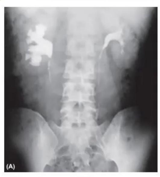
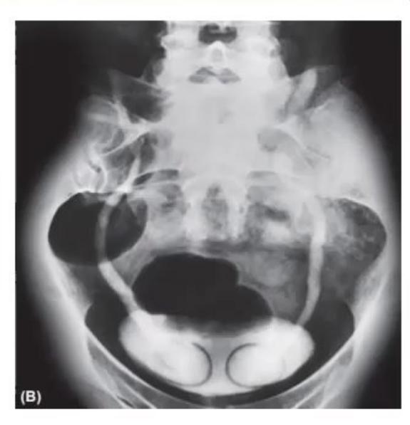
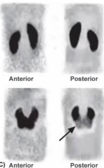
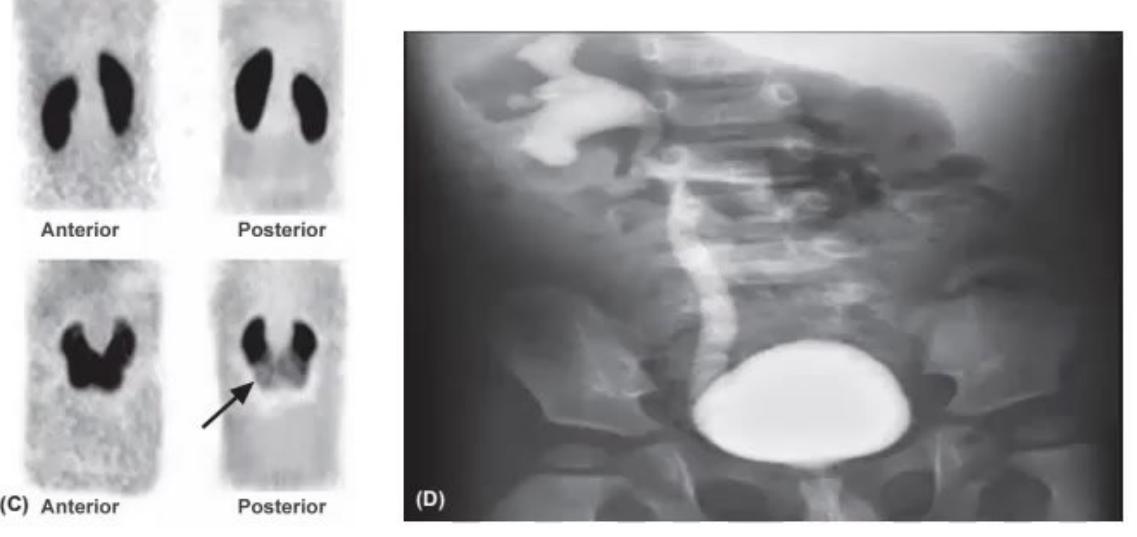
With regard to renal radioimaging, identify the following images and state the underlying diagnosis:
Answer
- Image A: Intravenous Urogram (IVU / IVP) showing Pelviureteric Junction (PUJ) Obstruction (marked hydronephrosis with abrupt cutoff at PUJ).
- Image B: Excretory Urogram showing "Cobra-head" / "Spring-onion" sign characteristic of an intravesical Ureterocele.
- Image C:
-DMSA cortical renal scintigraphy showing a Horseshoe Kidney (fusion of lower poles across midline). - Image D: Micturating Cystourethrogram (MCU) demonstrating High-Grade (Grade IV/V) Vesicoureteral Reflux (VUR) with tortuous, dilated ureters and blunted calyces.
MNJ31-014
A 13-year-old girl Radha with recurrent muscle cramps has ABG: pH: 7.65,
- Interpret the ABG.
- Is it compensated? What is the expected respiratory compensatory formula?
- What is the probable diagnosis?
- What is the basic molecular defect?
- Mention 3 associated metabolic abnormalities in this condition.
Answer
- Partially Compensated Metabolic Alkalosis with hypoxemia absent.
- Expected respiratory compensation in metabolic alkalosis:
Since actualis 46 mmHg, appropriate respiratory compensation is present. - Diagnosis: Gitelman Syndrome.
- Molecular defect: Inactivating mutation in the SLC12A3 gene on chromosome 16q13 encoding the Thiazide-sensitive
Cotransporter (NCCT) in the distal convoluted tubule. - Associated metabolic abnormalities:
- Severe Hypokalemia
- Severe Hypomagnesemia
- Hypocalciuria (decreased urinary calcium excretion)
- Secondary hyperreninemic hyperaldosteronism with normal/low blood pressure.
MNJ31-015
A 7-month-old male child Sanju is admitted with Tubercular Meningitis. Laboratory tests show serum Sodium: 124 mEq/L and Potassium: 4.1 mEq/L:
- What causes of hyponatremia will you consider in this child?
- How will you differentiate SIADH from Cerebral Salt Wasting (CSW)?
- How will you acutely manage hyponatremia in CSW vs SIADH?
Answer
- Etiologies: Cerebral Salt Wasting (CSW) vs Syndrome of Inappropriate ADH Secretion (SIADH).
- Differentiating features:
| Feature | SIADH | Cerebral Salt Wasting (CSW) |
|---|---|---|
| Volume Status | Euvolemic / Hypervolemic | Hypovolemic (Dehydrated) |
| Urine Output | Normal or Decreased | Profuse Polyuria |
| Urine Sodium | Elevated ( |
Massively Elevated ( |
| Central Venous Pressure (CVP) | Normal / High ( |
Low ( |
| Serum Uric Acid / Urea | Low / Diluted | Normal or High (prerenal azotemia) |
- Management:
- CSW: Volume and sodium replacement using Normal Saline (0.9% NaCl) or 3% Hypertonic Saline plus oral Fludrocortisone (
) to enhance renal sodium retention. (Fluid restriction is dangerous and contraindicated in CSW). - SIADH: Fluid restriction (
of maintenance). Hypertonic (3%) saline reserved only for acute symptomatic seizures.
- CSW: Volume and sodium replacement using Normal Saline (0.9% NaCl) or 3% Hypertonic Saline plus oral Fludrocortisone (
MNJ31-016
Match the imaging modality of choice with the given clinical condition:
| Imaging Modality | Clinical Renal Condition |
|---|---|
| 1. |
a. Detection of renal scarring / chronic pyelonephritis |
| 2. Plain Abdominal X-ray (KUB) | b. Pelviureteric junction obstruction / dynamic drainage |
| 3. Micturating Cystourethrogram (MCU) | c. Horseshoe kidney / duplex collecting system anatomy |
| 4. Renal Doppler Ultrasonography | d. Radiopaque calcium stones in urinary tract |
| 5. Intravenous Urogram (IVU) | e. Vesicoureteral Reflux (VUR) grading |
| 6. |
f. Renal artery / vein stenosis and vascular flow |
Answer
-
-DMSA Scan a. Detection of renal parenchymal scarring / acute pyelonephritis
-
- Plain Abdominal X-ray (KUB)
d. Radiopaque calcium oxalate/phosphate stones
- Plain Abdominal X-ray (KUB)
-
- Micturating Cystourethrogram (MCU)
e. Vesicoureteral Reflux (VUR) grading and PUV
- Micturating Cystourethrogram (MCU)
-
- Renal Doppler USG
f. Renal artery / vein thrombosis and stenosis
- Renal Doppler USG
-
- Intravenous Urogram (IVU)
c. Horseshoe kidney / anatomical duplication
- Intravenous Urogram (IVU)
-
-MAG3 Renogram b. Pelviureteric junction (PUJ) obstruction and GFR drainage kinetics
MNJ31-017
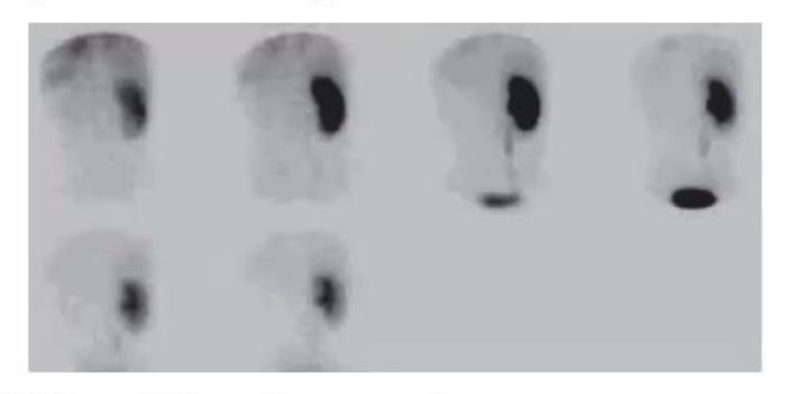
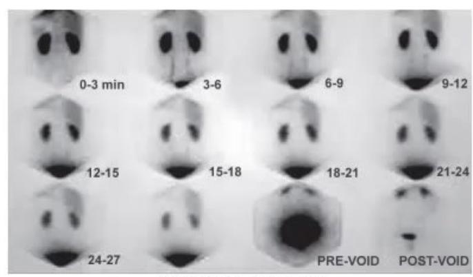
A 9-month-old child comes for follow-up of recurrent UTI. Renal USG showed unilateral pelvicalyceal dilatation. A dynamic diuresis renogram is performed with IV Furosemide (Lasix) administered at 20 minutes (
- What is this nuclear scan called?
- What are its clinical applications?
- Describe the three phases of a normal renogram curve.
- What abnormality does this child's renogram curve show?
Answer
-MAG3 (Mercaptoacetyltriglycine) Diuretic Dynamic Renogram (Furosemide renogram). - Clinical applications:
- Differentiating mechanical anatomical obstruction from non-obstructive hydronephrosis
- Evaluation of Pelviureteric Junction (PUJ) obstruction and Vesicoureteric Junction (VUJ) obstruction
- Measurement of differential (split) renal function and drainage half-time (
).
- Three phases of normal renogram curve:
- Phase 1 (Vascular / Transit Phase): Rapid initial upstroke (first 30–60 seconds) representing initial arrival and vascular perfusion of tracer.
- Phase 2 (Parenchymal / Secretory Phase): Gradual upward curve (peaking at 3–5 minutes) reflecting active tubular extraction of tracer from blood.
- Phase 3 (Elimination / Excretory Phase): Rapid downward descent as tracer washes out of the pelvicalyceal system into the bladder (
).
- Interpretation in this child: The left kidney shows a continuous rising curve with failure of clearance after Furosemide (
), diagnostic of Left Pelviureteric Junction (PUJ) Mechanical Obstruction.
MNJ31-018
Match each clinical presentation of acute scrotum with its most likely diagnosis:
| Clinical Scenario | Diagnosis |
|---|---|
| 1. A 6-year-old boy presents with acute, severe left scrotal pain of 3 hours duration. He is afebrile with negative urinalysis; left testis is high-riding, horizontally oriented, swollen, and exquisitely tender with absent cremasteric reflex. | a. Epididymo-orchitis |
| 2. A 3-year-old boy presents with palpable purpura on the lower extremities, arthralgia, microscopic hematuria, and painful acute scrotal swelling. | b. Testicular Torsion |
| 3. A 13-year-old boy presents with 2 days of gradual-onset scrotal pain, fever, dysuria, and pyuria. Tenderness is maximal over the posterolateral aspect of the testis with relief of pain on scrotal elevation (Prehn sign positive). | c. Henoch-Schönlein Purpura (IgA Vasculitis scrotal involvement) |
Answer
- 1
b. Testicular Torsion (Urological emergency requiring immediate surgical exploration and detorsion within 6 hours). - 2
c. Henoch-Schönlein Purpura (HSP / IgA Vasculitis) (Scrotal vasculitis). - 3
a. Epididymo-orchitis (Bacterial or viral infection treated with targeted antibiotics).
MNJ31-019
- Identify the nuclear medicine imaging modality.
- What is the diagnosis shown in this film?
- What is the time interval between radiotracer injection and image acquisition?
- Write down four major clinical indications for this investigation.
Answer
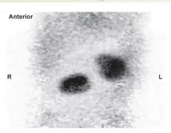
-DMSA (Dimercaptosuccinic Acid) Static Renal Cortical Scintigraphy. - Ectopic Right Kidney (Pelvic Kidney) with normal left orthotopic kidney.
- Time interval: 2 to 4 hours post-intravenous injection (to allow fixed cortical tubular binding and clearance of background tracer).
- Indications:
- Detection of Renal Cortical Scarring secondary to recurrent pyelonephritis / VUR (gold standard)
- Diagnosis of Acute Pyelonephritis (focal/multifocal photopenic areas of reduced uptake)
- Accurate measurement of Differential / Split Renal Function
- Detection of congenital anomalies: Ectopic kidney, Horseshoe kidney, Dysplastic kidney, or Single functioning kidney.
MNJ31-020
- Identify the nuclear investigation.
- What functional physiological information is obtained from this scan?
- How soon after tracer injection is imaging initiated?
- Write down four clinical indications for this investigation.
Answer
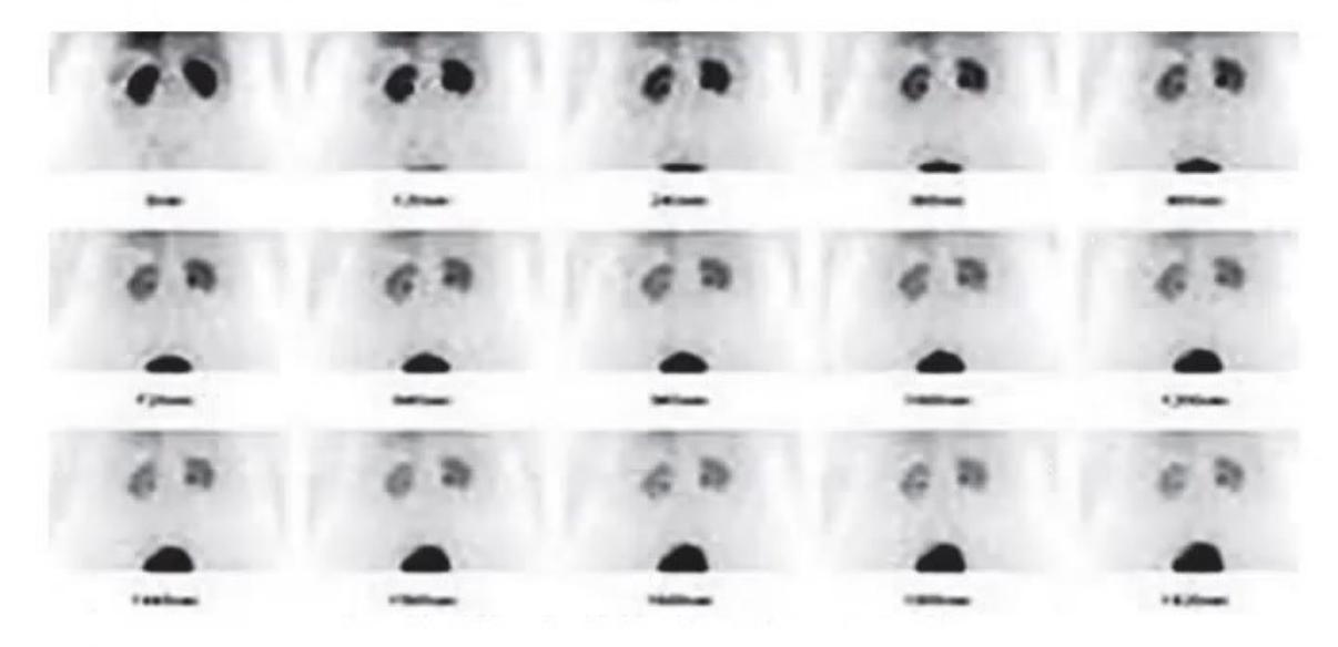
-DTPA (Diethylenetriaminepentaacetic Acid) Dynamic Renal Scintigraphy (Gates method). - Functional parameters obtained:
- Glomerular Filtration Rate (GFR) measurement (purely filtered by glomerulus without tubular secretion or reabsorption)
- Renal Perfusion / Blood Flow kinetics
- Differential (Split) Renal Function
- Upper urinary tract drainage and clearance dynamics.
- Timing of imaging: Immediate continuous dynamic sequential imaging starts concurrently with the IV bolus injection (0–60 seconds for perfusion, up to 20–30 minutes for excretion).
- Indications:
- Quantitative evaluation of individual and global GFR in CKD / acute kidney injury
- Evaluation of hydronephrosis and urinary tract obstruction
- Assessment of renal allograft function and detection of acute tubular necrosis vs graft rejection
- Captopril renography for screening Renovascular Hypertension (Renal Artery Stenosis).