THG21-001
An 8-year-old girl child was evaluated for abdominal pain. Her ultrasound KUB (kidney, ureter, and bladder) showed an abnormality for which the following investigation was done.
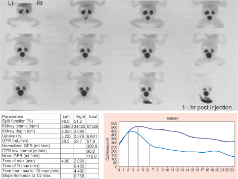
a. Identify the investigation.
b. What are the three different substances used for this study?
c. What is t½, intrarenal transit time (IRT), and its significance?
d. Interpret the investigation given in image below.
e. What is the next line of management?
Answer
- a. Diuretic renogram/dynamic renal scintigraphy
- b. Diethylenetriamine pentaacetate (DTPA), ethylene dicysteine (EC), and mercaptoacetyltriglycine (MAG3)
- c. T½: time taken for 50% of the trace to get cleared, IRT: time taken for trace to appear in medial pelvis
- d. Significance: T½ >20 minutes and IRT >4 minutes is suggestive of pelvic ureteric junction obstruction (PUJO)
- e. Refer to pediatric surgeon for left pyeloplasty
THG21-002
A 1-year-old male infant was evaluated for urinary tract infection (UTI). The following investigation was performed.
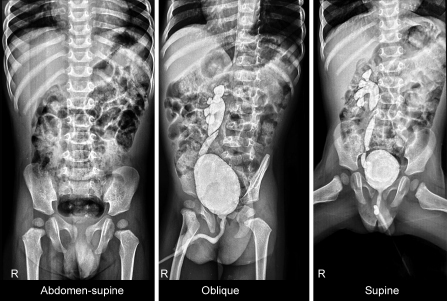
a. What is the investigation?
b. What is your diagnosis?
c. What are the three indications to perform the test in children with UTI as per current Indian Society of Pediatric Nephrology (ISPN) guidelines?
d. When should this test be performed after an episode of UTI?
e. Name the best alternative test for the investigation given in image here.
Answer
- a. Micturating cystourethrogram (MCU)
- b. Right grade IV VUR
- c. (i) Recurrent UTI, (ii) abnormal ultrasound, (iii) non-Escherichia coli UTI in children below 2 years of age
- d. Any time after completion of UTI treatment
- e. DRCG: Direct radionuclide cystography
THG21-003
A 2-year-old male infant was treated for pyelonephritis. During further evaluation, the following investigation was performed.
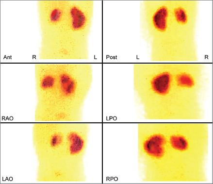
a. What is the investigation given in the image?
b. What is the substance used for this investigation?
c. What is your diagnosis?
d. When is this investigation performed after UTI according to current ISPN guideline?
e. What are the two indications for this investigation?
Answer
- a. Static renal scintigraphy
- b. DMSA
- c. Right kidney is small with photopenic areas in both poles suggestive of scarring
- d. 4–6 months after an UTI episode
- e. (i) High grade (grade III/IV) VUR and (ii) Recurrent UTI
THG21-004
A 3-year-old child was investigated for recurrent urinary tract infections with the following radionuclide study:
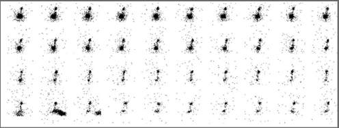
a. What is the investigation given in the above image?
b. What is the diagnosis?
c. Mention one indication for the above investigation.
d. Name two advantages of this investigation.
e. Name two disadvantages of the above investigation.
Answer
- a. DRCG: Direct radionuclide cystography
- b. Right grade III VUR
- c. Alternative for MCU during follow-up of VUR. Children to check for resolution of reflux
- d. Less radiation, urinary catheterization not required
- e. Grading and anatomical details are inadequate.
THG21-005
A 1-year-old male infant was evaluated for UTI and was found to have grade IV vesicoureteral reflux (VUR) on the right side. His dimercaptosuccinic acid (DMSA) scan showed of 42% function in the right kidney with scar in the upper pole. Child is on regular follow-up and has no breakthrough UTI.
a. Mention two indications for uroprophylaxis.
b. Name one drug for uroprophylaxis that can be used in this child and mention the dose.
c. When can you stop uroprophylaxis?
d. Is surgery indicated in this child?
e. Should we repeat the micturating cystourethrogram (MCU) in this child?
Answer
- a. (i) Recurrent febrile UTI and bowel bladder dysfunction (BBD); (ii) Infant with recurrent febrile UTI and lower grade VUR; (iii) High-grade VUR
- b. Cotrimoxazole 2 mg/kg and nitrofurantoin 1–2 mg/kg
- c. Child older than 2 years if they satisfy all the three criteria:
- i. Toilet trained
- ii. Absence of BBD
- iii. No febrile UTI in preceding 1 year
- d. Surgery is not recommended in children as there is no breakthrough UTI.
- e. According to the current ISPN guideline, repeat MCU is not routinely indicated. To check for resolution of reflux, it needs to be performed 4–8 years after initial diagnosis if deemed necessary by a treating physician.
THG21-006
A 11-year-old female child was brought with second episode of UTI. She had a history of lower urinary tract symptoms, such as dysuria, urgency, and frequency. She does not urinate in the school and on further probing the history; she was constipated and passes hard stools once in 2–3 days. Her ultrasound showed the right kidney size of 10.1 cm and left kidney size of 10.8 cm, bilateral pelvicalyceal dilatation with anteroposterior diameter (APD) 6 mm, and postvoid residue (PVR) of 43 mL.
a. What is your diagnosis?
b. Mention the imaging to be done in these children.
c. How to calculate bladder capacity and what is the capacity in this child?
d. How do you initially manage this child?
e. What drug will you use if the initial management fails?
Answer
- a. Recurrent UTI with BBD
- b. Ultrasound, MCU, and DMSA
- c. (Age in years + 1) × 30 mL = (11 + 1) × 30 = 360 mL
- d. She needs to be initiated on uroprophylaxis for recurrent UTI and initiate treatment for BBD.
- Management of BBD:
- Diet with increased fiber-rich food and water 6–8 cups per day
- Laxative for constipation
- Timed and double voiding
- Management of BBD:
- e. Tamsulosin, Doxazosin, and biofeedback therapy
THG21-007
Answer the following:
a. Mention two conditions for false-positive and negative in urine dipstick for albuminuria.
b. Define normal, subnephrotic and nephrotic range proteinuria according to spot urine protein creatinine ratio.
c. Define orthostatic proteinuria.
d. What precautions should a parent take while collecting a 24-hour urine sample.
e. Name two conditions in which transient proteinuria are seen.
Answer
- a. Urine dipstick albuminuria false results:
| False positive | False negative |
|---|---|
| Hematuria | Proteinuria other than albumin |
| High specific gravity | Low specific gravity |
| pH >8.0 | pH <4.5 |
- b. Spot urine protein-creatinine ratio (UPCR):
| Spot UPCR | Range |
|---|---|
| <0.2 | Normal |
| 0.2 to 2.0 | Subnephrotic range proteinuria |
| >2.0 | Nephrotic range proteinuria |
- c. Orthostatic proteinuria, also known as postural proteinuria, is a condition where an abnormally large amount of protein is excreted in the urine when the patient is in an upright position and normal protein excretion in the supine position.
- d. (i) Collection duration 24-hour without missing even a single void of urine; (ii) Empty bladder at start of collection; (iii) Empty bladder at the finish and add to the collected sample
- e. Fever, exercise, dehydration, seizures, and congestive heart failure (CHF).
THG21-008
Match the following in Table A and B:
| Table A | Table B (urine crystal) |
|---|---|
| Acyclovir | Diamond-shaped crystal |
| Cystinuria | Dumbbell-shaped crystal |
| Tumor lysis syndrome | Triple phosphates |
| Primary hyperoxaluria | Needle-shaped crystal |
| Recurrent UTI | Hexagonal-shaped crystal |
Answer
| Drug / Condition | Urine Crystal |
|---|---|
| Acyclovir | Needle-shaped crystal |
| Cystinuria | Hexagonal-shaped crystal |
| Tumor lysis syndrome | Diamond-shaped crystal |
| Primary hyperoxaluria | Dumbbell-shaped crystal |
| Recurrent UTI | Triple phosphates |
THG21-009
A 7-year-old male child presented with acute onset of gross painless cola colored urine, edema, and oliguria. He had skin lesions over the legs a week prior to the illness. On examination, his blood pressure (BP) was 120/90 mm Hg. He also had facial puffiness, ascites, and pedal edema.
a. What is the probable diagnosis?
b. What are the complications of the above condition?
c. When will you do renal biopsy in this child?
d. What is the best single antibody titer to document cutaneous streptococcal infection?
e. List any two other causes of hypocomplementemia.
f. What is the prognosis of this condition?
Answer
- a. The probable diagnosis is postinfectious glomerulonephritis (PSGN).
- b. Hypertension (60%)/hypertensive encephalopathy (10%)/uremia/congestive cardiac heart failure/dyselectrolytemia (hyperkalemia and metabolic acidosis)/hyperphosphatemia, and hypocalcemia.
- c. Indications for renal biopsy:
- Nephritic-nephrotic presentation
- Rapidly progressing renal failure
- Normal complement levels at onset
- Low C3 levels persisting for >2 months
- Systemic features suggestive of secondary glomerulonephritis
- d. Anti-deoxyribonuclease B (anti-DNase B) level
- e. Postinfectious glomerulonephritis, systemic lupus erythematosus (SLE), C3 glomerulopathy/membranoproliferative glomerulonephritis (MPGN)
- f. It carries very good prognosis (95% recover from the illness).
THG21-010
A 2-year-old boy was brought to outpatient department (OPD) with history of progressive abdominal distension for the past few months noticed by his mother while bathing him. No other significant history present except for oligohydramnios during antenatal period and neonatal intensive care unit (NICU) stay for respiratory distress. His physical examination revealed height and weight between 3rd to 25th centile, blood pressure in the 95th centile, hepatomegaly, and bilateral ballotable kidneys. Ultrasonography (USG) of abdomen revealed bright echogenic kidneys and liver.
a. What is your diagnosis?
b. What is the gene involved in this condition?
c. What is Caroli’s disease?
d. What liver complication will you expect in this child?
e. List three cystic kidney diseases.
f. List three extrarenal manifestations of autosomal dominant polycystic kidney disease (ADPKD).
g. What is the most common cause of an abdominal mass in a newborn?
h. Name two associated physical findings to suspect renal disease in a child.
Answer
- a. Autosomal recessive polycystic kidney disease (ARPKD)
- b. The polycystic kidney and hepatic disease 1 (PKHD1) gene
- c. Cystic dilatation of the intrahepatic bile ducts (Caroli’s disease)
- d. Portal hypertension with hepatosplenomegaly, gastroesophageal varices, ascending cholangitis, reversal of portal vein flow, and thrombocytopenia
- e. Polycystic kidney diseases (ARPKD and ADPKD), multicystic dysplastic kidney (MCDK), nephronophthisis, simple renal cyst, glomerulocystic kidney disease, tuberous sclerosis, and von Hippel–Lindau disease
- f. Cysts in other organs, such as liver, pancreas, spleen, and ovaries / intracranial aneurysms / mitral valve prolapse / aortic and coronary artery aneurysms, aortic valve insufficiency, and hernia / bronchiectasis, intestinal diverticula
- g. Multicystic dysplastic kidney (MCDK)
- h. Physical findings to suspect renal disease:
- i. Single umbilical artery
- ii. Abnormality of another organ system (congenital heart disease)
- iii. External ear anomalies
- iv. Imperforate anus
- v. Vertebral anomalies (scoliosis)
- Infants with these physical findings should undergo renal ultrasound.
THG21-011
A 3-year-old female child presented with faltering of growth, retarded motor milestones, and difficulty in getting up from sitting posture with bony deformities. Child weighed 7 kg, height was 78 cm, and had features of clinical rickets. Her laboratory values were:
- Serum creatinine: 0.3 mg/dL
- Serum electrolytes (sodium/potassium/chloride/bicarbonate): 133/2.8/109/9 mEq/L
- Urine pH: 7.5
- Urinary anion gap: +22
- USG abdomen revealed medullary nephrocalcinosis.
a. What is your diagnosis? What is the classical metabolic abnormality?
b. How will you calculate urine anion gap? What is the significance?
c. What are the other common metabolic defects that cause nephrocalcinosis?
d. What is Fanconi syndrome?
e. Write two causes of hyperkalemic renal tubular acidosis (RTA).
f. What is the line of management for the above child in the scenario?
g. What is Lowe syndrome?
h. Write two metabolic causes of Fanconi syndrome.
Answer
- a. Distal RTA (Type 1 RTA) / normal anion gap; hyperchloremic hypokalemic metabolic acidosis
- b. Urine anion gap =
. A positive gap suggests a deficiency of ammonia genesis and the possibility of distal RTA. - c. Hypercalciuria, hypocitraturia, furosemide intoxication, and vitamin D intoxication
- d. Global proximal tubular dysfunction causes Fanconi syndrome. It is characterized by low molecular weight proteinuria, glycosuria, phosphaturia, and aminoaciduria.
- e. Obstructive uropathy (posterior urethral valve, ureteropelvic junction obstruction); Acute pyelonephritis / hypoaldosteronism, pseudohypoaldosteronism
- f. Alkali therapy (bicarbonate) 2–4 mEq/kg/day
- g. Oculocerebrorenal syndrome characterized by congenital cataracts, mental retardation, and Fanconi syndrome
- h. Galactosemia / hereditary fructose intolerance / tyrosinemia / Wilson disease
THG21-012
A 5-day-old newborn boy with history of oligohydramnios in the antenatal period underwent MCU.
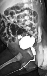
a. Describe the MCU?
b. What antenatal intervention would have helped in this child?
c. What is the cause of respiratory distress in newborn with this condition?
d. What is the surgical management of this condition?
e. What is the triad of Prune belly syndrome?
f. What are the favorable prognostic factors of this condition?
g. What are the unfavorable prognostic factors of this condition?
h. What is VURD syndrome?
i. What is Hinman syndrome?
Answer
- a. MCU shows narrow anterior urethral stream, dilated posterior urethra, and trabeculated bladder with diverticulae.
- b. Vesicoamniotic shunt (bladder decompression)
- c. Pulmonary hypoplasia
- d. Cystoscopic transurethral ablation of valve leaflets (fulguration)
- e. Triad of Prune belly syndrome: Deficient abdominal musculature, undescended testes (cryptorchidism), and urinary tract abnormalities
- f. Favorable prognostic factors:
- Normal prenatal ultrasonogram between 18 and 24 weeks of gestation
- Serum creatinine level <0.8–1.0 mg/dL after valve fulguration
- Preserved corticomedullary differentiation on USG
- g. Unfavorable prognostic factors:
- Oligohydramnios in antenatal scans
- Identification of hydronephrosis before 24 weeks of gestation
- Serum creatinine >1 mg/dL after valve fulguration
- Cortical cysts in both kidneys
- Persistence of diurnal incontinence beyond 5 years of age
- h. VURD syndrome: Valves, Unilateral Reflux and Dysplasia
- i. Hinman syndrome: Failure of the external sphincter to relax during voiding in children without any neurologic abnormalities (non-neurogenic neurogenic bladder)
THG21-013
A 4-year-old boy presented with periorbital puffiness, abdominal distension, leg swelling, and reduced urine output of 1 week duration. Examination was significant for anasarca and urine dipstick showed 4+ proteinuria.
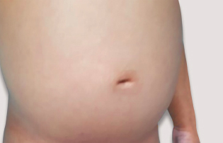
a. What is your diagnosis?
b. What are the drugs indicated in case of steroid resistance?
c. Which organism is most common to cause peritonitis in this child?
d. What are the indications for renal biopsy in this child?
e. What are the types of proteinuria?
f. What are the causes of transient proteinuria?
Answer
- a. The diagnosis is nephrotic syndrome.
- b. Calcineurin inhibitors (tacrolimus and cyclosporine) / anti-CD20 monoclonal antibody (rituximab)
- c. Streptococcus pneumoniae (pneumococcus)
- d. Indications for renal biopsy in nephrotic syndrome:
- Age of onset <1 year and >10 years
- AKI not attributed to hypovolemia
- Persistent microscopic or gross hematuria
- Systemic features, such as fever, rash, arthralgia, low C3
- Initial or late steroid-resistant nephrotic syndrome
- Prior to starting calcineurin inhibitors, or reduced kidney function during their use
- e. Types of proteinuria: Glomerular, tubular, orthostatic, and overflow proteinuria
- f. Causes of transient proteinuria: Fever, exercise, dehydration, cold exposure, seizures, stress, and CCF
THG21-014
A 2-year-old boy presented with failure to thrive. He had genu valgum and short stature. His investigations revealed elevated parathyroid hormone and phosphorus levels.
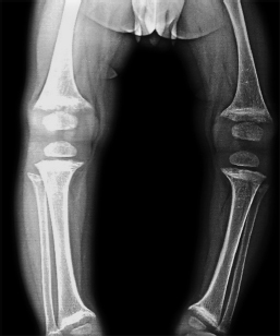
a. What is your diagnosis?
b. Write the X-ray findings of rickets.
c. When will you suspect nonnutritional rickets?
d. What is burosumab? In which condition is it used?
e. What is the treatment of vitamin D dependent rickets?
Answer
- a. Rickets due to CKD or mineral bone disease (clue: hyperphosphatemia)
- b. X-ray findings of rickets:
- Loss of provisional zone of calcification
- Widening and irregularity of the physes in long bones
- Fraying and cupping/broadening of the metaphyses
- Generalized demineralization of bones
- c. Suspect nonnutritional rickets when:
- Presentation before 6 months or after 2 years of age
- Failure to respond to standard vitamin D therapy
- Obvious clues in history, clinical examination, and initial investigations suggestive of renal/hepatic and oncogenic rickets
- d. Burosumab is a human recombinant anti-FGF23 monoclonal antibody, used in X-linked hypophosphatemic rickets.
- e. Active vitamin D (calcitriol / 1,25-dihydroxyvitamin D)
THG21-015
A 6-year-old girl child presented with dysuria, hematuria, urgency, and colicky abdominal pain for 2 days. Ultrasound abdomen revealed a stone at the mid ureter. Her urine microscopy showed the below image.
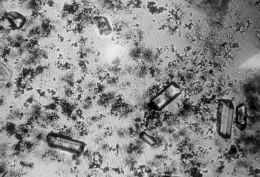
a. Describe the finding.
b. What urinary investigations are done to confirm the diagnosis?
c. What is the chemical reaction used in dipstick to find hematuria in a urine sample?
d. Define microscopic hematuria.
e. Name two types of hereditary nephritis with hematuria.
Answer
- a. Coffin-lid appearance—triple phosphate (struvite/magnesium ammonium phosphate) crystals
- b. 24-hour urine for crystalluria evaluation—calcium, oxalate, uric acid, cystine, and citrate
- c. Peroxidase chemical reaction: The dipstick uses a sensitive peroxidase chemical reaction between hemoglobin (or myoglobin) and a colorimetric indicator (benzidine/tetramethylbenzidine) impregnated on the strip pad. The pseudoperoxidase activity of hemoglobin catalyzes oxidation of the indicator, resulting in a color change proportional to red blood cells/hemoglobin present.
- d. Presence of at least 5 RBCs per high power field (HPF) in a centrifuged urine sample (or >5 RBCs/µL).
- e. Alport syndrome and thin basement membrane disease
THG21-016
Parents brought a 9-year-old boy with primary enuresis. As a pediatrician how do you counsel the parent about the diagnosis and management.
Answer
- Introduction & Rapport: Make eye contact, greet parents, confirm comfortable language, and assess understanding of the problem.
- Diagnosis & Overview: Primary nocturnal enuresis is defined as discrete episodes of involuntary urinary incontinence during sleep in children ≥5 years of age. Approximately 16% of children are incontinent at 5 years, dropping to 1–2% at ≥15 years.
- General Measures & Motivational Therapy:
- Restrict fluids 2 hours before bedtime.
- Empty bladder immediately before sleeping.
- Encourage the child to make bedtime resolution / avoid fluid-heavy evening meals.
- Maintain a bladder diary (calendar to mark dry and wet nights).
- Positive reinforcement and reward system (star charts) for dry nights; strictly avoid punishment or criticism.
- Conditioning & Pharmacotherapy:
- Enuresis alarm therapy (moisture sensor alarm) as most effective long-term conditioning device.
- Medications: Desmopressin (oral/melt) or anticholinergics (oxybutynin) for refractory cases.
- Check parental understanding, address queries, and schedule regular follow-up.
THG21-017
A 12-year-old girl came with history of fever for a month, weight loss, anorexia, arthralgia, arthritis, and rash over the malar area of face. She presented with hematuria and mild renal insufficiency. Diagnosis of systemic lupus erythematosus (SLE) nephritis was entertained.
a. International Society of Nephrology/Renal Pathology Society (ISN/RPS) classification of lupus nephritis (LN).
b. Renal involvement in SLE, American College of Rheumatology (ACR) criteria and immunological criteria (mention any two).
c. What is the serological marker for drug-induced lupus?
d. Mention three drugs which can cause the same?
Answer
- a. ISN/RPS Classification of Lupus Nephritis:
- Class I: Minimal mesangial LN
- Class II: Mesangial proliferative LN
- Class III: Focal LN (<50% of glomeruli)
- Class IV: Diffuse LN (≥50% of glomeruli)
- Class V: Membranous LN
- Class VI: Advanced sclerotic LN (≥90% of glomeruli globally sclerosed)
- b. Renal Involvement Criteria:
- i. Proteinuria: >500 mg/24-hour urine (or >3+ dipstick)
- ii. Active urine sediment (>5 RBC, >5 WBC, and cellular casts)
- iii. Class II–V in renal biopsy
- Immunological criteria (any two):
- Low C3 and C4 complement levels
- Positive antiphospholipid antibodies (anticardiolipin, anti-β2GP1, lupus anticoagulant)
- Anti-dsDNA antibodies or anti-Smith (Sm) antibodies
- c. Anti-histone antibodies
- d. Hydralazine, procainamide, penicillamine, isoniazid, methyldopa, chlorpromazine, phenytoin, ethosuximide, quinidine, antithyroid drugs, sulfonamides, and rifampicin
THG21-018
A 1.5-year-old male child, firstborn of third-degree consanguineous marriage, had a birth weight of 2.4 kg (present weight 6.5 kg) has failure to thrive, polyuria, polydipsia, and history of recurrent infections treated by local practitioner. Laboratory test showed Hb— 8.5 g/dL, blood urea nitrogen (BUN)—24, serum creatinine—0.4, serum Na—112, serum K—2.8, serum Cl—94, and serum HCO3—28, serum Mg—1.8. His blood pressure is normal. USG KUB is normal.
a. What is your probable diagnosis?
b. What is the channel involved in it?
c. What are the additional investigations you will do?
d. Mention appropriate management for the condition.
e. What is the long-term renal complication?
Answer
- a. Classical Bartter syndrome (Type 3)
- b. ClC-Kb chloride channel mutation
- c. Hearing assessment (audiometry) and urine calcium/creatinine ratio
- d. Potassium supplements (preferably potassium chloride), indomethacin / NSAIDs 1–4 mg/kg/day (if dyselectrolytemia not corrected by supplements alone)
- e. Chronic kidney disease (CKD) / focal segmental glomerulosclerosis in rare long-term cases
THG21-019
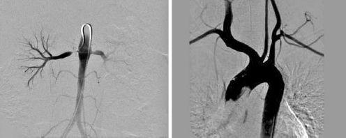
A 13-year-old girl referred for evaluation of dilated cardiomyopathy with features of congestive cardiac failure (CCF). Her BP is 180/120 mm Hg in right upper limb. Physical examination findings were significant with weak carotids, brachial and radial pulses, and bilateral carotid bruits. Renal USG with Doppler was suggestive of bilateral renal artery stenosis. Serum creatinine—0.35 mg/dL, urinalysis—normal, erythrocyte sedimentation rate (ESR)—80 mm/hour, and C-reactive protein (CRP)—27 mg/L.
a. How will you choose the appropriate cuff size for BP measurement?
b. What is the stage of hypertension in this child?
c. Classify hypertension in children between 1 to 12 years.
d. What is the likely diagnosis in this child?
e. Which group of antihypertensives should be avoided in bilateral renal artery stenosis?
Answer
- a. The inflatable bladder width should be at least 40% of arm circumference at midpoint between acromion and olecranon; bladder length should encircle 80–100% of arm circumference.
- b. Stage 2 hypertension (>95th percentile + 12 mm Hg or ≥140/90 mm Hg)
- c. AAP Definitions of BP Categories (1–12 years):
| Category | Definition |
|---|---|
| Normal BP | <90th percentile |
| Elevated BP | ≥90th percentile to <95th percentile (or 120/80 mm Hg to <95th percentile, whichever is lower) |
| Stage 1 HTN | ≥95th percentile to <95th percentile + 12 mm Hg (or 130/80 to 139/89 mm Hg, whichever is lower) |
| Stage 2 HTN | ≥95th percentile + 12 mm Hg (or ≥140/90 mm Hg, whichever is lower) |
- d. Takayasu arteritis
- e. Angiotensin-converting enzyme (ACE) inhibitors and Angiotensin receptor blockers (ARBs)
THG21-020
A 15-day-old girl baby born by normal delivery to second-degree consanguineous parents, borderline preterm (35 weeks), birth weight of 2.5 kg, antenatal period—uneventful, admitted for edema and decreased urine output since birth. No history suggestive of renal disease in family. General examination revealed sacral and vulval edema, vitals stable, massive ascites, anterior fontanel (AF)—wide open; posterior fontanel (PF)—open; and widened cranial sutures. No syndromic features were present. TORCH [toxoplasmosis, rubella cytomegalovirus (CMV), herpes simplex, and HIV] screening was negative, and USG abdomen revealed normal-sized kidney, increased echotexture, and free fluid abdomen.
a. What is the most probable diagnosis and how do you define it?
b. What are the common genes involved in this condition?
c. Mention at least two secondary causes of this condition.
d. What is the method of choice for precise diagnosis of the above condition?
e. What is the management of this condition?
Answer
- a. Congenital nephrotic syndrome (CNS), defined as onset of nephrotic syndrome within the first 3 months of life.
- b. Genes involved in congenital nephrotic syndrome:
- NPHS1 (Nephrin mutation - Finnish type)
- NPHS2 (Podocin mutation)
- WT1 (Wilms’ tumor 1 gene)
- LAMB2 (Laminin β2 gene - Pierson syndrome)
- PLCE1 (Phospholipase C epsilon 1 gene - NPHS3)
- c. Secondary causes: Congenital syphilis, toxoplasmosis, cytomegalovirus (CMV), rubella, hepatitis B, HIV, malaria, and maternal SLE
- d. Genetic testing (next-generation sequencing)
- e. Management of CNS:
- Conservative: High-calorie diet (130 kcal/kg/day), high-protein diet (4 g/kg/day), IV albumin infusions with diuretics, ACE inhibitors / indomethacin to reduce proteinuria, thyroxine and fat-soluble vitamin supplements, prophylactic anticoagulation.
- Surgical: Unilateral/bilateral nephrectomy (medical or surgical) to control severe protein loss, followed by peritoneal dialysis and eventual renal transplantation.
THG21-021
A 3-year-old child had acute kidney injury (AKI), gross hematuria, and documented to have hypertension following bloody diarrhea.
a. What is the most probable diagnosis and its triad?
b. Classify the causes of this condition.
c. Where can you have a positive Coombs test in this condition?
d. What is the specific treatment for the typical and atypical varieties of this condition?
Answer
- a. Diagnosis: Hemolytic uremic syndrome (HUS)
- Triad:
- Microangiopathic hemolytic anemia (Hb <10 g/dL, schistocytes >2%, elevated LDH >450 IU/L, low haptoglobin)
- Thrombocytopenia (platelets <150,000/µL)
- Acute kidney injury (serum creatinine elevation ≥50% from baseline)
- Triad:
- b. Classification of Causes:
- Infection-induced (typical/D+ HUS): Shiga toxin-producing E. coli (STEC O157:H7), Shigella dysenteriae type 1, Streptococcus pneumoniae, HIV
- Complement dysregulation (atypical/aHUS): Mutations in CFH, CFI, CFB, MCP, or anti-factor H autoantibodies
- Cobalamin C defect
- Medication-induced: Calcineurin inhibitors, quinine, chemotherapeutic agents
- Secondary HUS: SLE, antiphospholipid antibody syndrome, malignancy, pregnancy
- c. Pneumococcal-induced HUS (Streptococcus pneumoniae producing neuraminidase causing Thomsen-Friedenreich antigen exposure)
- d. Specific Treatment:
- Typical (STEC-HUS): Supportive therapy (judicious fluid/electrolyte management, dialysis if indicated, avoidance of antibiotics and antimotility agents)
- Atypical HUS: Plasma therapy (plasmapheresis / fresh frozen plasma infusion), terminal complement inhibitor (Eculizumab), and hemodialysis
THG21-022
A 6-year-old child who is a diagnosed case of focal segmental glomerulosclerosis FSGS, progressed to chronic kidney disease (CKD), now on intermittent hemodialysis for past 2 years. His height was 100 cm and weight was 20 kg, BP of 140/90 mm Hg. He had bony deformities. Investigations showed a serum creatinine of 6.8 mg/dL, serum K of 6.0 mEq/L, serum HCO3 of 8.0 mEq/L with serum Ca of 7.0 mg/dL and a serum PO4 of 8.2 mg/dL. His parathyroid hormone (PTH) was 321 pg/mL.
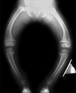
a. What is the stage of CKD and bone X-ray diagnosis?
b. Mention Kidney Disease Improving Global Outcomes (KDIGO) staging of CKD.
c. What are the steps in management?
d. What is the pathogenesis of this bony condition?
Answer
- a. CKD Stage 5 (G5) with renal osteodystrophy
- b. KDIGO Staging of CKD:
| Stage | Description | GFR (mL/min/1.73 m²) |
|---|---|---|
| G1 | Normal or high GFR | ≥90 |
| G2 | Mildly decreased | 60–89 |
| G3a | Mildly to moderately decreased | 45–59 |
| G3b | Moderately to severely decreased | 30–44 |
| G4 | Severely decreased | 15–29 |
| G5 | Kidney failure (End-stage renal disease) | <15 |
- c. Management Steps:
- Dietary phosphate restriction and non-calcium phosphate binders (e.g., sevelamer) or calcium-based binders
- Calcium supplementation and active vitamin D (calcitriol/alphacalcidol)
- Correction of metabolic acidosis with sodium bicarbonate
- Management of hyperkalemia and fluid balance
- Adequate renal replacement therapy (hemodialysis/peritoneal dialysis) and preparation for renal transplantation
- d. Pathogenesis: Decreased renal mass leads to reduced 1α-hydroxylase activity and low 1,25-(OH)₂D₃ levels
hypocalcemia and hyperphosphatemia (phosphate retention) secondary hyperparathyroidism increased bone resorption. Chronic metabolic acidosis also directly leaches bone minerals.
THG21-023
An 8-year-old boy weighing 24 kg with the height of 130 cm is admitted following a road traffic accident with crush injury of the left upper limb. His renal function test shows a BUN of 168 mg/dL and serum creatinine of 8.3 mg/dL and needs hemodialysis.
a. Calculate the estimated glomerular filtration rate (eGFR) for this child by modified Schwartz formula.
b. What is the KDIGO classification of AKI?
c. What is the stage of AKI in this child?
d. Mention at least four indications for kidney replacement therapy (KRT) in AKI.
e. List two urinary indices which are useful to differentiate intrinsic renal from prerenal AKI
Answer
-
a. eGFR Calculation (Modified Schwartz formula):


-
b. KDIGO Classification and Criteria for AKI:
| Stage | Serum Creatinine Criteria | Urine Output Criteria |
|---|---|---|
| 1 | 1.5–1.9 times baseline OR |
|
| 2 | 2.0–2.9 times baseline | |
| 3 |
- c. Stage 3 AKI
- d. Indications for KRT in AKI:
- Volume overload / pulmonary edema refractory to diuretics
- Refractory hyperkalemia (
with ECG changes) - Severe refractory metabolic acidosis (
) - Uremic complications (encephalopathy, pericarditis, neuropathy, bleeding)
- Severe calcium-phosphate imbalance / tumor lysis syndrome
- e. Urinary Indices Differentiating Prerenal vs Intrinsic AKI:
| Parameter | Prerenal AKI | Intrinsic Renal AKI |
|---|---|---|
| Urine microscopy | Normal / Hyaline casts | Granular / cellular / muddy brown casts |
| Urine Specific Gravity | >1.020 | <1.010 |
| Urine Osmolality (mOsm/kg) | >500 | <300 |
| Urinary Sodium ( |
<20 | >40 |
| Fractional Excretion of Sodium ( |
<1% | >2% |
| Fractional Excretion of Urea ( |
<35% | >35% |
THG21-024
A 2.5-month-old child with a weight of 3.8 kg and coexisting transposition of great arteries (TGA) with a baseline urea and creatinine of 35/0.4 mg/dL, underwent cardiopulmonary bypass for 2 hours. Creatinine postoperative 0.8 mg/dL. Urine output is 0.3 mL/kg/hour. Child is on inotropic support (adrenaline, dopamine, and noradrenaline). Initially child was given IV Lasix 0.1 mg/kg/hour. But urine output failed to improve. Below are mentioned laboratory parameters (creatinine and urea on increasing trend) and child was started on peritoneal dialysis (PD).
| Parameter | Value |
|---|---|
| Creatinine | 1.2 mg/dL |
| Na | 142 mEq/L |
| K | 6.6 mEq/L |
| Cl | 99 mEq/L |
| HCO3 | 14 mEq/L |
| SGOT | 24 IU/L |
| Ammonia | 15 µmol/L |
| SGPT | 30 IU/L |
| Serum albumin | 3.2 g/dL |
a. Explain the principles of PD?
b. How to investigate a child with suspected peritonitis?
c. What is the glucose concentration of acute PD?
d. What are the absolute contraindications for PD (mention any four)?
Answer
- a. Principles of Peritoneal Dialysis:
- Diffusion: Movement of solutes across peritoneal membrane down concentration gradient
- Convection / Ultrafiltration: Movement of water and dragged solutes driven by osmotic gradient created by dextrose
- Absorption: Fluid and solute reabsorption via lymphatic stomata
- b. Investigating Suspected PD Peritonitis:
- Dialysis effluent cell count and differential (WBC >100/µL with >50% polymorphonuclear leukocytes)
- Dialysis effluent Gram stain and culture (bacterial/fungal)
- c. 1.7% (or 1.5%–2.5%) dextrose concentration
- d. Absolute Contraindications for PD:
- Omphalocele / Gastroschisis
- Bladder exstrophy
- Congenital diaphragmatic hernia
- Obliterated peritoneal cavity / severe peritoneal adhesions / peritoneal membrane failure
THG21-025
A 4-year-old male child has undergone MCU.
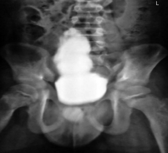
a. Describe the findings in this MCU?
b. What is Hinman syndrome?
c. What step done on daily basis with sterile precautions will prevent or retard the progression of CKD in this child?
d. Is there any need for chemoprophylaxis?
Answer
- a. Christmas tree (or fir-tree) appearance of the bladder with trabeculations and diverticula, suggestive of neurogenic bladder.
- b. Hinman syndrome: Non-neurogenic neurogenic bladder characterized by functional bladder outlet obstruction due to voluntary external sphincter dyssynergia in the absence of neurological or anatomical lesions.
- c. Clean intermittent catheterization (CIC)
- d. Chemoprophylaxis is not routinely recommended for all patients on CIC unless there are recurrent symptomatic febrile UTIs or high-grade VUR.
THG21-026
A 13-day-old term baby girl with antenatally diagnosed renal anomaly was now brought for follow-up. She had stable vitals. Other systems are normal. Evaluation showed serum urea/creatinine—20/0.4, serum Na—138, serum K—4.8, serum HCO3—22, and urinary protein-creatinine ratio (UPCR)—0.5.
- USG: Right kidney (RK)—6.2 cm (compensatory hypertrophy); Left kidney (LK)—2.8 cm, multiple noncommunicating cysts seen.
- DMSA scan: RK (100%), no scars; LK—not visualized.
- MCU: Grade III reflux on RK. No documented UTI, blood pressure was normal, and follow-up renal function test (RFT)—normal.
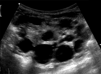
a. What is the possible diagnosis in this antenatally detected cystic kidney disease which involves only one kidney?
b. Mention any three complications of the above mentioned condition.
c. What is the common association in contralateral kidney?
d. Mention at least two syndromes associated with cystic kidney disease.
Answer
- a. Multicystic dysplastic kidney (MCDK)
- b. Complications of MCDK:
- Vesicoureteral reflux (VUR)
- Urinary tract infections (UTI)
- Hypertension
- c. Contralateral vesicoureteral reflux (VUR in up to 30%), ureteropelvic junction obstruction (UPJO), or compensatory hypertrophy
- d. Syndromes associated with cystic kidney disease:
- Tuberous sclerosis
- Bardet–Biedl syndrome
- Beckwith–Wiedemann syndrome
- Meckel–Gruber syndrome
- Joubert syndrome
- Zellweger syndrome
THG21-027
An 8-year-old male child presented with complaints of lower abdomen pain for 5 days. On evaluation, urine routine showed urine albumin—nil, RBCs—10–15/hpf, pus cells—1–2/hpf, and spot calcium/creatinine—0.8.
a. What is age-specific definition of hypercalciuria in spot urine sample?
b. What is the normal 24 hours urine calcium value?
c. What is the drug of choice for hypercalciuria?
d. What is the type of RTA associated with nephrocalcinosis?
Answer
- a. Age-specific spot urine calcium/creatinine (Ca/Cr) ratio thresholds:
- <6 months: >0.8 mg/mg
- 6 months to 2 years: >0.5 mg/mg
-
2 years: >0.2 mg/mg
- b. Normal 24-hour urine calcium: <4 mg/kg/day
- c. Hydrochlorothiazide (thiazide diuretics promote distal tubular calcium reabsorption) along with potassium citrate/alkali therapy
- d. Distal renal tubular acidosis (Type 1 RTA)
THG21-028
An ambulatory monitoring investigation is shown:
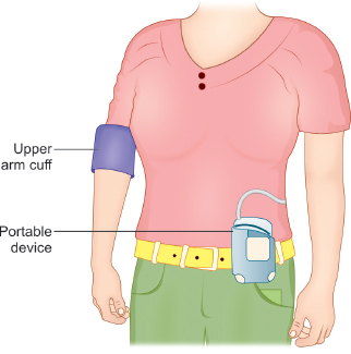
a. What procedure does the figure depict and define the same?
b. Mention at least two indications of this procedure.
c. What is white-coat hypertension?
d. What percentage of total BP readings measured by this device above the threshold is suggestive of hypertension?
Answer
- a. Ambulatory blood pressure monitoring (ABPM): Diagnostic modality to measure BP continuously over 24 hours at regular intervals during daily wakefulness and sleep.
- b. Indications for ABPM:
- Confirming Stage 1 hypertension documented over multiple office visits
- High-risk children (CKD, coarctation of aorta, diabetes mellitus, obesity, obstructive sleep apnea)
- Differentiating masked hypertension from white-coat hypertension
- Monitoring response to antihypertensive therapy in CKD
- c. White-coat hypertension: BP >95th percentile in clinic/office setting but persistently normal (<95th percentile) on ABPM/home measurements.
- d. BP load >25% (readings above 95th percentile) is suggestive of hypertension.
THG21-029
A 6-year-old female child presented with complaints of fever, joint pain in both legs, diffuse abdominal pain, and rashes noted in both lower limbs since 5 days. She also complaints of passing cola colored urine for past 2 days.
- On examination: Palpable petechiae/purpura noted, BP—130/88 mm Hg
- Laboratory test: Urine albumin—3+, red blood cells (RBCs) cast present, serum urea—12, serum creatinine—0.3 mg/dL
a. What is your diagnosis?
b. What are the diagnostic criteria?
c. What is the finding expected in skin biopsy?
d. What are the expected renal manifestations in this condition (mention any four)?
Answer
- a. Henoch–Schönlein purpura (IgA vasculitis) with nephritis
- b. EULAR/PRINTO/PRES Diagnostic Criteria: Palpable purpura (mandatory) plus at least one of:
- i. Diffuse abdominal pain
- ii. Arthritis or arthralgia
- iii. Renal involvement (hematuria and/or proteinuria)
- iv. Biopsy showing leukocytoclastic vasculitis with predominant IgA deposition or proliferative glomerulonephritis with IgA deposits
- c. Leukocytoclastic vasculitis with predominant IgA immune complex deposition on direct immunofluorescence
- d. Renal Manifestations:
- Microscopic / gross hematuria
- Subnephrotic proteinuria
- Nephrotic syndrome
- Acute nephritic syndrome / reduced eGFR
- Rapidly progressive glomerulonephritis (RPGN)
THG21-030
An 8-year-old male child presented with pain abdomen. On taking a detailed history, the parents revealed that the mother was told to have RBCs in urine and his maternal uncle aged 48 years has deafness and is on dialysis. On examination, his BP is 120/90 mmHg. On evaluation, his urine routine showed albumin 2+, RBC—15–20/hpf.
a. What is the most likely diagnosis in this child?
b. What other tests would you do in this child to support the diagnosis?
c. How will you confirm the diagnosis?
d. What is the likely mode of inheritance in this child?
e. What is the characteristic electron microscopy findings in renal biopsy?
Answer
- a. Alport syndrome
- b. Audiological evaluation (pure tone audiometry for high-frequency sensorineural hearing loss) and ophthalmological evaluation (anterior lenticonus, macular flecks)
- c. Genetic testing (mutations in COL4A5, COL4A3, or COL4A4)
- d. X-linked dominant (most common, COL4A5 mutation) / X-linked pattern
- e. Basket-weave appearance of the glomerular basement membrane (irregular thinning, thickening, and lamellation/splitting of the lamina densa)