MNJ30-001
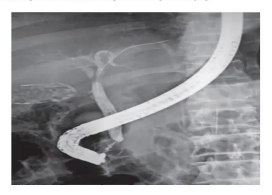
- Identify the radiological imaging modality.
- Mention two indications for this procedure.
- Mention two precautions / complications to be aware of prior to doing this imaging.
Answer
- Endoscopic Retrograde Cholangiopancreatography (ERCP).
- Indications:
- Obstructive jaundice (choledocholithiasis, biliary stricture, choledochal cyst)
- Chronic pancreatitis (ductal stones, strictures, pseudocyst drainage)
- Biliary / pancreatic tumors or biliary leaks post-surgery
- Sphincter of Oddi dysfunction.
- Precautions / Complications:
- Risk of acute post-ERCP pancreatitis (most common complication)
- Risk of duodenal perforation and cholangitis / sepsis
- Hemorrhage following endoscopic sphincterotomy
- Ensure pre-procedure correction of coagulopathy and antibiotic prophylaxis if biliary obstruction is present.
MNJ30-002
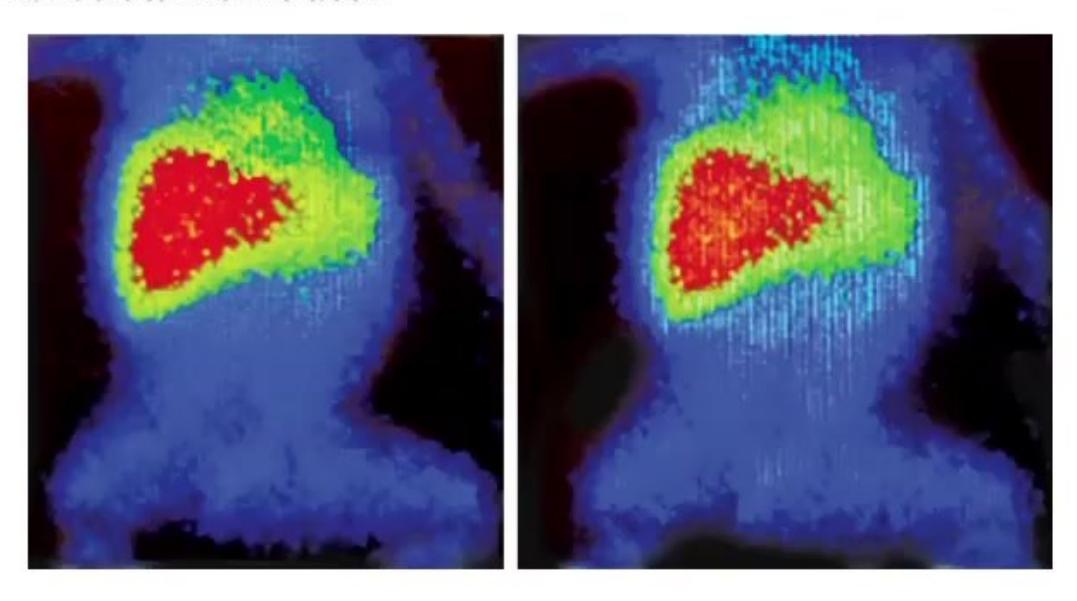
- What is this modality of investigation?
- Name one common pediatric indication.
Answer
- Hepatobiliary Scintigraphy (
-HIDA / Disofenin / Mebrofenin scan). - Key indication: Evaluation of neonatal cholestasis to differentiate Biliary Atresia (absence of tracer excretion into bowel at 24 hours despite prompt hepatic uptake) from Neonatal Hepatitis (delayed uptake with eventual bowel excretion).
MNJ30-003

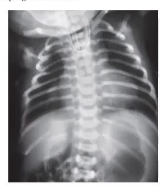
- What is the radiological diagnosis?
- What is the typical clinical presentation in the newborn?
- What is the requirement of echocardiography before surgery?
- What are poor surgical prognostic factors (Waterston / Spitz criteria)?
Answer
- Tracheoesophageal Fistula with Esophageal Atresia (Type C: proximal blind pouch with distal TE fistula; showing coiled red rubber catheter in proximal pouch and gas in stomach/bowel).
- Clinical presentation: Excessive frothy salivation/drooling, choking, coughing, and cyanosis with the first feed.
- Echocardiography requirement:
- To rule out associated congenital heart disease (VACTERL association, present in ~35%)
- To determine the laterality of the aortic arch (right-sided aortic arch in ~5% requires left thoracotomy instead of standard right thoracotomy).
- Poor prognostic factors:
- Birth weight
- Major congenital cardiac anomaly
- Long-gap atresia (
or vertebral bodies) - Severe preoperative pneumonia / ventilator dependence.
- Birth weight
MNJ30-004
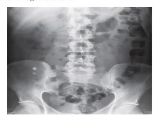
An 8-year-old female child Ritu presents with acute pain in the abdomen, fever, and vomiting for 12 hours. Child looks dehydrated on examination with tenderness at the right iliac fossa. X-ray and USG are shown:
- What is the clinical diagnosis?
- What does the X-ray show?
- What are the USG criteria for this disease?
- What is the most specific physical sign on clinical examination?
- What is the investigation of choice and gold standard?
Answer
- Acute Appendicitis.
- Radio-opaque calcified density in right lower quadrant representing an Appendicolith (fecalith).
- USG criteria (graded compression):
- Non-compressible, blind-ended tubular structure in RLQ
- Outer appendiceal diameter
- Appendiceal wall thickness
(target / bull's-eye sign on cross-section) - Hyperemia on color Doppler ("ring of fire") and periappendiceal hyperechoic fat / fluid collection.
- Point of maximal tenderness over McBurney's point (with localized rebound tenderness and guarding; Rovsing sign / Psoas sign).
- Ultrasonography is first-line; Contrast-Enhanced Computed Tomography (CECT) of the abdomen is the gold standard imaging modality.
MNJ30-005
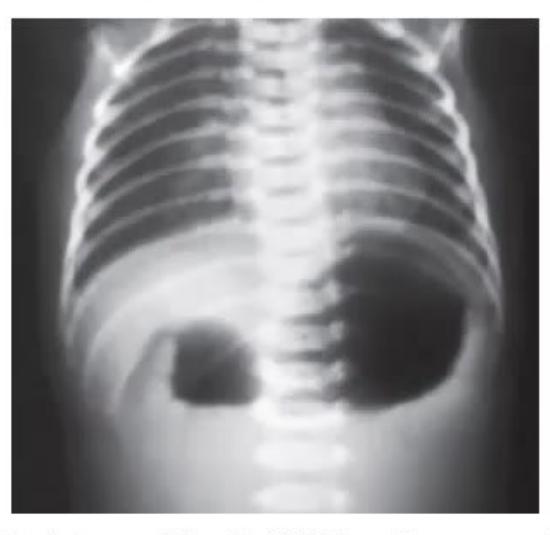
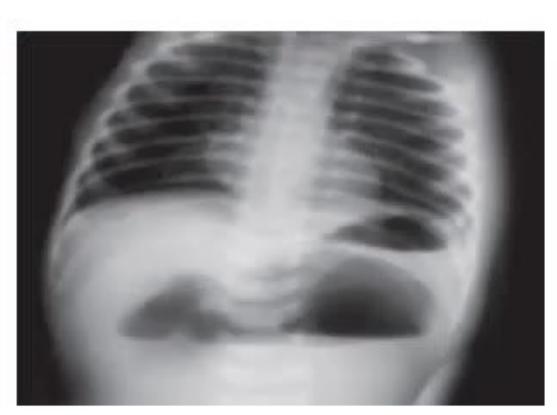
- Describe the signs and their causes in Image A and Image B.
- What syndrome is strongly associated with Image A?
- What associated anatomical anomaly is seen with Image A?
Answer
- Radiological signs:
- Image A: "Double bubble" sign (gas-filled distended stomach and first part of duodenum with absence of distal gas), diagnostic of Duodenal Atresia.
- Image B: "Triple bubble" sign (gas in stomach, duodenum, and proximal jejunum), diagnostic of Jejunal Atresia.
- Associated syndrome: Down syndrome (Trisomy 21, present in ~30% of duodenal atresia cases).
- Associated congenital anomalies: Annular pancreas, malrotation of gut, esophageal atresia, congenital heart defects (AVSD), and biliary atresia.
MNJ30-006
An 11-month-old male child Neeraj presents with severe constipation since birth. Mother gave a history of delayed passage of meconium beyond 48 hours:
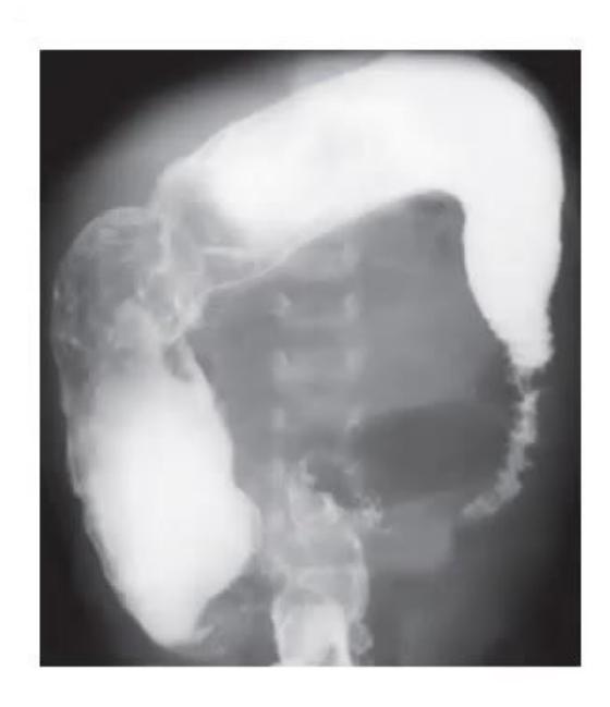
- What is this study, and what is the radiological interpretation?
- What is the diagnosis?
- How will you differentiate it clinically from functional constipation?
- What is the gold standard investigation of choice?
- What is the definitive treatment?
Answer
- Contrast (Barium) enema study: Shows a narrow, constricted aganglionic rectosigmoid segment with a funnel-shaped "transition zone" leading into a markedly dilated, hypertrophied proximal colon.
- Hirschsprung Disease (Congenital Aganglionic Megacolon).
- Differentiation from Functional Constipation:
- Onset: Hirschsprung presents in neonatal period with delayed meconium (
); functional constipation presents after 1–2 years (during toilet training). - Digital Rectal Exam: In Hirschsprung, rectum is empty, tight, followed by an explosive gush of liquid stool and gas upon withdrawal; in functional constipation, rectal ampulla is loaded with hard stool.
- Soiling/encopresis: Common in functional; absent in Hirschsprung.
- Growth: Failure to thrive common in Hirschsprung; normal in functional.
- Onset: Hirschsprung presents in neonatal period with delayed meconium (
- Investigation of choice: Full-thickness or suction Rectal Biopsy (taken at least
above dentate line) demonstrating absence of ganglion cells in submucosal (Meissner) and myenteric (Auerbach) plexuses, with hypertrophied nerve bundles and increased acetylcholinesterase (AChE) staining. - Definitive treatment: Surgical pull-through procedure (e.g., Swenson, Duhamel, Soave, or transanal endorectal pull-through [TEPT]).
MNJ30-007
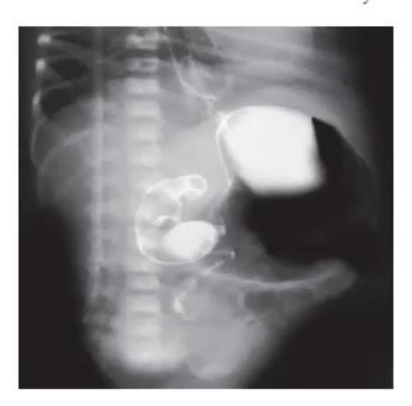
- What is the radiological investigation?
- What sign is demonstrated?
- What is the diagnosis?
- What is the commonest age group in which this condition occurs?
- What are other conditions associated with this abnormality?
Answer
- Upper Gastrointestinal (UGI) Barium contrast series / follow-through.
- "Corkscrew" sign (spiral appearance of twisted, obstructed duodenum and proximal jejunum on the right side of the spine with low/abnormal ligament of Treitz).
- Midgut Malrotation with Volvulus.
- Peak age of presentation: First month of life (
present in the first month/year of life with sudden bilious emesis). - Associated anomalies: Congenital diaphragmatic hernia, omphalocele/gastroschisis, duodenal atresia, annular pancreas, heterotaxy / polysplenia / asplenia syndromes.
MNJ30-008
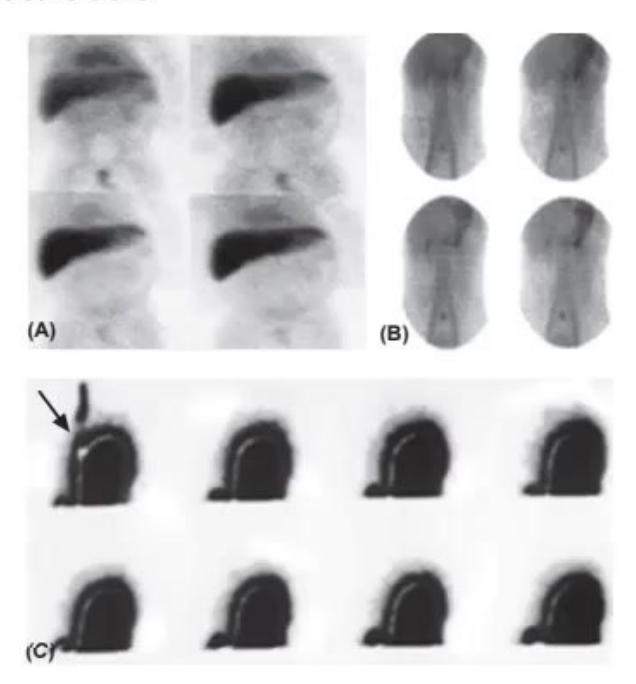
- Write down the names of the three diagnostic radionuclide studies.
- Give the diagnostic indication for each condition.
Answer
- Radionuclide studies:
- a.
-Mebrofenin / HIDA Scan (Hepatobiliary scintigraphy) - b.
-Pertechnetate Scan (Meckel's scan) - c.
-Sulfur Colloid / Milk Gastroesophageal Scintigraphy (Milk scan / Gastric emptying scan)
- a.
- Indications:
- a. HIDA scan
Diagnosis of Biliary Atresia vs neonatal hepatitis. - b. Meckel's scan
Detection of ectopic gastric mucosa in Meckel Diverticulum presenting with painless lower GI bleeding. - c. Milk scan
Evaluation of Gastroesophageal Reflux Disease (GERD), delayed gastric emptying, and pulmonary aspiration.
- a. HIDA scan
MNJ30-009
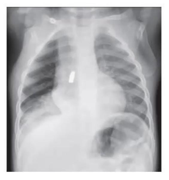
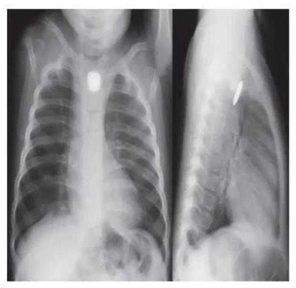
- Identify these two images and give the anatomical location and diagnosis.
- How will you differentiate a tracheal foreign body from an esophageal foreign body by plain X-ray?
Answer
- Image A: Foreign body (coin) in the Esophagus (coronal orientation in AP view). Image B: Foreign body in the Trachea / Right Main Bronchus (sagittal orientation in AP view).
- X-ray differentiation of flat radiopaque foreign bodies (e.g., coins):
- Esophageal Foreign Body: Lies in the coronal plane
Appears flat / circular ("face-on") on AP/PA view and end-on / edge-on on lateral view (because the flexible esophagus is compressed anteroposteriorly by the trachea). - Tracheal / Airway Foreign Body: Lies in the sagittal plane
Appears edge-on / end-on on AP view and flat / circular on lateral view (due to the rigid U-shaped cartilaginous tracheal rings with flexible posterior trachealis muscle).
- Esophageal Foreign Body: Lies in the coronal plane
MNJ30-010
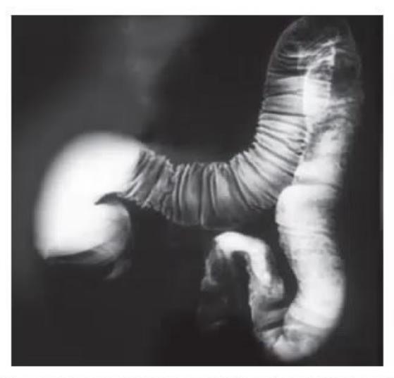
A 4-day-old neonate born to Anita presents with failure to pass meconium since birth, abdominal distension, and bilious vomiting. Plain radiograph and contrast enema are shown:
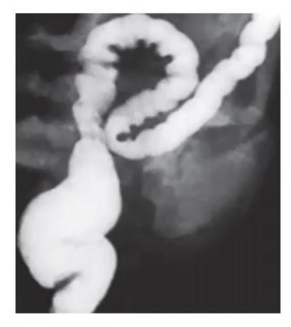
- What is the diagnosis based on these images?
- What is the associated underlying genetic disorder and responsible gene?
- What is the characteristic operative finding in the dilated ileal segment during laparotomy?
Answer
- Meconium Ileus (showing soap-bubble / Neuhauser sign on plain radiograph and microcolon of disuse with inspissated meconium pellets in terminal ileum on Gastrografin enema).
- Cystic Fibrosis (Autosomal recessive; mutations in the CFTR gene on chromosome 7q31.2, most commonly
). - Operative findings: Dilated distal ileum impacted with thick, tenacious, rubbery, dark-green tarry / inspissated mucus-rich meconium pellets causing complete intraluminal obstruction.
MNJ30-011
- Identify the clinical condition from the imaging.
- Write down the classical clinical triad.
- What is the classical finding on contrast / air enema?
- What is the initial treatment of choice?
Answer
- Ileocolic Intussusception (showing classical "coiled-spring" appearance and "claw sign" on contrast study; "target/doughnut" sign on USG).
- Classic triad:
- Paroxysmal colicky abdominal pain (with crying and drawing up of legs)
- Palpable sausage-shaped abdominal mass (typically in right hypochondrium / epigastrium with emptiness in RIF [Dance sign])
- Passage of "red currant jelly" stools (blood mixed with mucus).
- Contrast / Barium enema: "Coiled spring" appearance (contrast tracking around the intussusceptum) and meniscus/cupping filling defect.
- Treatment: Non-operative reduction under fluoroscopic/ultrasound guidance via Air (pneumatic) or Hydrostatic (saline/barium) enema; emergency laparotomy if perforation, peritonitis, or failed enema reduction.
MNJ30-012
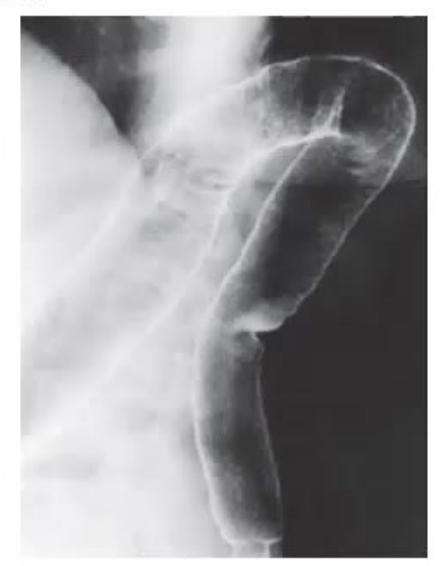
A 14-year-old girl has a 2-month history of bloody diarrhea and 10 kg weight loss. On examination she looks pale. A barium enema examination is performed:
- What abnormal features are shown on this X-ray?
- What is the diagnosis?
- What major complications can develop? (Write any 4)
- Name drugs used to induce and maintain remission in this condition.
Answer
- Loss of normal colonic haustrations, mucosal granularity, luminal narrowing, and featureless tubular rigid colon ("lead-pipe" colon sign).
- Ulcerative Colitis (Inflammatory Bowel Disease).
- Complications:
- Toxic megacolon (fulminant colonic dilatation
) - Colonic perforation and peritonitis
- Severe lower gastrointestinal hemorrhage
- Colorectal adenocarcinoma (risk increases after 8–10 years of extensive colitis)
- Extraintestinal manifestations: Primary Sclerosing Cholangitis (PSC), erythema nodosum, pyoderma gangrenosum, uveitis/episcleritis, arthritis.
- Toxic megacolon (fulminant colonic dilatation
- Pharmacotherapy:
- 5-Aminosalicylates (5-ASA): Mesalamine, Sulfasalazine (first-line for induction and maintenance in mild-moderate disease)
- Corticosteroids: Oral Prednisolone or IV Methylprednisolone (for acute flares; not for maintenance)
- Immunomodulators: Azathioprine, 6-Mercaptopurine
- Biologics: Anti-TNF agents (Infliximab, Adalimumab), Vedolizumab, Tofacitinib.
MNJ30-013
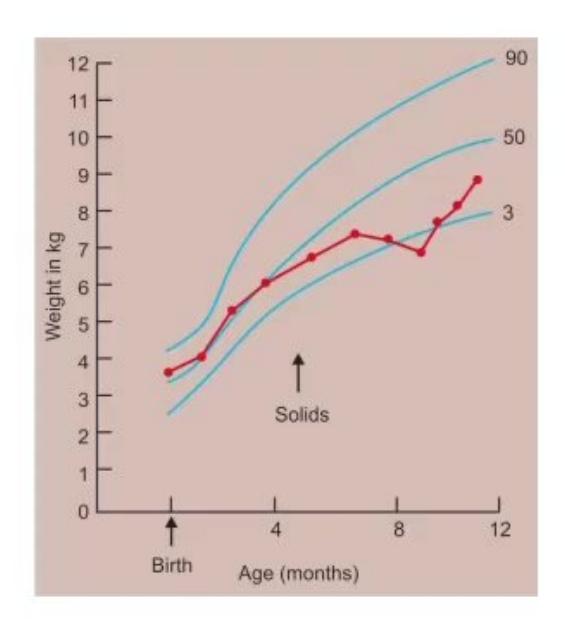
A baby born at term (birth weight 3.6 kg) was exclusively breastfed for 4.5 months when solid cereals were introduced. She presents at 8 months with poor weight gain, chronic diarrhea, vomiting, and irritability. On examination: pale, abdominal distension, and muscle wasting. Sweat test is normal:
- What is the most likely diagnosis?
- What is the gold-standard confirmatory investigation?
- What immediate therapeutic intervention resulted in the catch-up growth seen on the chart?
- State whether the following foods are Permitted (Yes) or Prohibited (No):
- a. Cornflakes
- b. Maggi noodles (wheat flour)
- c. Sago (sabudana)
- d. Oats cereal
- e. Bajra roti (pearl millet)
- f. Dalia (broken wheat)
Answer
- Celiac Disease (Gluten-Sensitive Enteropathy).
- Gold standard: Upper GI Endoscopy with multiple (at least 4–6) duodenal/jejunal mucosal biopsies showing Marsh classification III changes (total/subtotal villous atrophy, crypt hyperplasia, and increased intraepithelial lymphocytes
enterocytes). Supported by serology: Anti-tissue transglutaminase (anti-tTG IgA) and anti-endomysial antibody (EMA IgA). - Initiation of a strict, lifelong Gluten-Free Diet (GFD).
- Food permissions:
- a. Cornflakes
Yes (Permitted if gluten-free certified) - b. Maggi noodles
No (Prohibited, contains wheat gluten) - c. Sago (sabudana)
Yes (Permitted) - d. Oats cereal
No (Prohibited unless certified pure uncontaminated gluten-free oats) - e. Bajra roti
Yes (Permitted, pearl millet is naturally gluten-free) - f. Dalia
No (Prohibited, broken wheat contains gluten)
- a. Cornflakes
(Permitted: a, c, e; Prohibited: b, d, f)
MNJ30-014
- What is the diagnosis?
- What is the embryological basis of these defects?
- In an otherwise healthy infant, what is the optimal timing of surgical repair?
Answer
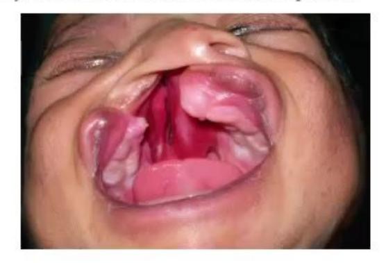
- Bilateral Cleft Lip and Complete Cleft Palate.
- Embryological basis:
- Cleft Lip: Failure of fusion between the medial nasal prominence and maxillary prominence on one or both sides (during 4th to 7th weeks of gestation).
- Cleft Palate: Failure of the secondary palatal shelves (lateral palatine processes) to fuse with each other and with the primary palate/nasal septum (during 7th to 12th weeks of gestation).
- Timing of surgical repair:
- Cleft Lip Repair (Cheiloplasty): Around 3 months of age (Rule of 10s: Weight
, , , age ). - Cleft Palate Repair (Palatoplasty): Around 9–12 months of age (before development of normal speech and phonation).
- Cleft Lip Repair (Cheiloplasty): Around 3 months of age (Rule of 10s: Weight
MNJ30-015
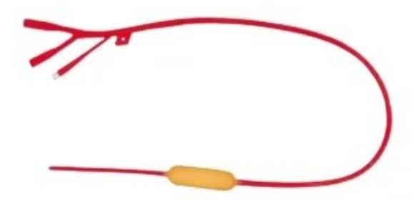
- Identify the instrument.
- What are the clinical indications for its use?
- What is the mechanism of its action?
Answer
- Sengstaken-Blakemore (SB) Tube (or Minnesota tube).
- Indication: Temporary emergency balloon tamponade for acute, life-threatening gastroesophageal variceal hemorrhage refractory to initial pharmacological therapy (octreotide/terlipressin) and endoscopic intervention (EVL/sclerotherapy).
- Mechanism: Triple-lumen tube with two inflatable balloons:
- Gastric balloon: Inflated with
of air and retracted against the gastroesophageal junction to compress submucosal feeding vessels and fundal varices. - Esophageal balloon: Inflated to a pressure of
to provide direct mechanical compression against bleeding esophageal varices.
- Gastric balloon: Inflated with
MNJ30-016
- Describe the nutritional norms for the Mid-Day Meal (PM POSHAN) scheme in primary school children.
- According to national standards, what is the minimum number of feeding days per year required to achieve a meaningful nutritional impact?
Answer
- PM POSHAN (Mid-Day Meal) Nutritional Norms:
- Primary (Classes I–V): At least 450 calories and 12 g protein per child per day (100 g food grains, 20 g pulses, 50 g vegetables, 5 g oil).
- Upper Primary (Classes VI–VIII): At least 700 calories and 20 g protein per child per day (150 g food grains, 30 g pulses, 75 g vegetables, 7.5 g oil).
- Micronutrient supplementation: Weekly Iron and Folic Acid (WIFS), biannual deworming (Albendazole).
- Minimum feeding days: At least 200 days/year for primary schools and 220 days/year for upper primary schools.
MNJ30-017
A 2-year-old child is evaluated 18 months after an unsuccessful Kasai portoenterostomy for extrahepatic biliary atresia. He is jaundiced with signs of cirrhosis and portal hypertension. He is drowsy, has an ataxic gait, absent deep tendon reflexes, and loss of proprioception. What specific nutrient deficiency has led to this neuromuscular disorder?
Answer
Severe Vitamin E deficiency (due to chronic severe fat malabsorption and cholestasis leading to spinocerebellar ataxia, posterior column loss, peripheral neuropathy, and absent DTRs).
MNJ30-018
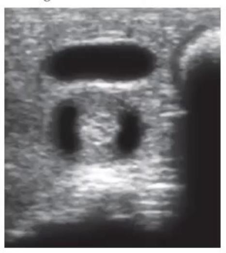
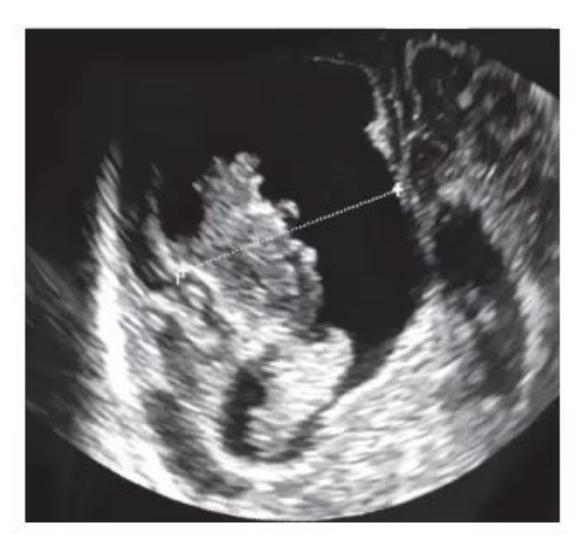
- Identify the ultrasound findings and write down the sonographic criteria for diagnosis of this disease.
- What medical treatment can be attempted in non-surgical candidates?
- What is the classic triad of gastric volvulus (Borchardt triad)?
Answer
- Infantile Hypertrophic Pyloric Stenosis (IHPS) (showing elongated pyloric canal and "doughnut / target" sign on transverse view, "cervix sign" on longitudinal view).
- USG diagnostic criteria:
- Pyloric muscle wall thickness
(single muscular wall) - Pyloric canal length
- Transverse pyloric diameter
- Pyloric muscle wall thickness
- USG diagnostic criteria:
- Medical therapy: Oral or IV Atropine sulfate (
titrated) with incremental feeds (reserved for cases where surgery is contraindicated). - Borchardt's Triad of Acute Gastric Volvulus:
- Severe, sudden epigastric pain and distension
- Intractable retching with minimal or no emesis
- Inability to pass a nasogastric / orogastric tube into the stomach.
MNJ30-019
Match each clinical scenario with the most likely etiology:
- An 8-month-old female has recent-onset chronic diarrhea, recurrent otitis media, frequent respiratory infections, and failure to thrive. No fever or vomiting.
- An 18-month-old boy receiving Amoxicillin-Clavulanate for otitis media develops watery loose stools and erythematous diaper rash. No fever.
- A 3-year-old male presents with high fever (
), chills, febrile seizures, and bloody mucoid diarrhea. Other daycare children are also affected. - A 12-year-old female presents with postprandial bloating, flatulence, cramping abdominal pain, and watery diarrhea 1–2 hours after drinking milk.
Answer
- Primary Immunodeficiency / Selective IgA Deficiency / Cystic Fibrosis (Giardiasis / recurrent mucosal infections).
- Antibiotic-Associated Diarrhea (Clavulanate-induced hypermotility
secondary Candida diaper dermatitis). - Shigella dysentery (Shigellosis) (produces Shiga toxin inducing neurotoxicity/febrile seizures and invasive bloody diarrhea).
- Lactose Intolerance (Primary / Secondary lactase deficiency) (undigested lactose causes osmotic diarrhea and hydrogen gas fermentation).
MNJ30-020
A 13-year-old boy develops right upper-quadrant pain, fever with chills and rigors. Abdominal ultrasound reveals hyperechoic liver parenchyma, saccular dilatation of multiple intrahepatic bile ducts, and bilateral medullary renal cysts:
- What is the diagnosis?
- What is the cause of fever with chills in this patient?
- What are the key components of this syndrome?
- Which gene has been implicated in this condition?
Answer
- Caroli Disease (isolated ductal ectasia) / Caroli Syndrome (ductal ectasia associated with Congenital Hepatic Fibrosis).
- Recurrent bacterial Ascending Cholangitis (due to bile stasis, intrahepatic lithiasis, and bacterial superinfection).
- Components of Caroli Syndrome:
- Non-obstructive congenital segmental saccular dilatation of intrahepatic bile ducts
- Congenital Hepatic Fibrosis (portal hypertension, splenomegaly, varices)
- Autosomal Recessive Polycystic Kidney Disease (ARPKD / medullary sponge kidney / renal tubular ectasia).
- PKHD1 gene on chromosome 6p12 (encoding Fibrocystin / Polyductin).
MNJ30-021
Mention five key differences between Osmotic Diarrhea and Secretory Diarrhea:
| Feature | Osmotic Diarrhea | Secretory Diarrhea |
|---|---|---|
| Stool Volume | ||
| Response to Fasting (NPO) | ||
| Stool Sodium ( |
||
| Stool Reducing Substances | ||
| Stool Osmotic Gap | ||
| Stool pH |
Answer
| Feature | Osmotic Diarrhea | Secretory Diarrhea |
|---|---|---|
| Stool Volume | Usually |
Large volume, |
| Response to Fasting | Stops / Diarrhea resolves | Continues unabated |
| Stool Sodium | Low ( |
High ( |
| Stool Reducing Substances | Positive (in carbohydrate malabsorption) | Negative |
| Stool Osmotic Gap | Elevated ( |
Low ( |
| Stool pH | Acidic ( |
Neutral / Alkaline ( |
MNJ30-022
With regard to hormone-secreting tumors causing diarrhea:
- Mention 5 hormones/peptides involved.
- Mention the associated tumor/disease for each.
- Mention one specific pharmacological or surgical treatment for each.
Answer
- Vasoactive Intestinal Peptide (VIP): VIPoma (Verner-Morrison / WDHA syndrome)
Somatostatin analogs (Octreotide), tumor resection. - Gastrin: Gastrinoma (Zollinger-Ellison syndrome)
High-dose PPIs (Omeprazole), surgical excision. - Serotonin / Tachykinins: Carcinoid tumor (Carcinoid syndrome)
Octreotide, Cyproheptadine, surgical resection. - Calcitonin: Medullary Thyroid Carcinoma (MTC)
Total thyroidectomy with central neck dissection, Vandetanib/Cabozantinib. - Histamine: Systemic Mastocytosis / Mastocytoma
antihistamines, Cromolyn sodium.
MNJ30-023
- Mention three types of Progressive Familial Intrahepatic Cholestasis (PFIC) with the molecular/transporter defect in each.
- Mention the dosage of fat-soluble vitamins required for supplementation in a child with prolonged cholestasis.
Answer
- PFIC Types:
- PFIC Type 1 (Byler disease): Inactivating mutation in ATP8B1 gene (encoding FIC1 flippase); normal/low serum GGT.
- PFIC Type 2 (Byler syndrome): Mutation in ABCB11 gene (encoding BSEP - Bile Salt Export Pump); normal/low serum GGT, high risk of early HCC.
- PFIC Type 3: Mutation in ABCB4 gene (encoding MDR3 - Multidrug Resistance Protein 3 / phosphatidylcholine translocator); High serum GGT.
- Fat-Soluble Vitamin Supplementation in Cholestasis:
- Vitamin A: 5,000–15,000 IU/day orally (water-miscible / Aquasol A)
- Vitamin D: 3,000–8,000 IU/day of
or of Alfacalcidol / Calcitriol - Vitamin E: 50–200 IU/kg/day of d-
-tocopheryl polyethylene glycol-1000 succinate (TPGS) - Vitamin K: 2.5–5 mg orally 2–7 times weekly or
IV/SC monthly.
MNJ30-024
- Mention 5 conditions that present with a clinical/pathologic picture resembling Reye syndrome (Reye-like metabolic disorders).
- Outline the clinical staging of Reye syndrome (Lovejoy staging).
Answer
- Conditions mimicking Reye syndrome:
- Medium-Chain Acyl-CoA Dehydrogenase (MCAD) deficiency and other Fatty Acid Oxidation Disorders (FAODs)
- Urea Cycle Disorders (e.g., OTC deficiency)
- Organic acidemias (Methylmalonic, Propionic, Isovaleric acidemia)
- Carnitine palmitoyltransferase (CPT I/II) deficiency
- Mitochondrial disorders / Glutaric aciduria type II
- Toxic ingestion: Aspirin toxicity, Valproate hepatotoxicity, Jamaican vomiting sickness (hypoglycin A).
- Lovejoy Clinical Staging of Reye Syndrome:
- Stage I: Vomiting, lethargy, sleepiness, lab evidence of liver dysfunction (hyperammonemia, elevated transaminases).
- Stage II: Delirium, combativeness, disorientation, hyperventilation, hyperactive reflexes.
- Stage III: Light coma, obtundation, decorticate rigidity, intact pupillary light reflexes.
- Stage IV: Deepening coma, decerebrate rigidity, loss of oculocephalic reflexes, fixed dilated pupils, liver dysfunction improving.
- Stage V: Flaccidity, loss of DTRs, respiratory arrest, isoelectric EEG, death.
MNJ30-025
Mention five distinct patterns of Drug-Induced Liver Injury (DILI) with one representative drug for each:
| Pattern of Hepatic Injury | Representative Causative Drug |
|---|---|
| Centrilobular Necrosis | |
| Microvesicular Steatosis | |
| Hepatic Fibrosis / Cirrhosis | |
| Cholestasis / Cholestatic Hepatitis | |
| Biliary Sludge / Pseudolithiasis | |
| Hepatic Veno-Occlusive Disease (SOS) |
Answer
| Pattern of Hepatic Injury | Representative Causative Drug |
|---|---|
| Centrilobular Necrosis | Acetaminophen (Paracetamol overdose), Halothane |
| Microvesicular Steatosis | Valproic acid, Tetracycline, Aspirin |
| Hepatic Fibrosis / Cirrhosis | Methotrexate, Vitamin A toxicity |
| Cholestatic Hepatitis | Amoxicillin-Clavulanate, Chlorpromazine, Erythromycin estolate |
| Biliary Sludge / Pseudolithiasis | Ceftriaxone |
| Sinusoidal Obstruction Syndrome (VOD) | Busulfan, Cyclophosphamide, Actinomycin-D |
| Hepatic Vein Thrombosis (Budd-Chiari) | Oral contraceptive pills (Estrogens/Androgens) |
MNJ30-026
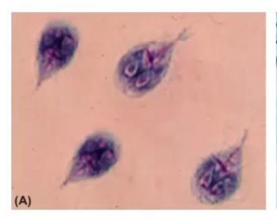
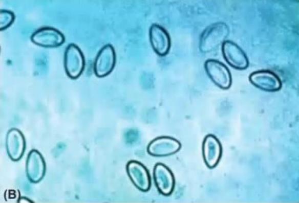
- Identify the parasite shown in Image A and Image B.
- What is the drug of choice for each?
Answer
- Image A: Giardia lamblia (trophozoite / cyst; characteristic teardrop-shaped binucleated flagellate). Image B: Enterobius vermicularis (Pinworm egg; planoconvex, D-shaped translucent ovum).
- Treatment:
- For Giardia: Metronidazole (
for 5–7 days), Tinidazole, or Nitazoxanide. - For Enterobius: Albendazole (
single dose, repeated after 2 weeks) or Mebendazole / Pyrantel pamoate (treat all household contacts).
- For Giardia: Metronidazole (
MNJ30-027
A 10-day-old neonate (born at 31 weeks preterm on formula feeds) presents with lethargy, feed intolerance, and abdominal distension:
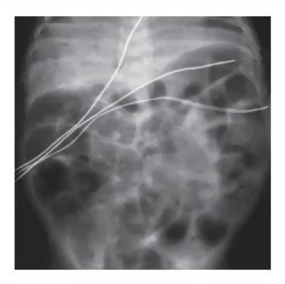
- What stage of Necrotizing Enterocolitis (NEC) is depicted on the X-ray?
- What is the hallmark radiological feature of Bell Stage II NEC vs Stage III NEC?
- Name two other pediatric conditions associated with pneumatosis intestinalis.
Answer
- Bell Stage II NEC (showing distinct intramural gas bubbles / Pneumatosis intestinalis and branching portal venous gas).
- Radiological criteria (Bell's Staging):
- Stage II (Definite NEC): Pneumatosis intestinalis
Portal venous gas. - Stage III (Advanced / Perforated NEC): Pneumoperitoneum (free intraperitoneal air under diaphragm / "football sign" / Rigler's double wall sign indicating bowel perforation requiring emergency surgical laparotomy/drainage).
- Stage II (Definite NEC): Pneumatosis intestinalis
- Other conditions with pneumatosis intestinalis: Hirschsprung-associated enterocolitis, severe rotavirus enteritis, ischemic bowel injury, immunosuppressed graft-versus-host disease (GVHD).
MNJ30-028
A 5-week-old infant presents with cholestatic jaundice and develops a tender swelling of the left thigh with an undisplaced femoral fracture:
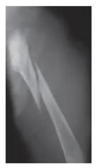
- What is the likely cause of the pathological femur fracture?
- How can this metabolic complication be prevented?
- How do you pharmacologically manage pruritus in infantile cholestasis?
- An infant with cholestasis, triangular facies, butterfly vertebrae, and a pulmonary stenosis murmur has what syndrome?
Answer
- Cholestatic Metabolic Bone Disease / Severe Hepatic Rickets (secondary to malabsorption of fat-soluble Vitamin D and calcium due to absence of intraluminal bile salts).
- Prevention: High-dose water-miscible Vitamin
(5,000–8,000 IU/day) or Calcitriol ( ), medium-chain triglyceride (MCT) enriched formulas, and elemental calcium supplementation ( ). - Medical management of pruritus:
- Ursodeoxycholic acid (UDCA):
- Rifampicin:
(enzyme inducer) - Cholestyramine:
(bile acid sequestrant) - Antihistamines (Hydroxyzine) / Opioid antagonists (Naltrexone).
- Ursodeoxycholic acid (UDCA):
- Alagille Syndrome (Arteriohepatic Dysplasia, autosomal dominant JAG1 or NOTCH2 mutation).
MNJ30-029
A two-hour-old neonate presents with scaphoid abdomen and severe respiratory distress since birth:
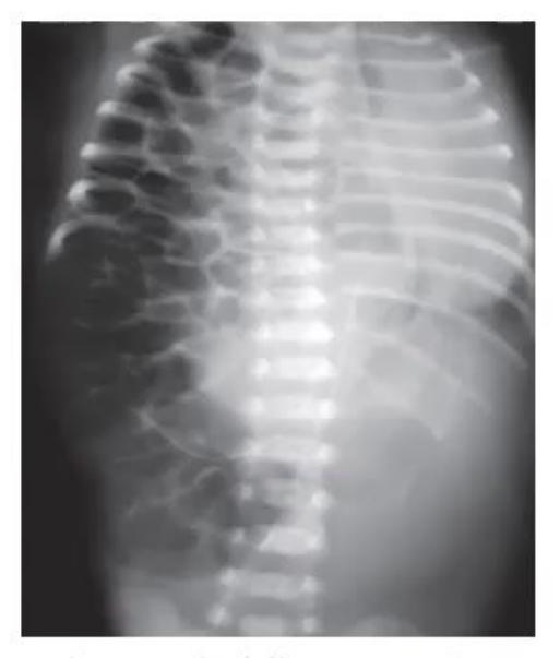
- What is the diagnosis?
- What embryological event leads to this development?
- What are three pathophysiological mechanisms causing respiratory failure in this newborn?
Answer
- Left-sided Congenital Diaphragmatic Hernia (Bochdalek Hernia) with mediastinal shift to the right.
- Embryological defect: Failure of fusion/closure of the posterolateral pleuroperitoneal membrane with the septum transversum and dorsal mesentery between the 8th and 10th weeks of gestation.
- Pathophysiological causes of respiratory distress:
- Ipsilateral and contralateral Pulmonary Hypoplasia (decreased bronchial branching and alveolar surface area)
- Severe Persistent Pulmonary Hypertension of the Newborn (PPHN) with right-to-left shunting
- Space-occupying compression of the lung parenchyma and mediastinum by herniated abdominal viscera.
MNJ30-030
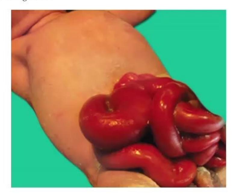
- What is the diagnosis?
- What is the primary differential diagnosis?
- What are the key differentiating features between the two conditions?
- What is the immediate neonatal management?
Answer
- Gastroschisis (full-thickness abdominal wall defect to the right of the intact umbilical cord, with evisceration of bowel loops devoid of a covering membrane).
- Differential diagnosis: Omphalocele (Exomphalos).
- Differentiating features:
| Feature | Gastroschisis | Omphalocele |
|---|---|---|
| Location | Paraumbilical (usually right) | Central (through umbilical ring) |
| Covering Sac | Absent (exposed edematous bowel) | Present (peritoneum + amnion) |
| Umbilical Cord Insertion | Normal to the left of defect | Onto the apex of the sac |
| Associated Anomalies | Rare ( |
Very Common (50–70%): Cardiac, Beckwith-Wiedemann, Trisomies |
- Immediate management:
- Wrap exposed bowel in sterile, warm saline-soaked gauze and place infant in a sterile bowel bag ("silo") up to the axillae to prevent heat and fluid loss.
- Insert NG/OG tube for gastric decompression.
- IV hydration (1.5–2 times maintenance due to fluid evaporative loss) and broad-spectrum IV antibiotics.
- Primary or staged surgical closure.
MNJ30-031
A 7-year-old girl with a family history of chronic liver disease presents with jaundice, Coombs-negative hemolytic anemia, and choreiform abnormal hand movements for 7 days:
- What bedside clinical examination will clinch the diagnosis?
- State the probable diagnosis.
- List three diagnostic laboratory tests with cutoff values.
- Outline the management of this child.
Answer
- Slit-lamp examination of the cornea for Kayser-Fleischer (KF) rings (copper deposition in Descemet's membrane).
- Wilson Disease (Hepatolenticular Degeneration) (autosomal recessive, ATP7B gene on chr 13q14).
- Diagnostic tests:
- Serum Ceruloplasmin: Low (
or ) - 24-hour Urinary Copper excretion: Elevated (
or ) - Hepatic copper concentration on liver biopsy:
dry weight (gold standard).
- Serum Ceruloplasmin: Low (
- Management:
- Copper chelators: D-Penicillamine (
with Pyridoxine/Vit ) or Trientine - Maintenance / Presymptomatic: Oral Zinc sulfate / acetate (
elemental zinc/day to induce intestinal metallothionein) - Dietary restriction: Avoid copper-rich foods (shellfish, nuts, chocolate, mushrooms, liver)
- Orthotopic Liver Transplantation for acute fulminant hepatic failure or decompensated cirrhosis.
- Copper chelators: D-Penicillamine (
MNJ30-032
Match each pathophysiology mechanism with its representative clinical condition:
| Pathophysiological Mechanism | Clinical Condition |
|---|---|
| 1. Secretory Diarrhea | a. Celiac disease (Villous atrophy) |
| 2. Osmotic Diarrhea | b. Vibrio cholerae infection |
| 3. Increased Intestinal Motility | c. Blind loop syndrome / SIBO |
| 4. Decreased Motility / Stasis | d. Thyrotoxicosis |
| 5. Decreased Absorptive Surface Area | e. Lactase deficiency |
Answer
-
- Secretory Diarrhea
b. Vibrio cholerae infection (enterotoxin-mediated CFTR stimulation)
- Secretory Diarrhea
-
- Osmotic Diarrhea
e. Lactase deficiency (carbohydrate malabsorption)
- Osmotic Diarrhea
-
- Increased Motility
d. Thyrotoxicosis
- Increased Motility
-
- Decreased Motility / Stasis
c. Blind loop syndrome / SIBO
- Decreased Motility / Stasis
-
- Decreased Absorptive Surface Area
a. Celiac disease (villous atrophy) / Short bowel syndrome
- Decreased Absorptive Surface Area
(Match: 1-b, 2-e, 3-d, 4-c, 5-a)
MNJ30-033
- Write down 4 metabolic causes of chronic constipation in children.
- Write down 4 clinical/metabolic complications of persistent severe vomiting.
- Calculate the Stool Osmotic (Anion) Gap given: Stool Osmolality = 290 mOsm/kg, Stool
, Stool . - What is the clinical importance of the stool osmotic gap?
Answer
- Metabolic causes of constipation: Hypothyroidism, Hypercalcemia, Hypokalemia, Diabetes insipidus, Renal tubular acidosis (RTA), Multiple Endocrine Neoplasia (MEN 2B).
- Complications of severe vomiting:
- Hypokalemic, hypochloremic metabolic alkalosis
- Dehydration and prerenal azotemia
- Mallory-Weiss mucosal tear / Boerhaave esophageal rupture
- Aspiration pneumonia / chemical pneumonitis
- Reflux esophagitis / tooth enamel erosion.
- Calculation:
- Clinical significance:
Osmotic Diarrhea (unabsorbed solutes present in stool). Secretory Diarrhea (pure electrolyte secretion).
MNJ30-034
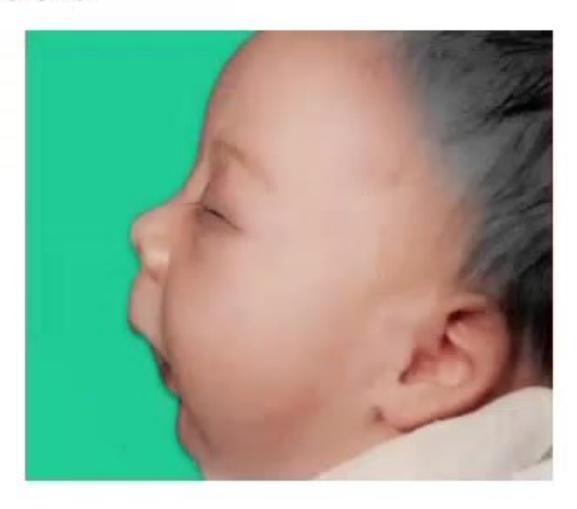
- Identify the clinical condition in this infant.
- Write down its classical clinical features (triad).
- Is this classified as a syndrome, sequence, or association?
- Write two other pediatric clinical examples sharing the same pattern of pathogenesis.
- What is Stickler syndrome?
Answer
- Pierre Robin Sequence (PRS).
- Classic triad:
- Severe Micrognathia / Retrognathia (hypoplastic mandible)
- Glossoptosis (posterior displacement of tongue causing airway obstruction)
- U-shaped (or V-shaped) Cleft Palate.
- It is a Sequence (a primary embryological defect—mandibular hypoplasia—initiates a cascade of secondary developmental abnormalities: preventing the tongue from descending, thereby obstructing midline fusion of the palatal shelves).
- Other clinical sequences:
- Potter Sequence (renal agenesis
oligohydramnios pulmonary hypoplasia + facial compression + clubfoot) - Prune Belly Sequence (urethral obstruction
massive megacystis abdominal muscle deficiency + cryptorchidism).
- Potter Sequence (renal agenesis
- Stickler Syndrome: An autosomal dominant connective tissue disorder (collagen COL2A1, COL11A1 mutations) characterized by Pierre Robin sequence associated with severe myopia, retinal detachment, sensorineural hearing loss, and premature spondyloepiphyseal degenerative arthropathy.
MNJ30-035
State True or False regarding Celiac Disease:
- Gliadins (wheat), hordeins (barley), and secalins (rye) are the toxic prolamins triggering disease.
- HLA-DQ6 is the primary human leukocyte antigen association.
- Isolated short stature and refractory iron deficiency anemia are common extraintestinal presentations.
- Bilateral occipital calcifications and intractable epilepsy can occur (Glaser syndrome).
- Enteropathy-associated T-cell lymphoma (EATL) is the most feared long-term neoplastic complication.
- A flat mucosal surface on duodenal biopsy is pathognomonic and seen only in celiac disease.
- Most patients tolerate
of gluten without mucosal damage, but universal safe threshold varies.
Answer
- True (Prolamin fractions of wheat, barley, and rye trigger the autoimmune mucosal response).
- False (Strongly associated with HLA-DQ2 [
] and HLA-DQ8 [ ]). - True (Non-classical celiac disease frequently presents without diarrhea as isolated short stature or refractory IDA).
- True (Celiac disease with epilepsy and bilateral occipital calcifications - CEC syndrome).
- True (Enteropathy-Associated T-Cell Lymphoma [EATL] is a severe malignancy associated with chronic untreated celiac disease).
- False (Total villous atrophy / flat mucosa is also seen in severe cow's milk protein allergy, autoimmune enteropathy, tropical sprue, and post-gastroenteritis enteropathy).
- True (Strict gluten-free diet aims for
/ gluten intake).
(Answers: 1-True, 2-False, 3-True, 4-True, 5-True, 6-False, 7-True)
MNJ30-036
A 6-year-old male child Mahapatra presents with progressive ataxia, bulky foul-smelling stools for 1 month, and poor school performance. Blood triglyceride level is 15 mg/dL:
- What is the diagnosis of this condition?
- What will you find on retinal examination and peripheral blood smear?
- Which parts of the nervous system are primarily affected?
- What is the treatment?
Answer
- Abetalipoproteinemia (Bassen-Kornzweig syndrome) (autosomal recessive mutation in the MTTP microsomal triglyceride transfer protein gene; failure to synthesize ApoB-48 and ApoB-100).
- Diagnostic findings:
- Retinal examination: Atypical Retinitis Pigmentosa (pigmentary retinopathy / loss of night and color vision).
- Peripheral blood film: Acanthocytes ("spur cells", thorny star-shaped erythrocytes present in
).
- Nervous system involvement: Degeneration of spinocerebellar tracts, posterior columns (proprioception/vibration loss), peripheral neuropathy, and cerebellar ataxia.
- Treatment:
- Restriction of dietary long-chain saturated fats with medium-chain triglyceride (MCT) oil supplementation
- Massive doses of fat-soluble vitamins: Vitamin E (
orally to halt neurological and retinal damage) and Vitamin A ( ).
MNJ30-037
Match the clinical scenario with the most likely foodborne pathogen:
| Clinical Scenario | Likely Pathogen |
|---|---|
| 1. Vomiting starting 1–6 hours after ingesting reheated fried rice | a. Salmonella enterica |
| 2. Fever, abdominal cramps, and bloody diarrhea 2–5 days after unpasteurized milk / poultry | b. Bacillus cereus (emetic type) |
| 3. Fever, vomiting, and diarrhea 12–36 hours after ingesting raw eggs / poultry | c. Campylobacter jejuni |
| 4. Severe nausea and profuse vomiting starting 2–6 hours after mayonnaise/potato salad at a party | d. Staphylococcus aureus (enterotoxin) |
Answer
- 1
b. Bacillus cereus (preformed emetic heat-stable toxin cereulide in reheated fried rice) - 2
c. Campylobacter jejuni (unpasteurized milk, undercooked poultry; pseudoappendicitis/bloody diarrhea) - 3
a. Salmonella enterica (eggs, poultry; inflammatory gastroenteritis) - 4
d. Staphylococcus aureus (preformed enterotoxins A–E in dairy/mayonnaise salads)
MNJ30-038
Match the diagnostic stool test with its clinical utility:
| Stool Diagnostic Test | Clinical Application / Significance |
|---|---|
| 1. Fecal |
a. Pancreatic exocrine insufficiency (stable enzyme) |
| 2. Fecal Elastase-1 | b. Enteric protein loss (Protein-Losing Enteropathy) |
| 3. Fecal Calprotectin | c. Intestinal / colonic mucosal neutrophil inflammation (IBD) |
| 4. Stool Reducing Substances | d. Carbohydrate malabsorption ( |
| 5. Fecal Chymotrypsin | e. Pancreatic exocrine function |
| 6. Dual-sugar (Lactulose/Mannitol) test | f. Small intestinal mucosal permeability |
Answer
- 1
b. Protein-Losing Enteropathy (resists degradation by intestinal proteases) - 2
a. Pancreatic exocrine insufficiency ( indicates insufficiency; gold standard non-invasive test) - 3
c. Neutrophilic intestinal inflammation (differentiating IBD from IBS) - 4
d. Carbohydrate malabsorption (lactose, sucrose) - 5
e. Pancreatic exocrine function - 6
f. Intestinal mucosal permeability
MNJ30-039
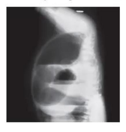

- What is this radiographic investigation called, and what is the diagnosis?
- What is the initial surgical management for low vs high lesions?
- State True or False:
i. Low anorectal malformations are typically associated with a perineal fistula and well-formed gluteal muscles/buttocks.
ii. High lesions are frequently associated with a rectobulbar / rectoprostatic urethral fistula and flat buttocks with sacral agenesis.
iii. Classical invertogram has been largely replaced by the cross-table prone lateral radiograph (taken after 18–24 hours of life).
Answer
- Wangensteen-Rice Invertogram (or Cross-Table Lateral Prone Radiograph), demonstrating Imperforate Anus / Anorectal Malformation (ARM).
- Surgical management:
- Low ARM: Single-stage Primary Anoplasty / Cutback Anoplasty in the neonatal period (without diverting colostomy).
- High / Intermediate ARM: Initial emergency Divided Sigmoid Colostomy with mucus fistula, followed at 3–6 months by definitive Posterior Sagittal Anorectoplasty (PSARP / Peña procedure).
- True/False:
i. True (Low lesions have good sphincter development and good prognosis for continence).
ii. True (High lesions have poor pelvic musculature and frequent genitourinary fistulae).
iii. True (Prone cross-table lateral film after 18–24 hours allows rectal gas to reach the distal-most pouch without infant inversion distress).
MNJ30-040
State the underlying pathophysiological mechanism for each of the following peripheral stigmata of chronic liver disease:
- Palmar erythema and spider nevi
- Leukonychia (Muehrcke's lines / Terry's nails)
- Dupuytren's contracture
- Fetor hepaticus
- Bilateral parotid enlargement
Answer
- Palmar erythema & Spider nevi: Impaired hepatic clearance and metabolism of Estrogens, resulting in hyperestrogenemia and peripheral arteriolar vasodilation.
- Leukonychia: Chronic Severe Hypoalbuminemia (
) altering the vascular bed of the nail matrix. - Dupuytren's contracture: Palmar fascial fibrosis and myofibroblast proliferation mediated by free radicals, alcohol toxicity, and fibroblast growth factors.
- Fetor hepaticus: Exhalation of volatile Dimethyl sulfide / Mercaptans resulting from severe portosystemic shunting and impaired hepatic clearance of methionine metabolites.
- Parotid swelling: Chronic sialadenosis due to autonomic neuropathy, malnutrition, and protein deficiency.
MNJ30-041
A 10-year-old male child presents with jaundice, irritability, poor school performance, and severe anemia. On examination, the child has wing-beating tremors and choreoathetosis:
- What is the probable diagnosis?
- What endocrine abnormalities can occur in this disease?
- What is the gold-standard diagnostic investigation?
- Name 4 drugs used in pharmacological management.
- What is the mechanism of anemia in this patient?
Answer
- Wilson Disease (Hepatolenticular Degeneration).
- Endocrine manifestations: Hypoparathyroidism, delayed puberty, primary amenorrhea / infertility, gynecomastia, and renal tubular acidosis (RTA with nephrocalcinosis).
- Gold standard test: Hepatic copper quantification by liver biopsy (
dry tissue weight) or biallelic pathogenic mutations in the ATP7B gene. - Pharmacotherapy:
- D-Penicillamine
- Trientine hydrochloride
- Zinc acetate / sulfate
- Ammonium tetrathiomolybdate (for neurological disease).
- Mechanism of anemia: Coombs-negative acute intravascular hemolytic anemia caused by acute massive release of free non-ceruloplasmin-bound copper from necrotic hepatocytes, inducing lipid peroxidation of red cell membranes and glutathione depletion.
MNJ30-042
Interpret the Hepatitis B serological profiles:
| Profile | HBsAg | Anti-HBc IgM | Total Anti-HBc | Anti-HBs | Clinical Interpretation |
|---|---|---|---|---|---|
| a | + | - | - | - | Early acute infection / Incubation period |
| b | + | + | + | - | Acute Hepatitis B infection |
| c | - | - | - | + | Vaccinated (Immunity via immunization) |
| d | + | - | + | - | Chronic Hepatitis B infection |
| e | - | - | + | + | Natural infection (Resolved / Recovered with immunity) |
Answer
- a: Early acute infection / window period prior to core antibody seroconversion.
- b: Acute Hepatitis B infection (High infectivity; positive HBsAg and Anti-HBc IgM).
- c: Vaccine-induced Immunity (Isolated anti-HBs positivity with negative core antibodies).
- d: Chronic Hepatitis B infection (Persistence of HBsAg
months with IgG anti-HBc). - e: Past resolved natural infection with immunity (Positive anti-HBs and positive IgG anti-HBc).
MNJ30-043
A mother of two children (aged 3 years and 9 months) seeks advice prior to traveling for 4 months to a region where Hepatitis A is highly endemic. Outline the pre-exposure Hepatitis A prophylaxis recommendations for both children:
Answer
- Older Child (3 years of age,
months): - Administer Hepatitis A Inactivated Vaccine (single dose of 0.5 mL IM prior to travel; provides
protection within 2–4 weeks). - Complete the 2-dose series by administering the second booster dose 6–12 months later.
- Administer Hepatitis A Inactivated Vaccine (single dose of 0.5 mL IM prior to travel; provides
- Younger Infant (9 months of age,
months): - Inactivated HAV vaccine is not routinely licensed for infants
months. - Administer Hepatitis A Standard Immune Globulin (IG):
IM (since anticipated travel duration is 3–5 months). - When the infant reaches 12 months of age, standard Hepatitis A vaccination can be initiated.
- Inactivated HAV vaccine is not routinely licensed for infants
MNJ30-044
A 7-month-old female baby presents with persistent cough, failure to thrive, and a full-thickness prolapsing rectal mass:
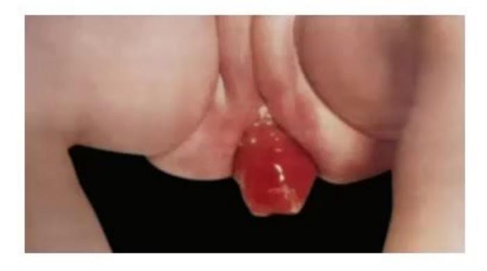
- What is the most likely diagnosis?
- What is the genetic basis and inheritance of this condition?
- What is the pathophysiological mechanism leading to rectal prolapse in this disease?
- How is this disease screened in the neonatal period?
Answer
- Cystic Fibrosis presenting with Rectal Prolapse and chronic suppurative lung disease / exocrine pancreatic insufficiency.
- Genetic basis: Autosomal recessive mutation in the CFTR (Cystic Fibrosis Transmembrane Conductance Regulator) gene located on chromosome 7q31.2 (commonest mutation:
). - Pathophysiology of rectal prolapse:
- Exocrine pancreatic insufficiency causes severe malabsorption, steatorrhea, and bulky voluminous stools.
- Chronic increased intra-abdominal pressure from persistent paroxysmal coughing.
- Malnutrition leading to loss of perirectal ischiorectal fat pads and hypotonic pelvic floor musculature.
- Neonatal Screening: Dried blood spot measurement of Immunoreactive Trypsinogen (IRT) on Guthrie card, followed by second-tier DNA mutation panel (IRT/DNA protocol) and confirmatory Quantitative Pilocarpine Iontophoresis Sweat Chloride Test (
is diagnostic).