MNJ27-001
An 8-year-old child Mahesh presented with bilateral sudden loss of vision, motor weakness and seizures following upper respiratory infection. There is no significant family history. MRI finding of the child is shown here:
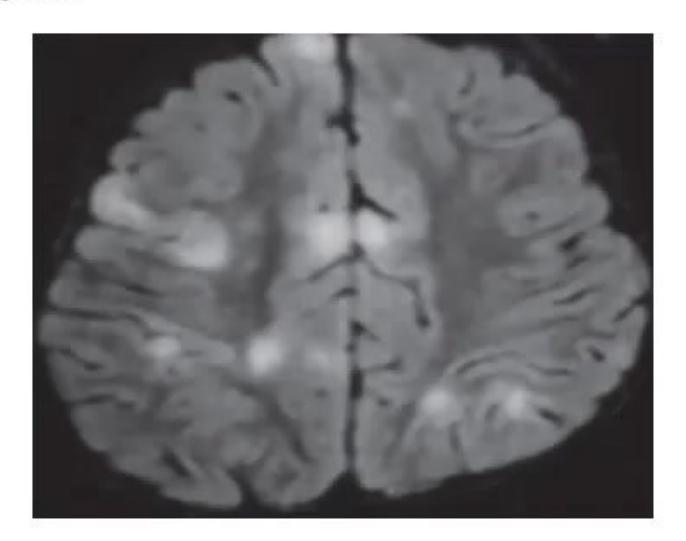
- Describe the abnormality and diagnosis.
- Give a differential diagnosis.
- How will you differentiate between the two?
- What is the CSF finding?
- What is the line of treatment?
- What is the prognosis?
Answer
- Large, patchy areas of subcortical and deep white matter hyperintensity in the bilateral corona radiata suggestive of ADEM (Acute Disseminated Encephalomyelitis).
- Multiple sclerosis (MS).
- Differentiating features between ADEM and MS:
| Feature | ADEM | MS |
|---|---|---|
| Age | <10 years | >10 years |
| Stupor / coma | + | - |
| Fever / vomiting | + | - |
| Family history | No | 20% |
| Sensory complaints | + | - |
| Optic neuritis | Bilateral | Unilateral |
| Manifestations | Polysymptomatic | Monosymptomatic |
| MRI imaging | Widespread lesions: Basal ganglia, thalamus, cortical gray-white junction | Isolated lesions: periventricular white matter, corpus callosum |
| CSF | Pleocytosis (lymphocytosis) | Oligoclonal bands |
| Response to steroids | + | + |
| Follow-up | No new lesions | New lesions |
(+ = more likely to be present, - = less likely to be present)
- Lymphocytic pleocytosis.
- Intravenous methylprednisolone 30 mg/kg/day for 3–5 days followed by oral prednisolone 1 mg/kg/day for 10 days.
- 70 percent will recover without any residual disability in 6 months.
Note: Oligoclonal bands are also found in: Multiple sclerosis, Devic's disease, Systemic lupus erythematosus, Neurosarcoidosis, SSPE, Subarachnoid hemorrhage, Syphilis, CNS lymphoma.
MNJ27-002
A 6-month-old child came to your clinic. Mother is complaining that child still not able to support his head, no smile and not able to sit. MRI brain was done:
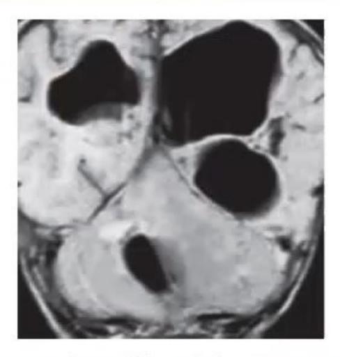
- What are the MRI findings?
- What could be the etiology?
Answer
- Multifocal cerebral and cerebellar encephalomalacia.
- Sequel of ischemic injury in the perinatal period (Hypoxic Ischemic Encephalopathy).
MNJ27-003
A 12-year-old child has complaints of fever for last three months and backache and now he has problem in bending forward. MRI spine was done:
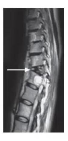
- What is the likely diagnosis?
- Which is the commonest site?
Answer
- TB Spine (Pott's spine / Tubercular spondylodiscitis).
- Thoracolumbar junction—area of maximum stress and valveless venous drainage (Batson venous plexus).
MNJ27-004
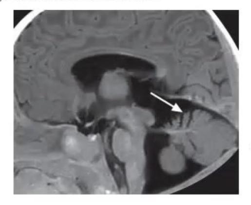
- What is the diagnosis?
- By what gestational age would this defect occur?
- What is preventive treatment, in what dose and started when?
- What is a risk factor for this disorder?
Answer
- Occipital encephalocele.
- 26 days post-conception (closure of cranial neuropore).
- Folic acid: 0.4 mg/day (4 mg/day for high-risk), started periconceptionally (before pregnancy) and continued till 12 weeks of gestation.
- Maternal folic acid deficiency, maternal diabetes, periconceptional hyperthermia, valproate/anticonvulsant exposure.
MNJ27-005
Parents of a 2-year-old child came to you that their child is not able to walk properly, he sways to one side when walking and for the last 2 months he started to lose his previously acquired milestones and has abnormal hand movements throughout the day (myoclonic jerks). He had six episodes of respiratory infection in the last one year. You examine the child and found some positive findings in his eyes:
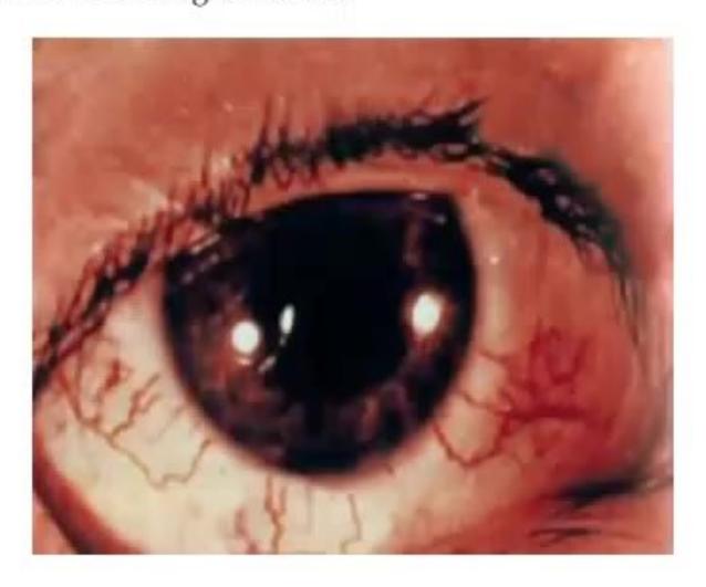
- Give the diagnosis of given condition.
- State mode of inheritance.
- Name five important components.
- Name most frequent immunological defect.
Answer
- Ataxia-telangiectasia (Louis-Bar syndrome).
- Autosomal recessive (ATM gene mutation on chr 11q22.3).
- Important components:
a. Cerebellar ataxia
b. Oculocutaneous telangiectasia
c. Immunodeficiency
d. Sinopulmonary infections
e. Lymphoreticular malignancies - Selective absence / deficiency of IgA (and IgE / IgG subclasses).
MNJ27-006
A 6-year-old child admitted in hospital with complaints of intractable epilepsy, mental retardation and lesions on the face:
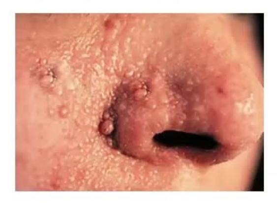
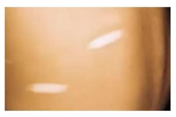
- What is the diagnosis of this condition?
- Describe the lesions given in both images.
- What is the mode of inheritance and which chromosomes are involved?
- What is the CNS association?
Answer
- Tuberous Sclerosis Complex (TSC / Bourneville disease).
- Image A: Adenoma sebaceum (facial angiofibromas). Image B: Ash-leaf macule (hypomelanotic macule).
- Autosomal dominant; TSC1 (chromosome 9q34, hamartin) and TSC2 (chromosome 16p13, tuberin).
- Characteristic brain lesions include cortical tubers in convolutions of cerebral hemispheres, subependymal nodules, and subependymal giant cell astrocytomas (SEGA) typically in subependymal region.
MNJ27-007
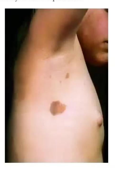
- Name the neurocutaneous marker seen.
- In NF-1, what is the diagnostic criteria with respect to this marker?
- Name an X-linked dominant neurocutaneous syndrome.
- What are the stages of the syndrome in question 3?
Answer
- Café-au-lait macule (CALM).
- Six or more CAL macules larger than 5 mm in greatest diameter in prepubertal individuals, and larger than 15 mm in greatest diameter in postpubertal individuals.
- Incontinentia pigmenti (Bloch-Sulzberger syndrome).
- Stages:
- Stage 1: Vesicular stage
- Stage 2: Verrucous stage
- Stage 3: Hyperpigmented stage
- Stage 4: Hypopigmented / atrophic stage
MNJ27-008
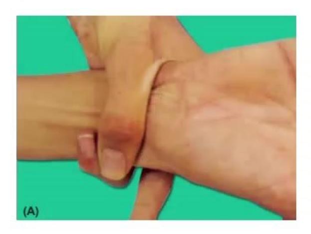
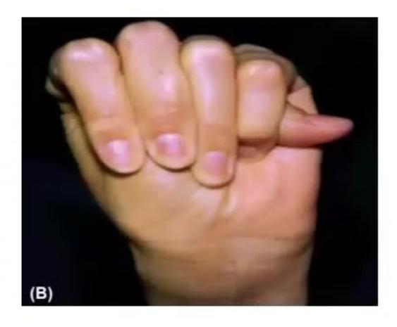
- What is exhibited in the photographs A and B?
- What is the diagnosis?
- What is the mode of inheritance?
- What is the major ocular criteria for diagnosis?
Answer
- Image A: Hypermobility of finger joints (arachnodactyly). Image B: Positive thumb sign (Steinberg sign).
- Marfan syndrome (FBN1 gene mutation).
- Autosomal dominant.
- Ectopia lentis (superior and temporal subluxation of the lens).
MNJ27-009
A 4-year-old child admitted in hospital with complaints of intractable epilepsy. You examine the child and found a birth lesion on face involving upper eyelid. Neuroimaging was done that is shown below:
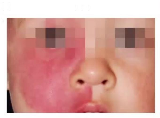
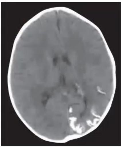
- Diagnose the condition according to clinical picture and images.
- What is the pattern of inheritance?
- What is the characteristic radiographic image called?
- What is the ophthalmologic complication?
Answer
- Sturge-Weber syndrome (Encephalotrigeminal angiomatosis).
- Sporadic (somatic activating mutation in GNAQ gene).
- "Tram-track" sign (gyriform cortical calcification).
- Buphthalmos / Glaucoma / Choroidal hemangioma.
MNJ27-010
What is the classic triad of infantile spasms (West syndrome)?
Answer
Classic triad of West syndrome:
- Epileptic spasms (infantile spasms)
- Hypsarrhythmia on EEG
- Developmental arrest or regression
MNJ27-011
The examination of a newborn reveals a lump of soft tissue of the size of a ₹2 coin overlying the lower spine. There is no neurological deficit and child is moving both lower limbs well, and knee and ankle reflexes are present:
- What is the likely diagnosis?
- What is the likely risk of recurrence of disorder in future pregnancies?
- What intervention, started at what time and for how long, can reduce the risk of recurrence?
- What are the options for managing bladder incontinence in this condition?
Answer
- Occult spinal dysraphism (Spina bifida occulta / Lipomyelomeningocele).
- Risk of recurrence:
- One affected child: 3–4%
- Two affected children: 10%
- Folic acid 4 mg/day (4000 µg/day) started periconceptionally (before pregnancy) and continued till 12 weeks of pregnancy. (400 µg/day in low-risk).
- Bladder incontinence management:
- Clean intermittent catheterization (CIC)
- Anticholinergics (e.g., Oxybutynin)
- Artificial urinary sphincter
- Surgical urinary diversion
- Augmentation cystoplasty
MNJ27-012
Match the following sign/symptom with the expected location of the lesion in the brain:
| Sign / Symptom | Possible Location / Sign |
|---|---|
| a. Deafferented pupil | 1. Midbrain tectum |
| b. Cerebellum | 2. Pinpoint pupils |
| c. Ipsilateral cortex | 3. Tonic deviation of the eye(s) |
| d. Midbrain tectum | 4. Horner's syndrome |
| e. Pons | 5. Midposition pupils |
| f. Cribriform plate of ethmoid | 6. Leakage of CSF from nose |
| 7. Marcus Gunn pupil | |
| 8. Nystagmus |
Answer
- a. Deafferented pupil
Marcus Gunn pupil (Afferent pupillary defect) - b. Cerebellum
Nystagmus - c. Ipsilateral cortex
Tonic deviation of the eye(s) - d. Midbrain tectum
Midposition pupils - e. Pons
Pinpoint pupils - f. Cribriform plate of ethmoid
Leakage of CSF from nose (CSF rhinorrhea)
MNJ27-013
A 3-year-old child Raju has complaints of weakness while playing, drooping of eyelids in evening and not able to sustain his grip. You think a provisional diagnosis of myasthenia gravis:

- What is the characteristic EMG in myasthenia?
- What is the chest X-ray finding in this condition?
- What is the clinical test for diagnosing myasthenia?
- Which antibiotics can worsen the condition?
- What drugs are used for treatment?
Answer
- Decremental response to repetitive nerve stimulation (RNS).
- Enlarged thymus / Thymoma / Thymic hyperplasia.
- Edrophonium (Tensilon) test / Neostigmine test / Ice pack test.
- Aminoglycosides (gentamicin, amikacin), fluoroquinolones, macrolides, polymyxins.
- Pyridostigmine / Neostigmine (oral: 0.4 mg/kg PO 4–6 hourly; parenteral: 0.04 mg/kg IM 4–6 hourly), corticosteroids, IVIg, plasma exchange.
MNJ27-014
Examination of the cerebrospinal fluid of a 6-year-old child Arjun, mildly febrile child with nuchal rigidity and intermittent stupor for 3 weeks shows the following:
- White blood cells: 100/µL (all lymphocytes)
- Gram stain: Negative
- Protein: 750 mg/dL
- Glucose: 25 mg/dL
- What is the likely differential diagnosis?
- What drugs can be used to reduce intracranial tension?
- Which cranial nerve is involved in false localizing sign?
Answer
- Tubercular meningitis (TBM), fungal (cryptococcal) meningitis, partially treated bacterial meningitis.
- Mannitol, glycerol, hypertonic saline, acetazolamide.
- Sixth cranial nerve (Abducens nerve).
MNJ27-015
Match the following:
| Clinical scenario | Diagnosis |
|---|---|
| 1. Eye blinking or throat-clearing noises in an otherwise healthy 5-year-old boy | a. Transient tic disorder of childhood |
| 2. A 6-year-old boy with eye twitching and echolalia | b. Tourette syndrome |
| 3. A 2-year-old infant who was born prematurely and is unable to walk or talk | c. Cerebral palsy |
| 4. A 14-year-old girl with a history of precocious puberty who now develops a large goiter | d. Tuberous sclerosis |
| 5. An infant with infantile spasms, a hypsarrhythmic EEG pattern, and ash-leaf depigmentation on her back | e. McCune-Albright syndrome |
Answer
1
2
3
4
5
MNJ27-016
A 9-year-old male child complaining of increasing weakness of all limbs
- What is the likely diagnosis?
- Mention three investigations to clinch diagnosis.
- What is the most sensitive measure of respiratory muscle involvement?
- Mention three modalities of treatment.
- What is the midline mass, explain its significance and how should it be managed?
Answer
- Guillain-Barré syndrome (GBS / AIDP).
- Investigations:
- CSF analysis: Albumino-cytological dissociation (elevated protein with normal cell count)
- Nerve Conduction Velocity (NCV): demyelinating pattern, prolonged F-latencies
- Electromyography (EMG)
- Vital capacity (VC). Normal VC
. If VC falls below of normal, endotracheal intubation is indicated. - IVIg (
total over 2–5 days), Plasmapheresis (plasma exchange), supportive/ventilatory care. - Palpable urinary bladder distension due to autonomic dysfunction (urinary retention). Manage with urinary catheterization (Foley catheter).
MNJ27-017
A 3-year-old boy was unconscious for 1 min following head injury after fall from bed. Now he is fully conscious but there is bruising on the left side of the head over the parietal bone. Skull X-rays are performed and he is admitted for neurological observations:
- What clinical features would suggest requirement of CT skull and/or neurosurgical opinion?
- What non-surgical temporary measures can be used to reduce raised intracranial pressure?
- What advice needs to be given to parents if after 24 hours the child has remained well and is ready for discharge?
Answer
- Indications for CT skull / neurosurgical opinion:
- Deteriorating level of consciousness / GCS < 15
- Focal neurological signs
- Depressed or compound skull fracture / signs of basal skull fracture
- Post-traumatic seizures
- CSF leak (rhinorrhea/otorrhea) or persistent vomiting
- Non-surgical measures to reduce raised ICT:
- Nursing in 30° head-up position in midline
- Osmotic diuretics (Mannitol 0.5–1 g/kg, 3% hypertonic saline)
- Controlled hyperventilation (
30–35 mmHg) - Sedation and analgesia
- Discharge advice (warning signs to return immediately):
- Persistent vomiting
- Drowsiness / altered sensorium
- Blurred / double vision
- Seizures or limb weakness
MNJ27-018

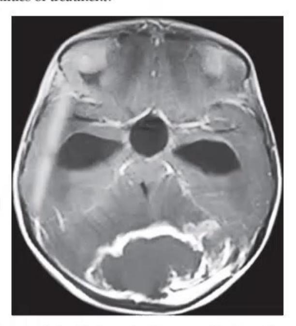
- What does the imaging show and what is probable diagnosis?
- What are the modalities of treatment?
Answer
- Space-occupying lesion (SOL) in the posterior cranial fossa / 4th ventricle with obstructive hydrocephalus; probable diagnosis is Medulloblastoma.
- Multimodal therapy: Surgical resection, craniospinal radiotherapy, chemotherapy, and CSF diversion (VP shunt/ETV) if indicated.
MNJ27-019
This newborn presented with hepatosplenomegaly, jaundice and convulsions. His mother's Sabin-Feldman dye test was positive:
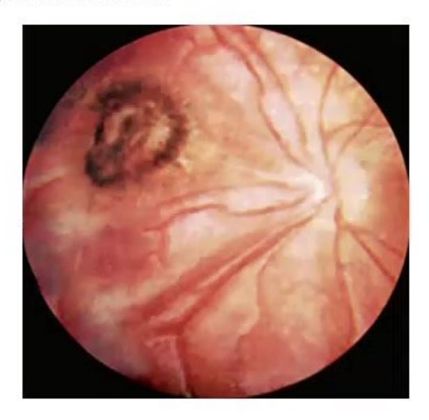
- What is the diagnosis?
- What is classic triad of signs in this condition?
- What are the drugs used in treatment?
Answer
- Congenital toxoplasmosis.
- Sabin triad:
- Chorioretinitis
- Intracranial calcifications
- Hydrocephalus
- Treatment (for 1 year):
- Pyrimethamine (2 mg/kg/day for 2 days, then 1 mg/kg/day for 2–6 months, then Mon/Wed/Fri)
- Sulfadiazine (100 mg/kg/day in 2 divided doses)
- Leucovorin / Folinic acid (5–10 mg 3 times weekly)
MNJ27-020
Nidhi is 5 months pregnant with her first child. Her 15-year-old brother Vijay began to have difficulty in climbing stairs at 4 years of age. He is now confined to a wheelchair. All other family members are normal. She is worried that her child may be affected and she is referred by a gynecologist to you for future advice:
- What is the most likely clinical diagnosis of Vijay?
- What is the inheritance pattern of this problem?
- What are the chances that Nidhi's child could be affected with the same disease?
- What would you recommend for antenatal diagnosis?
Answer
- Duchenne muscular dystrophy (DMD).
- X-linked recessive.
- If the fetus is male, the risk of being affected is 50% if mother is a carrier (overall risk
if brother is an isolated case); female fetuses will not be clinically affected (0%), but have a 50% carrier risk. - Chorionic villus sampling (CVS) at 10–12 weeks of gestation or amniocentesis at 15–18 weeks for fetal DNA analysis (MLPA for dystrophin gene).
MNJ27-021
10-year-old girl Shivani brought to emergency with history of fall from a height 4 hours back. After that she developed vomiting and GCS drops from 14/15 to 9/15. You advised an urgent CT scan in ER:
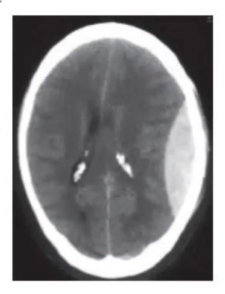
- Describe the abnormality shown in CT.
- What is the diagnosis of this condition?
- Which is the commonest site and vessel involved?
- What is the differential diagnosis and how will you differentiate the two?
- What is the management?
Answer
- High-density, biconvex (lentiform) extra-axial collection with mass effect.
- Extradural hematoma (Epidural hematoma, EDH).
- Temporoparietal region; Middle meningeal artery (MMA).
- Subdural hematoma (SDH):
- EDH: Biconvex (lenticular), limited by suture lines, arterial bleed.
- SDH: Crescent-shaped, crosses suture lines, venous bleed (bridging veins).
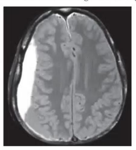
- Immediate airway protection / intubation due to falling GCS, emergency neurosurgical craniotomy and hematoma evacuation.
MNJ27-022
- A lumbar puncture is performed and the CSF is xanthochromic. What are four possible causes?
- CSF protein levels are 400 mg/dL. What are three possible causes for the same?
- CSF glucose is 200 mg/dL and blood glucose is 112 mg/dL. List causes for the same.
- CSF is also cloudy, what does it imply?
Answer
- Causes of xanthochromia:
- Subarachnoid hemorrhage
- Hyperbilirubinemia (jaundice)
- Markedly elevated CSF protein (>150–400 mg/dL, Froin's syndrome)
- Carotenemia / traumatic tap with delayed centrifugation
- Causes of CSF protein > 400 mg/dL:
- TB meningitis
- Guillain-Barré syndrome (GBS)
- Spinal block / spinal cord tumor (Froin's syndrome)
- Pyogenic meningitis
- Degenerative CNS disorders / vasculitis
- Causes of high CSF glucose:
- Systemic hyperglycemia / post-IV dextrose infusion
- Traumatic lumbar puncture with significant blood contamination
- Cloudy CSF implies:
- Elevated WBC count (pleocytosis, typically
seen in pyogenic meningitis) - High RBC count or dense bacterial/fungal load
- Elevated WBC count (pleocytosis, typically
MNJ27-023
Observe the images A and B given below and answer the following questions:
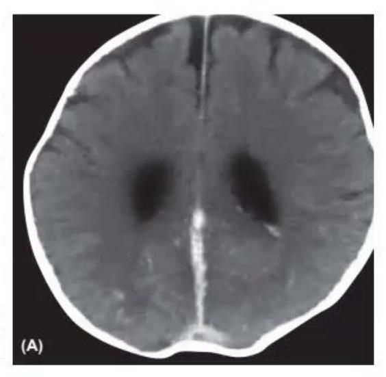
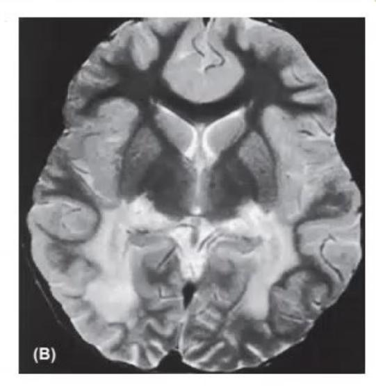
Image A:
- Identify image A / level.
- Give diagnosis.
- Differential diagnosis / causes (any four).
Image B:
- Identify image B / level.
- Give diagnosis.
- Mention other two diseases from the same group (metabolic / peroxisomal).
Answer
Image A:
- Contrast CT brain at the level of the lateral ventricles (showing empty delta sign / filling defect in superior sagittal sinus).
- Cerebral venous sinus thrombosis (CVST).
- Causes: Severe dehydration, sepsis, cyanotic congenital heart disease, nephrotic syndrome, protein C/S/antithrombin III deficiency, SLE/vasculitis.
Image B:
- Axial T2/FLAIR MRI brain at the level of internal capsule and basal ganglia.
- X-linked Adrenoleukodystrophy (ALD) (confluent symmetric parieto-occipital white matter demyelination).
- Other peroxisomal disorders: Zellweger syndrome, Refsum disease, Neonatal adrenoleukodystrophy.
MNJ27-024
These are three skin manifestations of the same disease:
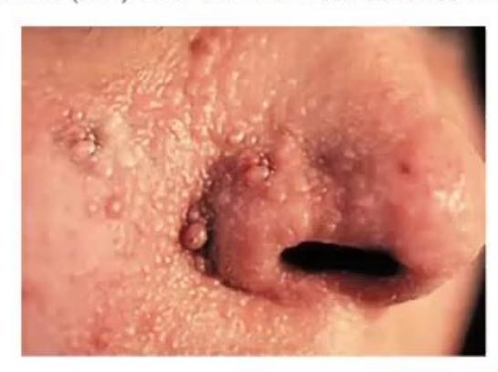
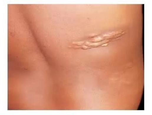
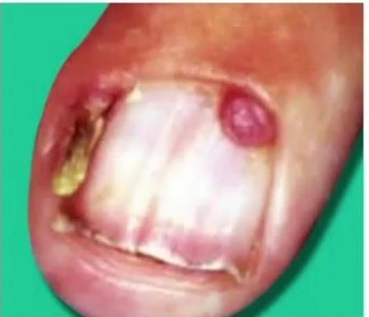

- Name all three skin manifestations.
- Give diagnosis of this disorder.
- Write two other skin manifestations seen in this condition.
Answer
- Skin manifestations:
- Adenoma sebaceum (facial angiofibromas)
- Shagreen patch (connective tissue nevus)
- Periungual / subungual fibroma (Koenen's tumor)
- Tuberous Sclerosis Complex (TSC).
- Other skin manifestations:
- Ash-leaf macules (hypomelanotic macules)
- Confetti skin lesions
- Forehead fibrous plaques
- Poliosis (tuft of white hair on scalp/eyelids)
- Café-au-lait spots
MNJ27-025
A 14-year-old female Anchal with history of recurrent seizures is admitted in PICU with stroke 12 hours back. She was evaluated for same twice before. You order MRA (shown below):
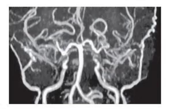
- Identify and describe the investigation.
- What is the diagnosis?
- What other disorders are associated with this disease?
- What is the treatment of choice?
- What are syndromes associated with contralateral hemiplegia in children?
Answer
- MRA / Cerebral Angiography: Shows stenosis/narrowing of distal internal carotid artery and proximal anterior/middle cerebral arteries with extensive basal collateral vessel formation ("puff of smoke").
- Moya-moya disease.
- Associated disorders: Down syndrome, Neurofibromatosis type 1 (NF-1), Sickle cell disease, Turner syndrome, previous cranial irradiation.
- Surgical revascularization: Direct bypass (STA-MCA bypass) or indirect bypass (EDAS / EMS).
- Brainstem syndromes: Weber syndrome, Benedikt syndrome, Millard-Gubler syndrome, Foville syndrome.
MNJ27-026
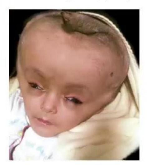
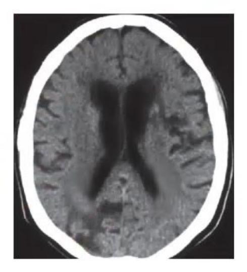
- Identify the clinical sign and neuroimaging sign.
- What abnormalities are likely on motor system examination?
- Name the treatment options (any two).
Answer
- Clinical sign: Setting sun sign (downward deviation of eyes). Neuroimaging sign: Ventriculomegaly / dilated ventricles (Hydrocephalus).
- Motor examination findings:
- Increased tone / spasticity (lower limbs > upper limbs)
- Hyperreflexia / brisk deep tendon reflexes
- Bilateral extensor plantar responses (Babinski sign)
- Scissoring of lower limbs
- Treatment options:
- Ventriculoperitoneal (VP) shunt
- Endoscopic third ventriculostomy (ETV)
- Ventriculoatrial (VA) shunt
MNJ27-027
An 8-year-old girl Priyanka came to emergency with history of rapidly progressive, bilateral lower limb weakness since 3 days. She was apparently normal except for a history of sore throat and bilateral neck swelling with fever about 20 days back. Now she is very depressed because of illness as she has exams in 7 days. She started with difficulty while walking and not being able to pass urine despite of the sensation being there. On examination: Normal sensorium and cranial nerves. Normal neurology findings in both upper limbs. Lower limbs: symmetrical flaccid paralysis; distal more than proximal. Deep tendon reflexes were exaggerated and plantars were extensor. There was a sensory discrepancy below T8 and temp/light touch were affected. Vibration and position were normal. Her urinary bladder was palpable:
- What is your differential diagnosis?
- What is the investigation of choice?
- What is the treatment?
- Name two causative factors for this condition.
Answer
- Differential diagnosis:
- Acute transverse myelitis (ATM)
- Spinal cord compression / SOL in spine (tubercular Pott's spine, epidural abscess/hematoma)
- Neuromyelitis optica spectrum disorder (NMOSD)
- Guillain-Barré syndrome (GBS)
- Investigation of choice: MRI spine with contrast (and MRI brain).
- Treatment: High-dose IV Methylprednisolone pulse therapy (30 mg/kg/day for 3–5 days) followed by oral prednisolone taper; IVIg or plasmapheresis in non-responders; supportive bladder catheterization.
- Causative factors: Post-infectious / para-infectious (viral/mycoplasma), post-vaccinal, systemic autoimmune disease (SLE), Lyme disease.
MNJ27-028
An 18-month-old male child Rahul presents with complaints of delayed milestones. On examination HC was 42 cm and he still has not developed milestones according to his age:
- What is definition of microcephaly?
- What are causes of primary microcephaly?
- What investigations would you advise in this child?
Answer
- Definition: Occipitofrontal head circumference (OFC)
(or for severe microcephaly) below the mean for age and sex. - Causes of primary microcephaly:
- Familial / genetic (microcephalia vera, autosomal recessive primary microcephaly)
- Chromosomal aneuploidies (Down syndrome, Edwards syndrome, Cri-du-chat syndrome)
- Neural tube defects / Lissencephaly / Holoprosencephaly
- Investigations:
- MRI / CT brain
- TORCH serology
- Genetic testing: Karyotype / Chromosomal microarray (CMA)
- Metabolic workup: Serum and urine amino acid profile, urinary organic acids
MNJ27-029
A 14-month-old male child Robert presents with cough, fever and one episode of generalized seizure for 4 minutes. He has history of a similar episode 2 months back:
- What is most probable diagnosis?
- What is risk for future epilepsy?
- What are major risk factors for recurrence?
- Will you do lumbar puncture in this patient?
- What are indications of neuroimaging in this disease?
- What is current status of the use of clonazepam/benzodiazepines?
Answer
- Simple febrile seizure (recurrent).
- Risk of future epilepsy:
after simple febrile seizure (compared to in general population; up to in complex febrile seizures). - Major risk factors for recurrence:
- Age
months at onset - Duration of fever
hours before seizure - Lower degree of fever (
) - Family history of febrile seizures
- Age
- Lumbar puncture: Recommended in infants
months (or 12–18 months if unimmunized for Hib/Pneumococcus or after prior antibiotics) and whenever there are meningeal signs or prolonged drowsiness. - Indications for neuroimaging: Complex febrile seizures (focal, prolonged
min, multiple within 24 hours), focal neurological deficit, abnormally large/small head, persistent altered consciousness. - Intermittent oral diazepam / clobazam / clonazepam given at onset of fever for 48 hours in children with recurrent febrile seizures or high parental anxiety; routine daily prophylactic AEDs are not recommended.
MNJ27-030
Match the common side effect of these drugs:
| Drug | Side Effect |
|---|---|
| 1. Carbamazepine | a. Irreversible visual field defect |
| 2. Lamotrigine | b. Stevens-Johnson syndrome |
| 3. Topiramate | c. Weight gain |
| 4. Valproic acid | d. Hirsutism / Gingival hyperplasia |
| 5. Vigabatrin | e. Hyperammonemia |
| 6. Phenytoin | f. Glaucoma / Oligohidrosis |
Answer
-
- Carbamazepine
b. Stevens-Johnson syndrome / c. Weight gain
- Carbamazepine
-
- Lamotrigine
b. Stevens-Johnson syndrome
- Lamotrigine
-
- Topiramate
f. Glaucoma / Oligohidrosis
- Topiramate
-
- Valproic acid
e. Hyperammonemia / c. Weight gain
- Valproic acid
-
- Vigabatrin
a. Irreversible visual field defect
- Vigabatrin
-
- Phenytoin
d. Hirsutism / Gingival hyperplasia
- Phenytoin
(Match: 1-c, 2-b, 3-f, 4-e, 5-a, 6-d)
MNJ27-031
A mother of a 6-year-old child with refractory seizures came to you saying that she found on Google that a special type of diet can stop seizures in children:
- What is this diet she is talking about?
- What are indications to use this diet?
- What are contraindications to use this?
- What is formulation of this diet?
Answer
- Ketogenic diet.
- Indications: Refractory/drug-resistant epilepsy, GLUT-1 deficiency syndrome, Pyruvate dehydrogenase (PDH) deficiency, Infantile spasms, Dravet syndrome, Lennox-Gastaut syndrome, Tuberous sclerosis.
- Contraindications: Primary carnitine deficiency, carnitine palmitoyl transferase (CPT I/II) deficiency,
-oxidation defects, medium-chain acyl-CoA dehydrogenase (MCAD) deficiency, pyruvate carboxylase deficiency, porphyria. - Formulation: 3:1 or 4:1 ratio of fat to combined carbohydrates and protein by weight.
MNJ27-032
A 7-year-old male child came to hospital with complaints of ataxia for the last 3 months along with dysarthric speech. On examination, Romberg test is positive, DTRs are absent, plantar response is extensor and there is loss of vibration sense. Nystagmus is also present:
- What is probable diagnosis and what is inheritance?
- Which parts of nervous system are involved in this disease?
- What are causes of recurrent ataxia?
- What is cause of death in this patient?
Answer
- Friedreich ataxia; Autosomal recessive (GAA trinucleotide repeat expansion on FXN gene).
- Spinocerebellar tracts, dorsal/posterior columns, corticospinal tracts, dorsal root ganglia, dentate nucleus of cerebellum.
- Brain tumor, anticonvulsant toxicity (phenytoin), basilar migraine, multiple sclerosis, ADEM, episodic ataxia, inborn errors of metabolism.
- Hypertrophic cardiomyopathy / Congestive heart failure / Arrhythmias.
MNJ27-033
A 9-year-old female child came with complaints of abnormal movements. Mother told that these movements are fast and increase when she is in stress and disappear in sleep. On examination, child has difficulty in keeping protruded tongue:
- What is probable diagnosis of this condition?
- What are hallmark features of this?
- What important investigation will you order?
- What is treatment for this?
Answer
- Sydenham chorea (Rheumatic chorea).
- Hallmark features: Chorea (involuntary purposeless movements), hypotonia, emotional lability, motor impersistence ("darting tongue").
- 2D Echocardiography (to evaluate carditis), throat swab / ASO titer / Anti-DNase B, ECG, inflammatory markers (ESR, CRP).
- Treatment:
- Secondary prophylaxis: Long-term penicillin (Benzathine penicillin G every 3–4 weeks)
- Symptomatic: Haloperidol, valproate, or carbamazepine
- Anti-inflammatory: Oral prednisolone (2 mg/kg/day for 2–4 weeks) or IVIg in refractory cases.
MNJ27-034
This 3-year-old female child Geeta came with neuro-regression from early infancy with abnormal movements and autism. Her head circumference is also below 3rd percentile:
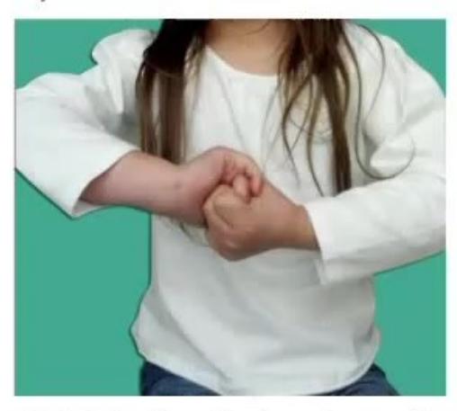
- What is the diagnosis?
- What is the hallmark of this disorder?
- What is age of onset of neuro-regression?
- Which are earliest neurological findings?
- Which milestones are delayed?
Answer
- Rett syndrome (MECP2 mutation, X-linked dominant).
- Stereotypic repetitive hand-wringing / hand-washing movements, loss of purposeful hand skills, acquired microcephaly.
- 6 to 18 months of age (usually 9–12 months).
- Deceleration of head growth (acquired microcephaly), hypotonia, autistic features, loss of social engagement, gait ataxia/apraxia.
- Motor milestones (loss of hand skills, abnormal gait) and expressive language/speech.
MNJ27-035
Write down the age of attaining the milestones in months:
| Milestone | Age of attaining (in months) |
|---|---|
| Follows moving object 180° | |
| Sustains social contact, listens to music | |
| Polysyllabic vowel sounds | |
| Creeps or crawls | |
| Plays simple ball game | |
| Hops on one foot | |
| Names 4 colors | |
| Makes tower of 4 cubes | |
| Dresses and undresses | |
| Puts 3 words together |
Answer
| Milestone | Age of attaining (in months) |
|---|---|
| Follows moving object 180° | 2–3 months |
| Sustains social contact, listens to music | 3 months |
| Polysyllabic vowel sounds | 7 months |
| Creeps or crawls | 10 months |
| Plays simple ball game | 12 months |
| Makes tower of 4 cubes | 18 months |
| Puts 3 words together | 24 months |
| Hops on one foot | 48 months (4 years) |
| Names 4 colors | 60 months (5 years) |
| Dresses and undresses | 60 months (5 years) |
MNJ27-036
A 32 weeks preterm baby with birth weight of 1.52 kg was born with HC of 26 cm (< 3rd centile). Otherwise stable baby has bilateral arthrogryposis with fixed flexion deformity. He has episode of seizure at 6 hours of life which was difficult to control after routine AED. His CBC showed platelets count 56,000 and low Hb. A cranial USG was done:
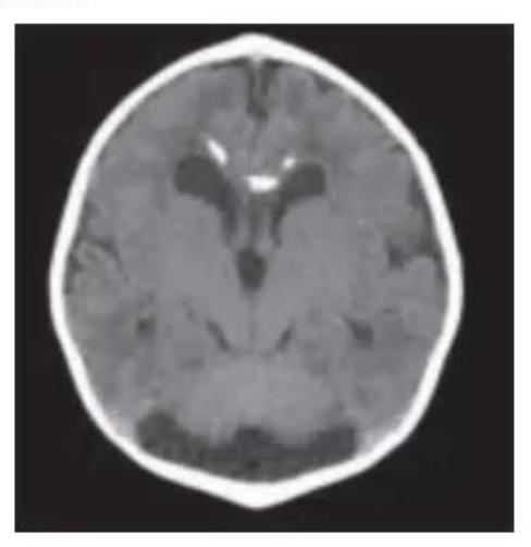
- What is most likely diagnosis?
- What three tests will you perform to confirm the diagnosis?
- Elaborate findings in USG brain.
- What are important sequelae?
Answer
- Congenital CMV (Cytomegalovirus) infection.
- Tests to confirm diagnosis:
- Urine / saliva CMV PCR (within first 2–3 weeks of life)
- Maternal and infant CMV IgM and IgG serology
- CSF PCR for CMV
- Ophthalmological and audiological (BERA) evaluation
- USG brain findings: Periventricular echodensities / calcifications, ventriculomegaly, subependymal cysts.
- Sequelae: Sensorineural hearing loss (SNHL), microcephaly, developmental delay, cerebral palsy, visual impairment (chorioretinitis / optic atrophy).
MNJ27-037
A 4-month-old male baby presented in OPD for a prominent forehead. According to mother, this has always been the case though to a lesser extent when he was born. The baby is otherwise well, weight following the 50th centile, length along 50th centile and head circumference is at 50th centile. On examination baby has very small anterior fontanelle and frontal prominence. X-ray head was done:
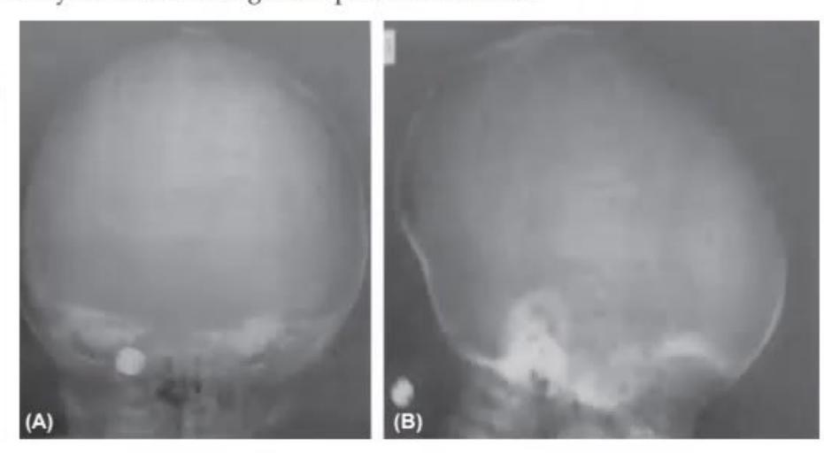
- What does the skull X-ray show (images A and B)?
- What is possible diagnosis?
- How will you make a management plan for this child?
Answer
- Image A: Fused / sclerosed sagittal suture. Image B: Dolichocephalic / scaphocephalic head shape.
- Sagittal craniosynostosis (Scaphocephaly).
- Management:
- Multidisciplinary assessment by craniofacial surgical and neurosurgical team
- 3D CT scan of skull to evaluate suture fusion and intracranial volume
- Surgical cranial vault remodeling or strip craniectomy (ideally between 3 to 9 months of age).