MNJ26-001
A 20-day-old male child came to you. The mother complains about his abnormal feet/legs shown below:
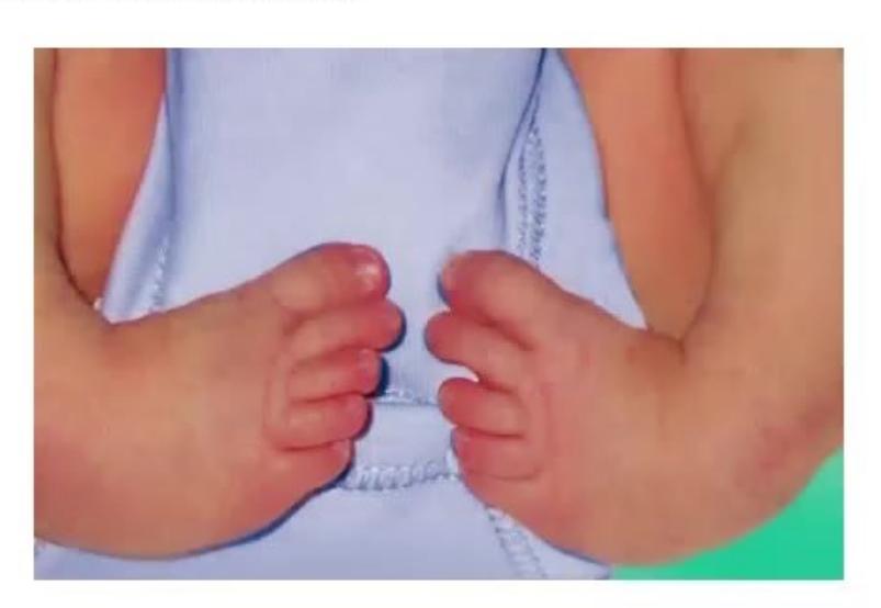
- Identify this problem.
- What is the mnemonic CAVE?
- What are common risk factors and associated conditions?
- What will you look for in the spinal examination of this child?
- When will you start intervention and what is the treatment of choice according to age?
- What is the Ponseti method of treatment?
Answer
-
Identification:
- Congenital Talipes Equinovarus (CTEV / Clubfoot).
-
Mnemonic CAVE (Components of deformity corrected in sequential order):
- C - Cavus (high medial arch due to pronation of forefoot in relation to hindfoot; corrected first by elevating 1st metatarsal / supinating forefoot).
- A - Adductus (forefoot and midfoot adducted relative to hindfoot; corrected by lateral abduction with counterpressure on lateral head of talus).
- V - Varus (hindfoot / calcaneus inverted; corrected simultaneously with abduction).
- E - Equinus (fixed plantarflexion at ankle; corrected last, usually via percutaneous Achilles tenotomy).
-
Risk factors & Associations:
- Idiopathic (most common, multifactorial inheritance, male:female ratio 2:1)
- Positive family history
- Intrauterine compression (oligohydramnios, multiple pregnancy)
- Neuromuscular conditions: Myelomeningocele / Spina bifida, Arthrogryposis multiplex congenita, Spinal Muscular Atrophy.
-
Spinal Examination:
- Examine the lumbosacral spine for cutaneous markers of Spinal Dysraphism / Spina Bifida Occulta (e.g., sacral dimple/sinus, hypertrichosis / hairy patch, subcutaneous lipoma, hemangioma, deviated gluteal cleft).
-
Treatment according to age:
- Timing: Non-operative treatment should start as early as possible, ideally in the first 1 to 2 weeks of life.
- Neonates to 2 years: Ponseti method (serial gentle manipulations and long-leg plaster casting followed by bracing).
- Late / Neglected cases (>2–3 years): Comprehensive posteromedial soft tissue release (Turco procedure), osteotomies, or Ilizarov external fixation.
-
Ponseti Method:
- Gold standard conservative method:
- Serial weekly long-leg plaster casts (correcting Cavus
Adductus Varus by abducting foot around a stabilized talar head). - Percutaneous Achilles tenotomy under local anesthesia in
of cases to correct residual equinus. - Final cast worn for 3 weeks post-tenotomy.
- Steenbeek / Denis Browne foot abduction brace (FAB) worn 23 hours/day for 3 months, then during naps and night-time until 4–5 years of age to prevent relapse.
- Serial weekly long-leg plaster casts (correcting Cavus
- Gold standard conservative method:
MNJ26-002
Mother of a 6-day-old child came to discuss the abnormal-looking foot of the baby shown in the image below:
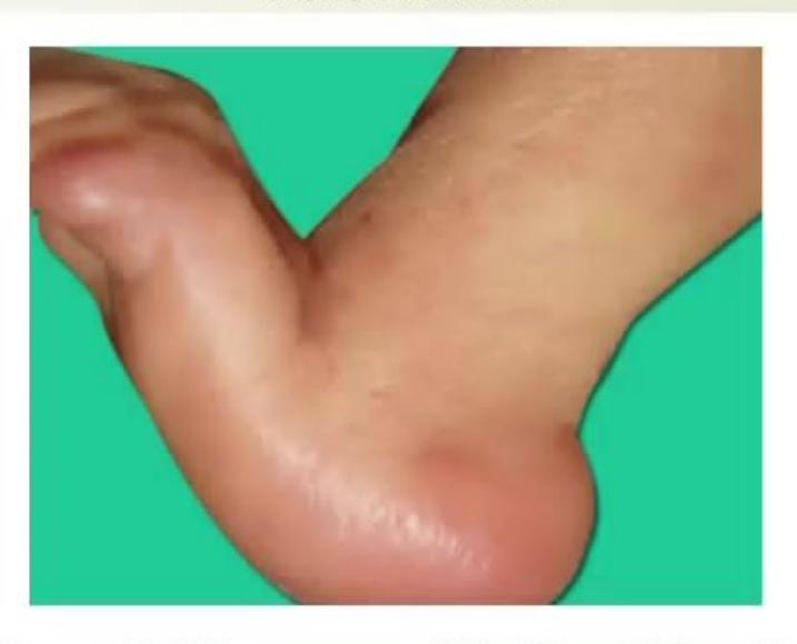
- Identify this foot problem.
- What are the common etiologies (at least four)?
- What are the associated anatomical problems/contractures with this?
- What is the treatment and when should it be started?
Answer
-
Identification:
- Congenital Vertical Talus (CVT / Rocker-Bottom Foot / Congenital Convex Pes Valgus).
-
Common Etiologies (at least 4):
- Idiopathic (isolated)
- Chromosomal trisomies: Trisomy 18 (Edwards syndrome), Trisomy 13 (Patau syndrome)
- Arthrogryposis multiplex congenita
- Myelomeningocele / Spinal dysraphism
- Spinal Muscular Atrophy (SMA)
- Neurofibromatosis Type 1 (NF-1).
-
Associated anatomical problems & contractures:
- Rigid dorsal dislocation of the navicular on the head of the vertically oriented talus.
- Severe contracture of the Achilles tendon (tendoachilles equinus).
- Severe contracture of anterior tibial tendons (tibialis anterior, extensor digitorum longus, extensor hallucis longus, peroneus brevis) holding the forefoot in rigid dorsiflexion against the anterior tibia.
-
Treatment:
- Start immediately in the early neonatal period (first weeks of life).
- Reverse Ponseti (Dobbs) technique: Serial manipulations and long-leg plaster casting in plantarflexion and inversion/adduction, followed by minimally invasive percutaneous/small-incision talonavicular pin fixation and percutaneous Achilles tenotomy.
MNJ26-003
Mother of a 3-year-old child came to your clinic with complaints of a large gap between the knee joints when the child stands. X-ray is given below:
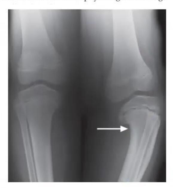
- What is this problem?
- What are important differential diagnoses of this?
- What is tibia vara (Blount disease)?
- How will you differentiate tibia vara from physiological bowing?
Answer
-
Problem:
- Genu Varum (Bow Legs) / Infantile Tibia Vara (Blount Disease).
-
Differential diagnoses:
- Physiological Genu Varum (Physiological bowing)
- Blount Disease (Tibia Vara - Infantile or Adolescent)
- Nutritional Rickets (Vitamin D deficiency) or Hypophosphatemic Rickets
- Skeletal dysplasias (Achondroplasia, Hypochondroplasia, Metaphyseal chondrodysplasia)
- Asymmetric physeal growth arrest post-trauma or infection.
-
Tibia Vara (Blount Disease):
- A progressive developmental disorder characterized by disturbed endochondral ossification of the posteromedial aspect of the proximal tibial physis, resulting in multiplanar varus, procurvatum, and internal rotation deformity of the tibia.
-
Differentiating Tibia Vara from Physiological Bowing:
- Clinical course: Physiological bowing is symmetrical, self-correcting by 2–3 years of age; Blount disease is progressive, often asymmetrical/unilateral, and worsens after age 2.
- Metaphyseal-Diaphyseal (Levine-Drennan) Angle:
- Physiological bowing:
- Tibia vara:
(often ).
- Physiological bowing:
- Radiographic features: Blount disease shows medial proximal tibial beaking, sloping/depression, and fragmentation of the medial metaphysis and epiphysis (Langenskiöld staging); physiological bowing shows smooth, generalized bowing of femur and tibia without localized focal metaphyseal beaking.
- Lateral knee thrust: Present during walking in Blount disease; absent in physiological bowing.
MNJ26-004
Study the image shown below:
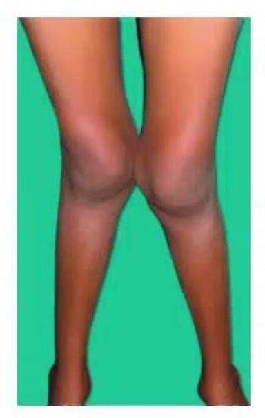
- Identify this condition.
- What is its definition and normal threshold?
- What are common causes?
- What is the indication for surgical correction?
Answer
-
Identification:
- Genu Valgum (Knock-Knees).
-
Definition & Normal Threshold:
- Deformity where knees touch each other while the medial malleoli of the ankles remain separated when standing with patellae facing directly forward.
- Quantified by measuring the Intermalleolar Distance (IMD).
- Physiological valgus peaks at 3–4 years of age (normal IMD up to
) and resolves spontaneously to adult alignment by 7–8 years of age (normal adult ). Pathological if or persists after 8 years.
-
Common Causes:
- Physiological genu valgum (resolves by 7 years)
- Nutritional Rickets / Renal Osteodystrophy
- Skeletal dysplasias (e.g. Spondyloepiphyseal dysplasia, Morquio syndrome)
- Post-traumatic asymmetric lateral tibial growth stimulation (Cozen's phenomenon)
- Obesity / mechanical overload.
-
Indications for Surgical Correction (Hemiepiphysiodesis / Guided Growth with 8-plates or Osteotomy):
- Intermalleolar distance
- Persistent deformity beyond
of age - Significant knee pain, gait instability, patellofemoral subluxation, or functional limitation
- Underlying metabolic bone disease or skeletal dysplasia causing progressive mechanical axis deviation.
- Intermalleolar distance
MNJ26-005
A 14-year-old athletic male child presents with complaints of pain and localized swelling over the anterior knee. The pain increases during running/jumping sports but persists during rest. X-ray of the knee joint was done:
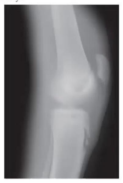
- What is the diagnosis based on this X-ray and clinical condition?
- What is the prognosis?
- What advice and management will you give to this boy?
Answer
-
Diagnosis:
- Osgood-Schlatter Disease (Traction Apophysitis of the Tibial Tuberosity).
- X-ray demonstrates soft tissue swelling, prominence, irregularity, and fragmentation of the tibial tubercle apophysis at the insertion of the patellar tendon.
-
Prognosis:
- Excellent; benign, self-limiting condition that resolves completely once skeletal maturity and physeal closure of the tibial tuberosity are reached (typically by 15–17 years of age).
-
Advice and Management:
- Activity modification: Relative rest; temporary avoidance of vigorous jumping, running, and kneeling sports that provoke pain.
- Cryotherapy: Ice application for 15–20 minutes after physical exertion.
- Analgesics: Short course of oral NSAIDs (Ibuprofen) during acute painful episodes.
- Physical therapy: Quadriceps and hamstring stretching and eccentric strengthening exercises.
- Infrapatellar / Patellar tendon strap: Decreases traction stress on the tibial tuberosity.
- Knee immobilizer / cast is rarely indicated (only for severe, refractory, acute disabling pain).
MNJ26-006
Study the clinical examination maneuvers and orthosis shown below:
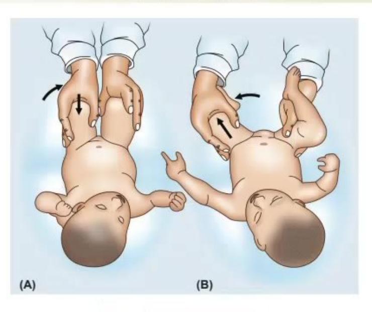
- What are the names of these two tests (A and B), and what are they used for?
- What is the incidence of this problem and which sex is more affected?
- What is the investigation of choice according to age?
- What is the treatment of choice in a newborn and in a 1-year-old child?
- What is the name of the orthosis shown in the picture?
Answer
-
Tests & Clinical Purpose:
- Test A: Barlow Test (Provocative dislocation test: Hip is flexed to 90° and adducted with gentle posterior pressure applied along the femoral shaft
dislocates an unstable femoral head over the posterior acetabular rim with a palpable "jerk of exit"). - Test B: Ortolani Test (Reduction test: Dislocated hip is flexed to 90° and gently abducted while lifting the greater trochanter anteriorly
reduces the femoral head back into the acetabular cup with a palpable "clunk of entry"). - Purpose: Neonatal screening and diagnosis of Developmental Dysplasia of the Hip (DDH).
- Test A: Barlow Test (Provocative dislocation test: Hip is flexed to 90° and adducted with gentle posterior pressure applied along the femoral shaft
-
Incidence & Gender:
- Incidence: 1 to 1.5 per 1,000 live births.
- Females are 4 to 8 times more commonly affected than males (due to higher sensitivity to maternal relaxin hormone).
-
Investigation of Choice:
- Age
months: Dynamic Ultrasonography of the Hip (Graf method) (femoral head is largely cartilaginous and radiolucent on plain X-rays). - Age
months: Plain Anteroposterior Pelvis X-ray (assesses Acetabular Index, Shenton's line, Perkins line, and Hilgenreiner line).
- Age
-
Treatment according to age:
- 0 to 6 months: Dynamic splinting with Pavlik Harness (maintains hips flexed
and abducted ). - 6 months to 18–24 months: Closed reduction under general anesthesia with hip spica cast immobilization (open reduction if closed reduction fails).
years: Open reduction with pelvic (Salter/Pemberton) and femoral shortening/derotation osteotomy.
- 0 to 6 months: Dynamic splinting with Pavlik Harness (maintains hips flexed
-
Orthosis Name:
- Pavlik Harness.
MNJ26-007
A 7-year-old male child came to your hospital with a limping gait and complaints of pain at the groin referred to the anteromedial thigh and knee. On examination, internal rotation and abduction are restricted at the hip joint. You ordered a pelvis X-ray shown below:
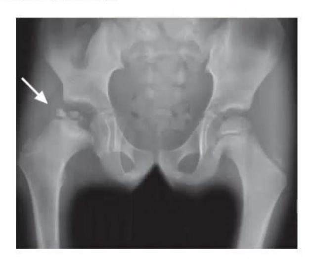
- What is the clinical and radiological diagnosis?
- What is the pathophysiology behind this?
- What treatment will you give to this boy?
Answer
-
Diagnosis:
- Legg-Calvé-Perthes Disease (LCPD / Coxa Plana) (Idiopathic avascular osteonecrosis of the capital femoral epiphysis).
- X-ray demonstrates increased radiodensity/sclerosis, subchondral fracture line (Crescent sign), fragmentation, and flattening of the capital femoral epiphysis on the affected hip.
-
Pathophysiology:
- Temporary idiopathic disruption/ischemia of the blood supply (lateral epiphyseal artery branch of medial circumflex femoral artery) to the growing capital femoral epiphysis
infarction/necrosis of bone and marrow subchondral fracture and bone resorption during weight-bearing collapse and flattening of the femoral head (coxa plana) revascularization, re-ossification, and remodeling.
- Temporary idiopathic disruption/ischemia of the blood supply (lateral epiphyseal artery branch of medial circumflex femoral artery) to the growing capital femoral epiphysis
-
Treatment:
- Goal: Relieve pain, preserve hip range of motion, and maintain "containment" of the femoral head within the acetabulum to ensure spherical remodeling.
- Non-operative: Temporary non-weight-bearing/bed rest, oral NSAIDs, physical therapy for maintaining abduction/internal rotation, abduction containment bracing (e.g. Scottish-Rite brace).
- Operative (for severe involvement / lateral extrusion / age
years): Containment surgery (Femoral varus derotation osteotomy or Salter innominate pelvic osteotomy).
MNJ26-008
A 14-year-old boy weighing 70 kg (obese) presents to the emergency department with complaints of limping and pain in the thigh and knee joint. On examination, there is marked restriction of internal rotation, flexion, and abduction. Pelvis X-ray is shown below:
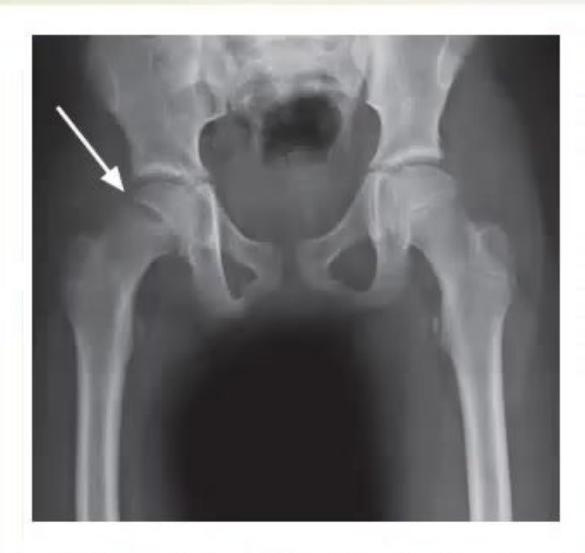
- What is the diagnosis of this condition?
- What is the underlying pathology?
- What is the treatment?
Answer
-
Diagnosis:
- Slipped Capital Femoral Epiphysis (SCFE / SUFE - Slipped Upper Femoral Epiphysis).
- X-ray shows postero-inferior displacement of the capital femoral epiphysis relative to the femoral neck (Trethowan's sign / Klein's line does not intersect the lateral epiphysis).
-
Underlying Pathology:
- Salter-Harris Type I physeal fracture through the hypertrophic zone of the proximal femoral growth plate, where excessive mechanical shear forces (due to adolescent growth spurt and obesity) overwhelm the weakened physis, causing the femoral neck to displace anteriorly, superiorly, and externally rotate relative to the femoral head.
-
Treatment:
- Strict non-weight bearing / emergency bed rest (avoid ambulation or manipulative reduction to prevent avascular necrosis).
- Urgent surgical stabilization: In situ percutaneous single cannulated screw fixation across the physis into the center of the femoral head.
MNJ26-009
A 6-year-old female child came to the hospital with neck pain. The doctor advised cervical spine flexion and extension radiographs shown below. On examination, a short neck, low posterior hairline, and restricted cervical motion were present:
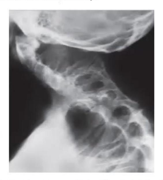
- What is the diagnosis of this condition?
- What is the classic clinical triad and radiological feature?
- What other associated syndromes or anomalies are seen?
Answer
-
Diagnosis:
- Klippel-Feil Syndrome (Congenital Brevicollis).
-
Classic Clinical Triad & Radiological Feature:
- Clinical Triad:
- Short neck ("webbed" appearance)
- Low posterior hairline
- Limitation/restriction of cervical spine mobility.
- Radiological Feature: Congenital failure of normal vertebral segmentation resulting in congenital fusion of
cervical vertebrae (most commonly C2–C3 or C5–C6), with potential hypermobility and instability at adjacent unfused motion segments.
- Clinical Triad:
-
Associated Anomalies & Syndromes:
- Sprengel's deformity (congenital elevation and hypoplasia of the scapula in 30% of cases)
- Scoliosis and kyphosis (in 60%)
- Congenital sensorineural or conductive hearing loss (in 30%)
- Genitourinary anomalies (unilateral renal agenesis)
- Congenital heart defects (VSD, PDA, Coarctation of Aorta)
- Associated syndromes: Goldenhar syndrome, VACTERL association.
MNJ26-010
Rajni, an 11-year-old girl, is referred to your clinic with complaints of intermittent pain in the left hip associated with limping. She stopped participating in school sports activities 1 month ago and prefers to stay in bed. Her mother has hypothyroidism. On examination, she is afebrile and her left limb is held in external rotation and flexion. She has restricted internal rotation, flexion, and abduction. Her weight is between the 92nd–98th percentile and height is below the 3rd percentile.
CBC, CRP, and ESR reports are normal, but thyroid function test shows TSH:
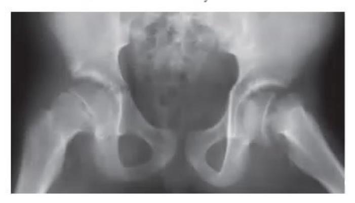
- What is the possible cause of this limping?
- What are the findings in this X-ray and which radiographic view is best to diagnose this disease early?
- What endocrinopathies or metabolic abnormalities are associated with this problem?
- What is the cause of her short stature and how will you treat it?
Answer
-
Cause of limping:
- Slipped Capital Femoral Epiphysis (SCFE / SUFE) occurring secondary to Severe Primary Juvenile Hypothyroidism (Hashimoto's Thyroiditis).
-
X-ray findings & Best View:
- Findings: Widening, blurring, and irregularity of the left proximal femoral physis with medial and posterior displacement of the femoral epiphysis relative to the femoral neck (positive Trethowan's sign).
- Best View: Frog-leg lateral (Lauenstein) view of the pelvis/hip (most sensitive view for detecting early posterior displacement of the epiphysis).
-
Associated Endocrinopathies / Metabolic Conditions:
- Primary Hypothyroidism
- Growth Hormone (GH) deficiency or Growth Hormone replacement therapy
- Hypogonadism / Panhypopituitarism
- Renal Osteodystrophy / Secondary Hyperparathyroidism
- Hypercortisolemia (Cushing's syndrome).
-
Cause of Short Stature & Treatment:
- Cause: Chronic severe untreated Juvenile Hypothyroidism, leading to profound impairment of endochondral ossification, delayed skeletal maturation (bone age retardation), and growth arrest.
- Treatment:
- Orthopedic management: Immediate non-weight-bearing and urgent in situ percutaneous cannulated screw fixation of the slipped capital femoral epiphysis.
- Endocrine management: Oral Levothyroxine (L-thyroxine) replacement therapy (starting dose
, titrating according to serum TSH and free T4 levels).
MNJ26-011
An 11-year-old girl Rani presents to your OPD with complaints of persistent cough for the last 4 months. She had non-productive cough, but no shortness of breath is present. Her past medical history is not significant. Her height and weight are within normal limits. On examination, all systemic examination is normal except you notice the abnormal lateral curvature of her spine shown below. There are no neurocutaneous markers:
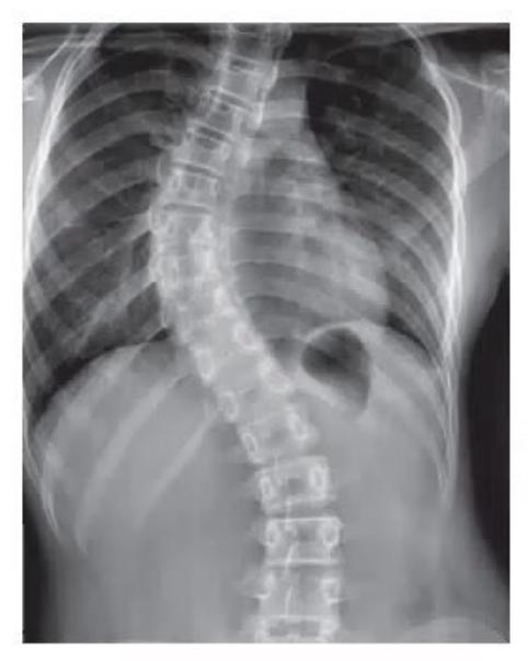
- What is this deformity called?
- What important anthropometrical/physical examination parameter must you evaluate in this child?
- What are the types of this deformity and which one is the most common?
- What are the treatment options available, and name 2 conditions in which early surgical treatment is recommended?
Answer
-
Deformity:
- Scoliosis (lateral spinal curvature with a Cobb angle
measured on standing whole-spine PA radiograph, accompanied by vertebral rotation).
- Scoliosis (lateral spinal curvature with a Cobb angle
-
Anthropometrical / Physical Parameter:
- True and Apparent Leg Length Discrepancy (LLD) (Pelvic obliquity from limb length inequality causes functional/non-structural scoliosis that resolves with an appropriate shoe raise).
- Also evaluate the Adam's Forward Bend Test with a scoliometer to measure the Angle of Trunk Rotation (ATR).
-
Types & Most Common:
- Non-structural (Functional) Scoliosis: Reversible, non-progressive curve without vertebral rotation (e.g. secondary to postural habit, muscle spasm, or leg length discrepancy).
- Structural Scoliosis: Fixed curvature with rotational deformity:
- Adolescent Idiopathic Scoliosis (AIS) — most common type (
of all structural scoliosis, commonly females 10–16 years). - Congenital Scoliosis (hemivertebrae, block vertebrae, unsegmented bars).
- Neuromuscular Scoliosis (Cerebral Palsy, Duchenne Muscular Dystrophy, SMA).
- Syndromic Scoliosis (Marfan syndrome, NF-1).
- Adolescent Idiopathic Scoliosis (AIS) — most common type (
-
Treatment Options & Early Surgery Indications:
- Treatment Options (based on Cobb Angle & Skeletal Maturity / Risser stage):
- Cobb angle
: Observation and serial clinical/radiographic monitoring every 6 months. - Cobb angle
in growing child: Bracing using a rigid Thoracolumbosacral Orthosis (TLSO / Boston brace) worn 18–23 hours/day. - Cobb angle
: Posterior spinal fusion with segmental instrumentation.
- Cobb angle
- Indications for Early Surgical Treatment:
- Duchenne Muscular Dystrophy (DMD) (early progressive neuromuscular curve with loss of sitting balance and impending pulmonary compromise).
- Congenital Scoliosis with unilateral unsegmented bar and contralateral hemivertebra (malignant, rapidly progressive curve refractory to bracing).
- Treatment Options (based on Cobb Angle & Skeletal Maturity / Risser stage):
MNJ26-012
A 6-year-old boy presents with complaints of multiple swellings over the back, arms, and legs for 6 months. The swellings are non-tender and hard in consistency. He experiences discomfort during sleep, but is otherwise active and playful. Evaluate the clinical and radiological images below:
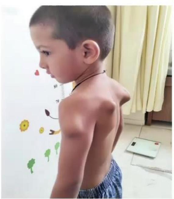
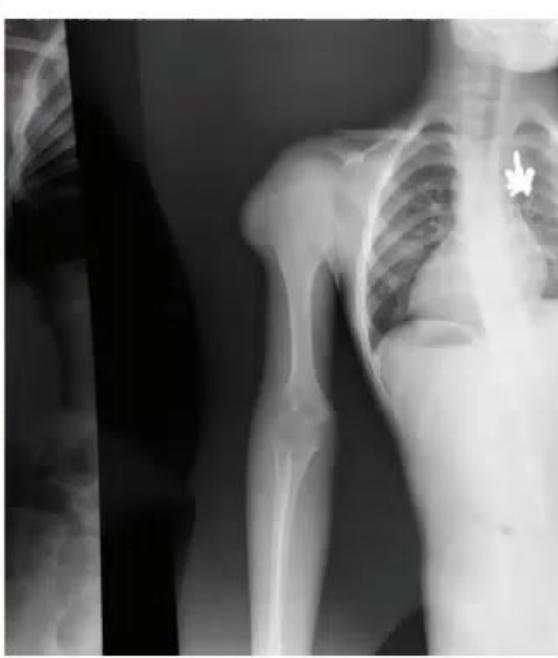
- What is the possible diagnosis?
- What are the clinical manifestations and complications?
- What are the treatment options?
Answer
-
Possible diagnosis:
- Hereditary Multiple Exostoses (HME / Hereditary Multiple Osteochondromas / Diaphyseal Aclasis) (Autosomal dominant disorder due to mutations in EXT1 or EXT2 genes).
-
Clinical Manifestations & Complications:
- Multiple painless, hard, bony juxta-epiphyseal projections (osteochondromas) capped with cartilage, predominantly at the metaphyses of long bones (distal femur, proximal tibia/fibula, proximal humerus).
- Angular limb deformities (genu valgum, ankle valgus), ulnar shortening with radial bowing (Madelung-like deformity), and limb length discrepancy.
- Local compression of adjacent nerves (peroneal, sciatic), blood vessels, and tendons causing pain, bursitis, and paresthesias.
- Malignant transformation: Risk of secondary Chondrosarcoma in adulthood (~1–5%), characterized by sudden rapid enlargement, new pain, or cartilage cap thickness
on MRI.
-
Treatment Options:
- Observation: Asymptomatic osteochondromas require periodic clinical and radiographic monitoring.
- Surgical Excision (Exostosectomy): Indicated for lesions causing severe pain, mechanical limitation of joint movement, vascular or nerve entrapment, progressive cosmetic/angular deformity, or suspicion of malignant transformation. Care is taken to protect adjacent growth plates in skeletally immature children.
- Corrective osteotomies and epiphysiodesis for limb length discrepancies and angular deformities.