MNJ36-001
Examine the abdomen of this child.
Answer
- Introduction & Consent: Introduces self, explains procedure, seeks verbal consent from parents/child, warms hands.
- Positioning & Exposure: Child in supine position with knees slightly flexed; completely expose abdomen from xiphisternum to midthigh (including groin/genitalia), maintaining modesty.
- Inspection:
- Abdominal contour/shape (scaphoid/flat/distended), symmetry.
- Movements with respiration (abdominal vs thoracoabdominal breathing).
- Umbilicus (position, eversion, discharge, herniation).
- Dilated superficial veins (note direction of blood flow), striae, visible peristalsis, visible pulsations, scars, sinuses.
- Palpation:
- Ask if and where there is pain (examine painful area last); look at the child's face for discomfort throughout.
- Light / Superficial Palpation: Assess local temperature, guarding, rigidity, hyperesthesia, superficial tenderness in all 9 quadrants.
- Deep Palpation:
- Liver: Palpate from right iliac fossa upwards towards right costal margin along midclavicular line; characterize edge, surface, consistency, tenderness; measure liver span by percussion (normal: 5–8 cm depending on age).
- Spleen: Palpate from right iliac fossa diagonally towards left costal margin; feel for splenic notch.
- Kidneys: Bimanual ballottement in both flanks.
- Percussion:
- Map out liver and splenic dullness.
- Percuss for ascites: Shifting dullness and fluid thrill (in massive ascites).
- Auscultation:
- Auscultate bowel sounds in all 4 quadrants (normal, sluggish, or hyperactive/borborygmi).
- Listen for vascular bruits (renal/aortic) and friction rubs.
- Hernial Orifices, External Genitalia, Spine & Anus: Check inguinal and femoral rings, palpate testes in scrotum, inspect perianal area and spine.
- Re-cover abdomen, thank child and parents, and summarize positive/negative findings to the examiner.
MNJ36-002
Measure the blood pressure (BP) of this 10-year-old child and record the findings.
Answer
- Introduction & Rapport: Introduces self, explains procedure to child and mother, ensures child is calm and rested for 5 minutes.
- Cuff Selection: Choose appropriate pediatric cuff size:
- Inflatable bladder width must cover
of mid-arm circumference (or 75–80% of upper arm length). - Bladder length must encircle 80–100% of arm circumference.
- Inflatable bladder width must cover
- Application: Apply cuff snugly on bare right upper arm with lower edge 2–3 cm above antecubital fossa; center bladder over brachial artery. Support arm at heart level.
- Palpatory Method (Estimate Systolic BP): Palpate radial pulse, inflate cuff rapidly until pulse disappears; inflate 20–30 mmHg further, then slowly deflate (2–3 mmHg/sec) to note pressure when pulse reappears.
- Auscultatory Method:
- Place diaphragm of stethoscope lightly over brachial artery (not under the cuff).
- Inflate cuff to 20–30 mmHg above estimated palpatory systolic BP.
- Deflate slowly at 2–3 mmHg per second.
- Systolic BP (SBP): Appearance of clear tapping sound (Korotkoff Phase I).
- Diastolic BP (DBP): Complete disappearance of sounds (Korotkoff Phase V; Phase IV muffling in young children).
- Measure in upper and lower extremities if evaluating for hypertension or coarctation.
- Plot SBP and DBP on age-, sex-, and height-percentile blood pressure charts (AAP/WHO).
- Assist child with clothing, thank child/mother, and report values and percentile to the examiner.
MNJ36-003
Demonstrate the step-by-step procedure for performing a Lumbar Puncture (LP) in a 1-year-old child on a manikin/model.
Answer
- Informed Consent & Pre-procedure Check: Explain indication and risks to parents, obtain written informed consent. Check platelet count and coagulation profile; verify absence of contraindications (signs of raised ICP, focal deficits, severe cardiorespiratory instability, local infection at puncture site).
- Monitoring & Equipment: Ensure continuous pulse oximetry, resuscitation equipment, and sterile LP kit ready (22G/24G spinal needle with stylet, sterile gloves/drape, collection tubes, manometer, antiseptic, local anesthetic).
- Positioning: Place child in left lateral decubitus position on a firm surface with spine flexed (fetal position — knees drawn to abdomen and neck gently flexed, taking care not to compromise airway). Assistant holds child securely.
- Landmark Identification: Draw an imaginary intercristal line connecting the highest points of both iliac crests (Tuffier's line), crossing the L3–L4 intervertebral space (safe level for LP in infants/children: L3–L4 or L4–L5).
- Asepsis & Local Anesthesia: Thorough surgical hand wash, sterile gloves, gown; clean skin with antiseptic solution in concentric circles; place sterile fenestrated drape. Infiltrate 1% lignocaine locally into skin and subcutaneous tissue.
- Needle Insertion:
- Insert 22G spinal needle with stylet in midline at L3–L4 space, bevel pointing cranially/laterally, angled slightly towards umbilicus (10°–15° cephalad).
- Advance slowly until a subtle "give-way / pop" sensation is felt as the ligamentum flavum and dura are pierced.
- Withdraw stylet partially to observe spontaneous clear CSF flow.
- CSF Collection & Pressure: Measure opening pressure with manometer if indicated; collect 1–2 mL of CSF into 3 sterile tubes (Tube 1: Biochemistry/glucose & protein, Tube 2: Microbiology/Gram stain & culture, Tube 3: Cell count & differential).
- Completion & Post-Care: Reinsert stylet, withdraw spinal needle swiftly, apply firm pressure with sterile gauze, apply dressing. Place child supine for 30–60 minutes; monitor vitals and puncture site. Dispose of sharps in designated puncture-proof bin.
MNJ36-004
A 13-year-old boy presents with ataxia. Demonstrate how you would systematically examine his gait and cerebellar coordination.
Answer
- Introduction & Safety: Introduces self, explains procedure, ensures a safe, well-lit, unobstructed walking path; stands close to prevent falls.
- Casual Walking: Ask child to walk barefoot in a straight line for 10–15 paces, turn around, and return:
- Observe stance width, stride length, speed, arm swing, trunk stability, balance during turning.
- Cerebellar ataxia: Broad-based, unsteady, staggering "drunken" gait with irregular steps and lurching toward side of cerebellar lesion.
- Sensory ataxia: Stamping gait with heavy heel-strikes, worse in dark / eyes closed.
- Spastic hemiparetic/diplegic: Circumduction / scissoring gait.
- Tandem Walking (Heel-to-Toe): Ask child to walk in a straight line placing heel directly in front of opposite toe (accentuates subtle midline vermian cerebellar ataxia).
- Toe and Heel Walking: Ask child to walk on toes (tests S1/calf strength) and heels (tests L4/L5 dorsiflexor strength).
- Romberg Test: Ask child to stand with feet together, arms at sides, eyes open, then close eyes for 30 seconds:
- Sensory ataxia (dorsal column lesion): Steady with eyes open, marked swaying/falling with eyes closed (Positive Romberg).
- Cerebellar ataxia: Unsteady with eyes open, no significant worsening with eyes closed (Negative Romberg / cerebellar instability).
- Check rapid turn test and perform other cerebellar signs (finger-nose, dysdiadochokinesia).
- Thanks child and parents; reports impression to examiner.
MNJ36-005
Perform a systematic motor system examination of the right upper limb in a child.
Answer
- Introduction & Consent: Introduces self, explains examination, warms hands, properly exposes right upper limb (and contralateral limb for comparison).
- Inspection:
- Muscle bulk and symmetry (look for wasting or pseudohypertrophy in deltoid, biceps, thenar, hypothenar, interossei muscles).
- Mid-upper arm circumference (MUAC) measurement if indicated.
- Involuntary movements (fasciculations, tremors, chorea, athetosis).
- Posture and deformities (wrist drop, claw hand, flexion posture).
- Palpation & Tone:
- Palpate muscle bulk/consistency.
- Assess tone by gentle passive movements across wrist, elbow, and shoulder joints (hypotonia vs spasticity/clasp-knife vs rigidity/lead-pipe).
- Power Assessment (MRC Scale 0 to 5):
- Shoulder: Abduction (C5), Adduction (C6/C7).
- Elbow: Flexion (C5/C6 - Biceps), Extension (C7 - Triceps).
- Wrist: Flexion (C7/C8), Extension (C6/C7).
- Hand & Fingers: Grip strength (C8/T1), Finger abduction/interossei (T1), Thumb opposition (T1).
- Deep Tendon Reflexes (DTRs):
- Biceps reflex (C5, C6)
- Supinator / Brachioradialis reflex (C5, C6)
- Triceps reflex (C7, C8)
- Coordination: Finger-to-nose test, rapid alternating movements (dysdiadochokinesia).
- Help child redress, thank child and parents, and summarize motor findings to the examiner.
MNJ36-006
Perform a clinical volume status assessment (evaluating for volume depletion / dehydration vs volume overload) in a patient.
Answer
- Introduction & Exposure: Introduces self, takes consent, exposes neck, chest, abdomen, and limbs appropriately.
- Tissue & Mucosal Signs:
- Oral mucosa and tongue: Moist (euvolemic) vs dry/parched (volume depletion).
- Axillae: Check axillary moisture.
- Eyes: Normal vs sunken eyes, presence/absence of tears on crying.
- Skin Turgor: Perform skin pinch on abdominal wall at level of umbilicus (normal: retracts immediately; slow
or very slow in dehydration).
- Peripheral Perfusion & Capillary Refill:
- Palpate peripheral extremities (warm vs cold/clammy).
- Capillary Refill Time (CRT): Press thumb nail bed or sternum for 5 seconds and release (normal
; indicates poor perfusion/shock).
- Cardiovascular / Hemodynamic Signs:
- Peripheral pulses: Rate, rhythm, volume (thready in hypovolemia, bounding in hypervolemia/high output).
- Blood Pressure & Postural Drop: Measure lying and standing BP (orthostatic drop in SBP
or DBP indicates volume depletion). - Jugular Venous Pressure (JVP): Inspect at 45° angle (elevated JVP
above sternal angle indicates volume overload/right heart failure).
- Signs of Fluid Overload:
- Dependent peripheral edema: Pitting edema over medial malleoli, pretibial area, and sacrum.
- Respiratory: Tachypnea, bilateral basilar fine crackles/crepitations, pleural effusion.
- Abdomen: Tender hepatomegaly, ascites.
- Report clinical volume status (dehydrated, euvolemic, or fluid-overloaded) to the examiner.
MNJ36-007
Demonstrate the correct preparation and administration of the MMR vaccine in a 16-month-old child.
Answer
- Introduction & Screening: Greet mother, verify child's identity and age (16 months), screen for contraindications (severe immunocompromise/immunodeficiency, anaphylaxis to neomycin/gelatin, recent IVIG/blood products within 3–11 months).
- Hand Hygiene & Vaccine Inspection:
- Wash hands thoroughly with soap and water.
- Inspect MMR lyophilized vaccine vial, sterile water diluent ampoule, expiry date, and Vaccine Vial Monitor (VVM) stage.
- Reconstitution:
- Draw entire volume of provided sterile water diluent with a mixing syringe.
- Slowly inject diluent into the MMR vial down the side of the glass; swirl gently to dissolve completely without foaming.
- Keep reconstituted vial on ice pack away from direct light; use within 4 hours.
- Dose & Syringe Loading: Draw exactly 0.5 mL of reconstituted vaccine into a sterile 0.5 mL auto-disable / tuberculin syringe with a 25G, 5/8-inch needle.
- Site & Administration:
- Site: Anterolateral aspect of upper thigh (or upper outer aspect of deltoid muscle).
- Clean skin with sterile water if visibly soiled (avoid spirit/alcohol wipes for live viral vaccines).
- Pinch subcutaneous tissue gently; insert needle at a 45° angle subcutaneously.
- Inject the full 0.5 mL dose smoothly; withdraw needle and apply gentle pressure with dry cotton (do not massage).
- Post-Vaccination Counseling:
- Advise waiting for 30 minutes in clinic to observe for acute allergic reactions.
- Counsel on expected delayed reactions (mild fever and rash 5–12 days post-vaccination).
- Dispose of sharps in puncture-proof sharps container; record in immunization card and schedule next dose (at 4–6 years).
MNJ36-008
Perform a focused pediatric musculoskeletal screening examination (pGALS) in an 8-year-old boy.
Answer
- Introduction & Consent: Introduce self, establish rapport with child and parents, ask if the child has pain anywhere.
- Gait:
- Observe the child walking 10 paces, turning, and returning.
- Walk on tiptoes and on heels (tests ankle and subtalar joints).
- Arms (Upper Limbs):
- Put hands out straight in front (palms down), inspect dorsum of hands, wrists, fingers.
- Turn hands over (palms up), inspect palmar aspect and thenar/hypothenar eminence.
- Make a tight fist (tests finger flexion) and touch thumb to each fingertip (fine dexterity).
- "Prayer sign" and reverse prayer sign (tests wrist extension and flexion).
- Touch back of neck with elbows back (shoulder abduction and external rotation).
- Reach hands high above head with arms straight (elbow extension and shoulder elevation).
- Legs (Lower Limbs):
- Inspect knees and ankles standing (alignment: valgus/varus).
- Lie on couch: Assess passive knee flexion and extension while palpating for joint effusion and joint line crepitus.
- Assess internal and external rotation of hips with hip and knee flexed at 90°.
- Inspect soles of feet for flat arches or deformities.
- Spine & Neck:
- Cervical spine: Look up at ceiling, chin to chest, turn head to left and right, ear to shoulder.
- Thoracolumbar spine: Inspect spine from behind and side for scoliosis/kyphosis/lordosis; palpate spinous processes.
- Forward bending test (Adams forward bend test) with knees straight to check spinal curvature and flexibility.
- Thank child and parents; report findings systematically to the examiner.
MNJ36-009
Demonstrate how to accurately measure the weight, length, and head circumference of a neonate.
Answer
- Introduction, Consent & Asepsis: Introduces self, explains procedure, ensures warm draft-free room, washes and warms hands.
- Weight Measurement (Electronic Infant Weighing Scale):
- Place clean disposable paper sheet on weighing pan.
- Zero/tare the digital weighing scale.
- Undress neonate completely (remove diaper).
- Place baby gently in center of pan; wait for infant to be still and read weight to nearest 5–10 grams.
- Report weight and immediately cover baby to prevent hypothermia.
- Length Measurement (Infantometer):
- Place infant supine on firm infantometer.
- Assistant holds baby's head firmly against fixed headboard in Frankfurt horizontal plane (line from lower margin of orbit to superior margin of auditory meatus is vertical).
- Examiner holds knees flat against baseboard with left hand and moves sliding footboard firmly against heels with toes pointing upwards.
- Read crown-to-heel length to the nearest 0.1 cm.
- Head Circumference (Occipitofrontal Circumference - OFC):
- Use a flexible, non-stretchable narrow measuring tape.
- Place tape across the widest part: anteriorly just above the eyebrows and supraorbital ridges, laterally above the ears, and posteriorly over the maximal prominence of the occiput.
- Pull tape snug to compress hair; take 3 measurements and record the largest reading to nearest 0.1 cm.
- Plot weight, length, and OFC on WHO/Fenton growth charts; report percentiles to the examiner.
MNJ36-010
Perform developmental milestone assessment on this child across all domains and conclude with the developmental age.
Answer
- Introduction & Rapport: Engage child in a relaxed, playful manner with colorful age-appropriate testing toys/cubes/paper.
- Gross Motor:
- Walks up and down stairs using alternate feet.
- Hops steadily on one foot for 2–3 hops.
- Throws ball overhand with coordination and catches a bounced ball.
- Fine Motor & Adaptive:
- Copies a square (
) and a cross ( ). - Draws a person ("Draw-a-Man") with
identifiable parts (head, trunk, limbs). - Builds a 3-step bridge with 5 wooden cubes; cuts along a straight line with blunt safety scissors.
- Copies a square (
- Language & Communication:
- Speaks in complete 5- to 6-word sentences; tells a coherent story/event.
- Identifies and names 4 primary colors (red, blue, green, yellow).
- Counts 4 objects accurately; understands prepositions (in front, behind, under).
- Personal & Social:
- Unbuttons buttons and dresses/undresses with minimal assistance.
- Washes and dries hands and face independently.
- Engages in cooperative group play and role-play games.
- Summary to Examiner: "All demonstrated milestones are consistent with a developmental age of 4 years."
MNJ36-011
- Demonstrate how to elicit the Amiel-Tison tone angles in an infant.
- What are the normal values for Amiel-Tison angles across infancy?
- What is the clinical utility of examining these passive tone angles?

Answer
-
Method of Elicitation:
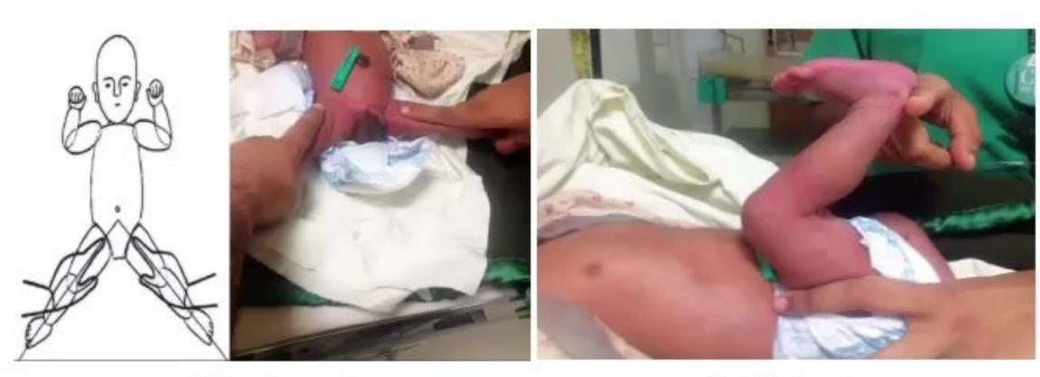
- Adductor Angle: Infant supine with legs extended; gently abduct both legs laterally as far as possible; measure angle formed between thighs.
- Popliteal Angle: Infant supine, pelvis flat; flex thighs onto abdomen, then extend lower legs at knees until resistance is felt; measure posterior angle formed at popliteal fossa.
- Heel-to-Ear Maneuver: Supine with pelvis flat; draw feet straight up towards ears without lifting pelvis off table; measure angle between thighs and trunk.
- Scarf Sign: Draw infant's hand across chest towards opposite shoulder; note position of elbow relative to midline.
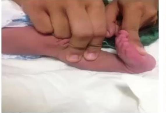
- Foot Dorsiflexion Angle: Flex foot dorsally against anterior lower leg; measure angle between dorsum of foot and anterior border of leg.
-
Normal Values by Age (Amiel-Tison Norms):
| Age Range | Adductor Angle | Popliteal Angle | Scarf Sign | Foot Dorsiflexion |
|---|---|---|---|---|
| 1–3 months | 40°–80° | 80°–100° | Elbow does not cross midline | 60°–70° |
| 4–6 months | 70°–110° | 100°–120° | Elbow reaches/crosses midline | 60°–70° |
| 7–9 months | 110°–140° | 120°–150° | Elbow passes beyond contralateral axillary line | 60°–70° |
| 10–12 months | 140°–160° | 150°–170° | Elbow easily passes beyond contralateral axillary line | 60°–70° |
- Clinical Utility:
- Evaluates passive muscle tone and maturation of the corticospinal pathways in infants (0–12 months).
- Early detection of hypertonia / spasticity (decreased angles / tight joints) in Spastic Cerebral Palsy.
- Early detection of hypotonia (excessively wide angles) in lower motor neuron or central hypotonia syndromes.
MNJ36-012
(a) Demonstrate how to elicit Soft Neurological Signs (Neurological Soft Signs - NSS) in a school-age child.
(b) What is the clinical significance and diagnostic utility of soft neurological signs?
Answer
(a) Elicitation of Soft Neurological Signs:
- Motor Coordination & Rapid Alternating Movements:
- Rapid finger tapping (thumb-to-fingers in sequence).
- Rapid alternating hand pronation-supination on thighs.
- Foot tapping (alternate heel-toe tapping on floor).
- Observe for motor persistence, dysmetria, and involuntary mirror movements (synkinesis) in the opposite hand.
- Motor Incoordination & Overflow:
- Fog's test: Ask child to walk on lateral borders of feet; observe for involuntary posturing/overflow movements in outstretched arms.
- Tandem walking and hopping on one foot.
- Sensory & Perceptual Integration:
- Double simultaneous tactile stimulation (extinction test / face-hand test).
- Graphesthesia: Identify numbers traced on palm with eyes closed.
- Astereognosis: Identify familiar objects (coin, key) by touch alone.
- Left-right orientation and finger agnosia.
- Spatial Orientation & Praxis:
- Constructive praxis: Copying geometric designs, tying shoelaces, buttoning/unbuttoning clothes.
(b) Clinical Utility of Soft Neurological Signs:
- NSS represent subtle non-focal abnormalities in motor control, sensory processing, and integration without focal neuroanatomical lesions.
- Found in higher prevalence in neurodevelopmental disorders: ADHD, Developmental Coordination Disorder (Dyspraxia), Specific Learning Disabilities (Dyslexia), and Autism Spectrum Disorder.
MNJ36-013
A child is admitted with suspected Infective Endocarditis (IE). Perform a systematic clinical examination for peripheral and cardiac signs of IE.


Answer
- Introduction & General Assessment: Introduces self, takes consent, measures temperature (fever), notes toxic appearance, pallor.
- Peripheral Stigmata of Infective Endocarditis:
- Hands & Nails:
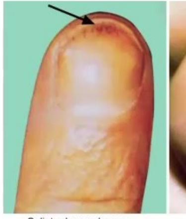
- Splinter hemorrhages (linear subungual hemorrhages).
- Osler nodes: Painful, tender, erythematous raised nodules on pads of fingers/toes (immunological).
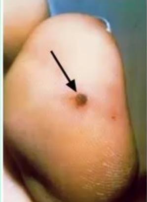
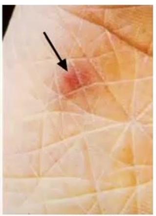
- Janeway lesions: Non-tender, flat, erythematous/hemorrhagic macules on palms and soles (micro-embolic).
- Digital clubbing.
- Eyes: Conjunctival petechiae/hemorrhages; ophthalmoscopy for Roth spots (pale retinal spots surrounded by hemorrhage).
- Skin: Generalized petechiae, purpuric rash.
- Hands & Nails:
- Cardiovascular Examination:
- Palpate pulses for rate, rhythm, and character.
- Precordial auscultation: Auscultate all 4 valve areas for new or changing murmurs (especially aortic or mitral regurgitation), prosthetic valve clicks, or friction rubs.
- Abdominal Examination: Palpate for splenomegaly (present in ~30–50% of chronic IE) and tender hepatomegaly (heart failure).
- Signs of Heart Failure / Complications: Raised JVP, pulmonary rales/crackles, peripheral pedal edema.
- Re-cover child, thank family, and present findings according to Duke criteria.
MNJ36-014
Examine an infant with Hydrocephalus, providing running commentary to the examiner.
Answer
- Introduction & Consent: Greet parents, introduce self, seek permission to examine the infant.
- Craniofacial Inspection & Palpation:
- Head Circumference: Measure maximal occipitofrontal circumference (OFC) with non-stretchable tape; plot on growth chart (OFC
/ percentile or crossing percentiles rapidly). - Cranial Shape & Forehead: Macrocephaly with frontal bossing.
- Scalp: Scalp is thin, shiny, and stretched with engorged, dilated, tortuous superficial scalp veins.
- Sutures & Fontanelles: Palpate cranial sutures (widely split/separated); palpate anterior fontanelle (large, tense, bulging, non-pulsatile).
- Percussion: Macewen's sign ("cracked-pot" sound on skull percussion).
- Transillumination: Transillumination test positive in severe hydrocephalus / hydranencephaly.
- Head Circumference: Measure maximal occipitofrontal circumference (OFC) with non-stretchable tape; plot on growth chart (OFC
- Ocular Signs:
- Setting-Sun Sign: Downward deviation of ocular globes with sclera visible above iris due to periaqueductal pressure on vertical gaze center.
- Assess pupillary reflexes and check for 6th cranial nerve (abducens) palsy (strabismus).
- Fundus examination for papilledema or optic atrophy.
- Neurological & Motor Examination:
- Hypertonia and spasticity in lower extremities (corticospinal tract stretching), exaggerated deep tendon reflexes, ankle clonus, extensor plantar response.
- High-pitched "cerebral" cry, irritability, poor feeding, vomiting.
- Spine Examination: Inspect and palpate spine for spina bifida / myelomeningocele or neurocutaneous markers.
- Re-dress baby, thank parents, and report findings to examiner.
MNJ36-015
Demonstrate systematic Palpation of the Respiratory System in a standing/seated child with running commentary.

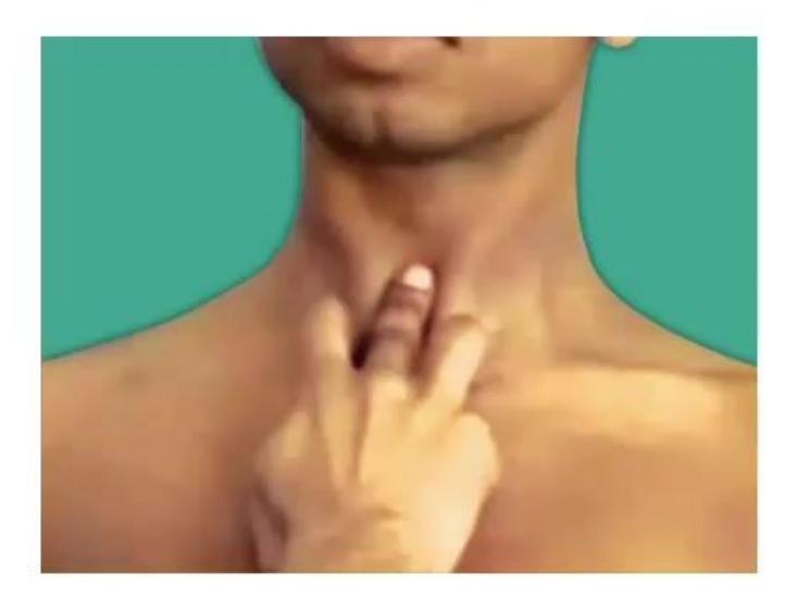
Answer
- Introduction & Position: Introduces self, warms hands, exposes chest properly, ensures child is comfortable seated or standing.
- Confirmation of Inspection Findings: Palpate for localized chest wall tenderness, rib deformities, palpable rhonchi, pleural friction rubs, or subcutaneous emphysema (crepitus).
- Tracheal Position:
- Place index and ring fingers on sternal heads of sternocleidomastoid muscles; gently insert tip of middle finger into suprasternal notch over trachea.
- Assess central position vs deviation to right/left.
- Apex Beat: Palpate apex beat (normal: 4th left ICS in
, 5th left ICS inside midclavicular line in older children); check for mediastinal displacement. - Chest Expansion (Upper & Lower Lobes):
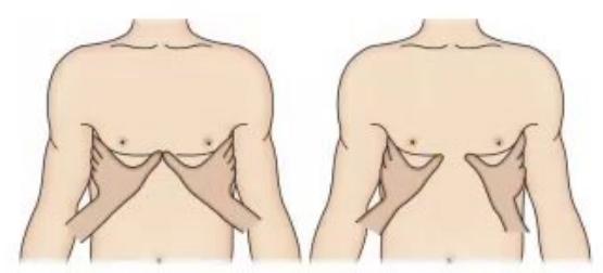
- Anterior Chest: Place palms on lower chest with fingers grasping lateral rib cages; extend thumbs midline until they meet over lower sternum without tension. Instruct child to inhale deeply; observe symmetric divergence of thumbs (normal: 3–5 cm separation).
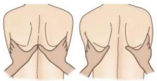
- Posterior Chest: Place hands around lateral chest walls with thumbs apposed midline over T10 spinous process; observe thumb separation on maximal deep inspiration.
- Tactile Vocal Fremitus (TVF):
- Place ulnar border of hand / palm flat on corresponding areas of anterior, lateral, and posterior chest walls sequentially.
- Ask child to repeat "99" or "one-one-one" each time; compare vibratory intensity between symmetrical lung zones (increased in consolidation/cavity, decreased in effusion/pneumothorax/collapse).
- Help child redress, thank child/mother, and report palpatory findings to examiner.
MNJ36-016
A 12-year-old child with Marfan Syndrome is diagnosed with Aortic Regurgitation (AR). Demonstrate the peripheral and cardiac physical signs of chronic AR with commentary.
Answer
- Introduction & Consent: Greet child and parents, take consent, warm hands, expose chest and extremities.
- Peripheral Arterial Signs of Wide Pulse Pressure in AR:
- Water-Hammer / Corrigan's Pulse: Palpate radial pulse, elevate child's forearm vertically above heart; feel rapid upstroke and sudden collapse (collapsing pulse).
- Corrigan's Sign: Visible vigorous pulsations of carotid arteries in the neck ("dancing carotids").
- de Musset's Sign: Rhythmical head bobbing / nodding synchronous with each heartbeat.
- Quincke's Sign: Alternating flushing and blanching of the nail bed when light pressure is applied to the fingernail tip.
- Müller's Sign: Systolic pulsation of the uvula and soft palate on mouth inspection.
- Landolfi's Sign: Systolic pupillary constriction and diastolic dilation.
- Lighthouse Sign: Alternating facial flushing and pallor.
- Rosenbach's Sign: Pulsatile liver on deep abdominal palpation.
- Gerhardt's Sign: Pulsatile spleen.
- Duroziez's Sign: Auscultate femoral artery with stethoscope diaphragm applying gentle compression (systolic murmur with proximal compression, diastolic murmur with distal compression).
- Traube's Sign ("Pistol-Shot Sounds"): Booming pistol-shot sounds auscultated over the femoral arteries.
- Hill's Sign: Systolic blood pressure in popliteal/lower extremity exceeds brachial/upper extremity SBP by
( in severe AR).
- Precordial Examination: Hyperdynamic displaced apex beat; early diastolic high-pitched decrescendo murmur best heard at 3rd left ICS (Erb's point) on leaning forward in end-expiration; Austin Flint murmur (apical mid-diastolic rumble).
- Redress child, thank family, and summarize findings.
MNJ36-017
A 13-year-old child is admitted with jaundice. Perform a focused physical examination for signs of acute liver failure / chronic liver disease with running commentary.
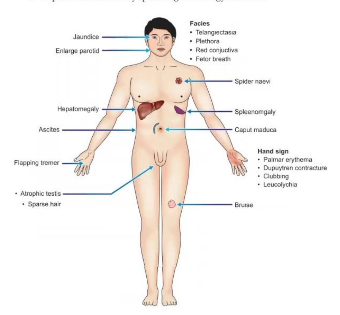
Answer
- Introduction & Exposure: Introduces self, takes consent, exposes child maintaining dignity.
- General Appearance & Neurological State:
- Scleral icterus / jaundice, fetor hepaticus (musty sweet breath).
- Assess Hepatic Encephalopathy: Grade I (sleep reversal, mild confusion), Grade II (drowsy, asterixis), Grade III (stuporous), Grade IV (coma).
- Asterixis (Flapping Tremor): Ask child to hyperextend arms, dorsiflex wrists, and spread fingers for 30 seconds with eyes closed; observe for irregular jerky lapses of posture.
- Constructional apraxia (inability to draw a 5-pointed star or simple shapes).
- Upper Limb & Cutaneous Stigmata:
- Palmar erythema (mottled erythema over thenar/hypothenar eminences).
- Leukonychia (Terry's white nails) and digital clubbing.
- Dupuytren's contracture.
- Multiple spider angiomas (spider nevi) in SVC territory (face, neck, upper chest, arms) — blanch with central pressure.
- Bruising / petechiae / purpura (coagulopathy).
- Head & Neck: Bilateral painless parotid gland enlargement, Kayser-Fleischer rings (slit lamp in Wilson disease).
- Abdominal Examination:
- Distended abdomen with tense ascites.
- Dilated abdominal wall collateral veins radiating from umbilicus (Caput Medusae — blood flows away from umbilicus).
- Liver: Shrunken, non-palpable liver in fulminant failure/cirrhosis (or hard, nodular enlarged liver in early cirrhosis).
- Splenomegaly (portal hypertension).
- Endocrine Stigmata: Gynecomastia, sparse body hair, testicular atrophy.
- Lower Extremities: Bilateral pitting pedal/pretibial edema.
- Redress child, thank family, and present structured summary to examiner.
MNJ36-018
Perform a systematic clinical assessment of Higher Mental Functions in a school-age girl with commentary.
Answer
- Introduction & Rapport: Greet child by name, establish warm, friendly communication to eliminate anxiety.
- General Appearance & Consciousness: Note alertness, behavior, cooperation, emotional affect, eye contact, and grooming.
- Orientation:
- Time: What time of day is it? What day, month, and year is it?
- Place: Where are we right now? What is the name of this hospital/city?
- Person: What is your name? Who is this person sitting next to you?
- Attention & Concentration:
- Digit Span Test: Ask child to repeat 3–4 numbers forward (e.g., 3-8-2-5) and 3 numbers backward.
- Serial subtraction: Subtract 3 or 7 from 100, or recite days of week/months backwards.
- Memory:
- Immediate Recall: Repeat 3 unrelated words (e.g., Apple, Table, Penny) immediately.
- Recent Memory: What did you have for breakfast today? Ask for the 3 words after 5 minutes.
- Remote Memory: When is your birthday? What festival did you celebrate last month?
- Speech & Language:
- Fluency, clarity, articulation, volume, and comprehension of 3-step commands ("Take this paper, fold it in half, put it on table").
- Naming common objects (pen, watch), repeating complex phrases, reading and writing.
- Intelligence & Abstract Thinking: Simple age-appropriate calculations (e.g.,
, ), explaining similarities (e.g., "How are an apple and an orange alike?"). - Thank child and parents; present concise assessment to examiner.
MNJ36-019
Demonstrate focused clinical examination of the motor and sensory components of the 7th Cranial Nerve (Facial Nerve).
Answer
- Introduction & Consent: Greet child and parents, explain procedure, ensure adequate lighting.
- Inspection at Rest:
- Facial symmetry, forehead wrinkle symmetry, palpebral fissure width, blink frequency.
- Symmetry of nasolabial folds, corners of mouth, and involuntary facial twitches.
- Motor Testing of Facial Muscles (Upper & Lower Face):
- Frontalis: Ask child to look up at ceiling / raise eyebrows; observe forehead wrinkling.
- Orbicularis Oculi: Ask child to close eyes tightly ("do not let me open them"); attempt to gently lift upper eyelids.
- Orbicularis Oris: Ask child to whistle, puff out cheeks against resistance (observe for air leak), and pout lips.
- Buccinator: Ask child to blow out a candle or hold water in mouth.
- Levator Anguli Oris / Zygomaticus: Ask child to show teeth / smile broadly; observe symmetry of mouth retraction.
- Platysma: Grimace or depress lower lip against resistance.
- Differentiating UMN vs LMN 7th Nerve Palsy:
- LMN Facial Palsy (Bell's Palsy): Ipsilateral upper AND lower facial paralysis (forehead wrinkles lost, inability to close eye with Bell's phenomenon, mouth deviates to opposite side).
- UMN Facial Palsy (Stroke/Cortical): Contralateral LOWER facial paralysis only with sparing of forehead wrinkling and eye closure (due to bilateral cortical representation of upper face).
- Sensory & Special Function (Taste):
- Test taste sensation on anterior 2/3 of tongue using sterile cotton swabs dipped in sweet (sugar), salty (salt), sour (lemon juice), and bitter solutions; child points to written chart without speaking. Rinse mouth between solutions.
- Parasympathetic Functions: Enquire about lacrimation (Schirmer's test) and salivation; check corneal reflex (afferent CN V1, efferent CN VII).
- Thank child and parents; report findings to examiner.
MNJ36-020
Demonstrate and localize the sensory dermatome segments from C2 to C8 and L1 to L5 on a child with your index finger, stating each anatomical landmark aloud to the examiner.
Answer
- Introduction & Explanation: Greet child and parents, explain that you will touch different areas lightly with your finger.
- Cervical Dermatomes (C2 to C8):
- C2: Occiput / behind the ear.
- C3: Supraclavicular fossa / high neck.
- C4: Acromioclavicular joint / top of shoulder.
- C5: Lateral aspect of upper arm (deltoid region).
- C6: Lateral forearm, dorsal base of the thumb.
- C7: Dorsum of the middle finger.
- C8: Medial border of the hand / little finger.
- Lumbar Dermatomes (L1 to L5):
- L1: Inguinal crease / upper groin area.
- L2: Anterior upper-to-mid thigh.
- L3: Medial femoral condyle / anterior knee.
- L4: Medial malleolus / medial side of great toe.
- L5: Dorsum of foot at the 3rd metatarsophalangeal joint / web space of 1st and 2nd toes.
- Re-cover/dress child, thank child and parents, and confirm completion to examiner.
MNJ36-021
Examine a 3-year-old child presenting with tremors. Provide your diagnostic approach and findings to the examiner.
Answer
- Differential Diagnoses Considered First:
- Drug-induced (Salbutamol/Asthalin nebulization, theophylline, valproate).
- Hypoglycemia, thyrotoxicosis, hypocalcemia.
- Cerebellar tremor (Intention tremor / ataxia).
- Post-infectious encephalitis / metabolic encephalopathy / Wilson disease.
- Physiological / shivering / infantile tremor syndrome (ITS).
- Clinical Examination Steps:
- Consent & Vitals: Temperature, PR (tachycardia in salbutamol/thyrotoxicosis), RR (acidotic breathing in metabolic disease), BP.
- Characterize Tremor:
- Rest tremor vs Postural tremor (with arms outstretched) vs Kinetic / Intention tremor (worsens approaching target in finger-nose test).
- Frequency, amplitude, symmetry, involvement of head/neck (titubation) or tongue.
- Neurological & Cerebellar Examination:
- Sensorium and interaction.
- Cranial nerves, nystagmus, ocular saccades.
- Tone (hypotonia in cerebellar disease, rigidity), DTRs, gait/ataxia.
- Neurocutaneous markers (cafe-au-lait spots, hypopigmented macules).
- Systemic Signs: Pallor, knuckle hyperpigmentation, sparse hypopigmented hair (Infantile Tremor Syndrome), hepatomegaly, splenomegaly.
- Thank child and parents; present structured differential diagnosis and examination findings to the examiner.
MNJ36-022
Perform a focused clinical examination on a 5-year-old female child presenting with Short Stature.
Answer
- Differential Diagnoses Considered:
- Normal variants: Familial (Genetic) Short Stature, Constitutional Delay of Growth and Puberty (CDGP).
- Endocrine: Growth Hormone Deficiency (GHD), Hypothyroidism, Cushing syndrome.
- Syndromic / Genetic: Turner syndrome, Down syndrome, Noonan syndrome, Skeletal dysplasias (Achondroplasia).
- Systemic / Chronic illness: Celiac disease, Malnutrition, CKD, Cyanotic CHD, Chronic liver disease.
- Anthropometry & Proportions:
- Measure standing height, weight; plot on WHO growth charts to confirm short stature (
). - Upper-to-Lower Segment (US:LS) Ratio: Measure lower segment (pubic symphysis to floor); upper segment =
. (Normal US:LS at 5 yr ; increased in skeletal dysplasias/hypothyroidism, normal in GHD/Turner). - Arm span measurement.
- Measure standing height, weight; plot on WHO growth charts to confirm short stature (
- General & Dysmorphic Examination:
- Turner syndrome stigmata: Webbed neck, low posterior hairline, cubitus valgus (increased carrying angle), broad shield chest with widely spaced nipples, short 4th metacarpal, high-arched palate.
- Endocrine signs: Truncal obesity, round moon facies, purple striae, buffalo hump (Cushing); dry skin, periorbital puffiness, delayed relaxation of deep tendon jerks, goiter (Hypothyroidism); microphallus/midline defects (GHD).
- Nutritional & Chronic Disease Signs: Pallor, rickets signs (wrist widening, rachitic rosary), clubbing, jaundice, abdominal distension, signs of malabsorption.
- Systemic Examination: Cardiac murmur (coarctation/aortic stenosis in Turner), chest rales, organomegaly.
- Thank family and report clinical findings to examiner.
MNJ36-023
Demonstrate systematic clinical examination of the inguinal, axillary, and epitrochlear lymph node groups in a 6-year-old child.
Answer
- Introduction & Consent: Introduce self, explain procedure to child and parents, ensure privacy, wash and warm hands.
- Inguinal Lymph Node Examination:
- Child supine with groin fully exposed; flex contralateral hip and knee slightly to relax abdominal/groin musculature.
- Palpate horizontal group (along and just below the inguinal ligament) and vertical group (along the proximal great saphenous vein in the femoral triangle) bilaterally using the pads of fingers.
- Axillary Lymph Node Examination (5 Groups):
- Support child's relaxed right forearm with your right forearm/hand.
- Insert left hand high into apex of right axilla with fingers flat against thoracic wall:
- Apical group: High in axillary apex.
- Central group: Against mid-chest wall (lateral to 4th rib).
- Anterior (Pectoral) group: Palpate behind anterior axillary fold (pectoralis major).
- Posterior (Subscapular) group: Palpate along posterior axillary fold (latissimus dorsi/subscapularis) from behind.
- Lateral group: Palpate along upper medial aspect of humerus.
- Reverse hands to examine left axilla (support left arm with left hand, examine with right hand).
- Epitrochlear Lymph Node Examination:
- Flex child's elbow to 90°; cup posterior elbow with your palm.
- Palpate groove 2–3 cm above and anterior to the medial epicondyle of humerus using the thumb/fingertips in an anteroposterior rolling motion.
- Characterize Lymph Nodes: Note exact anatomical site, size (diameter in cm), consistency (soft, firm, rubbery, hard/stony), tenderness, mobility (discrete/freely mobile vs matted/fixed to underlying tissue/overlying skin), overlying erythema/warmth.
- (Pathological cutoffs: Epitrochlear
, Inguinal , Axillary/Cervical ).
- (Pathological cutoffs: Epitrochlear
- Redress child, thank family, and present findings to the examiner.