MNJ16-001
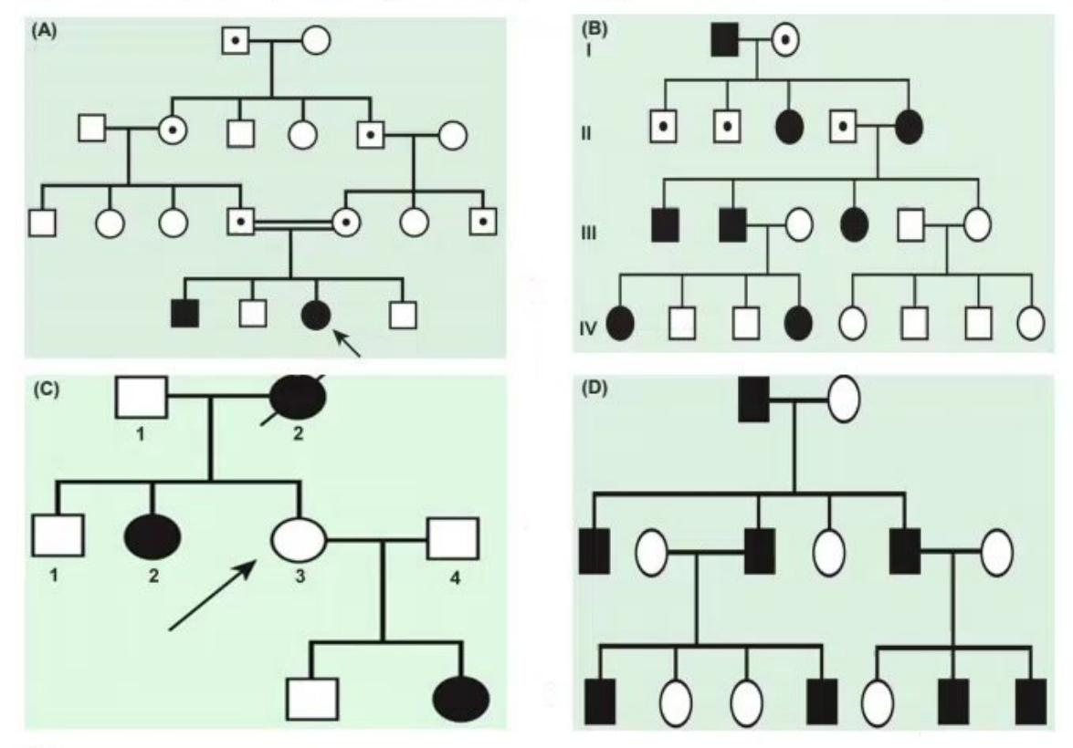
Identify the genetic inheritance pattern of pedigrees A, B, C, and D, and give 2 examples of each:
Answer
- A. Autosomal Recessive (AR): Cystic fibrosis, Thalassemia major, Wilson disease, Sickle cell anemia.
- B. Autosomal Dominant (AD) with Incomplete Penetrance: Familial retinoblastoma, Familial adenomatous polyposis (FAP), Hereditary non-polyposis colorectal cancer (HNPCC).
- C. X-Linked Dominant (XLD): Incontinentia pigmenti, Hypophosphatemic (Vitamin D-resistant) rickets, Rett syndrome, Ornithine transcarbamylase (OTC) deficiency.
- D. Y-Linked (Holandric): SRY gene defects / Hypertrichosis pinnae, Langer mesomelic dysplasia / Léri-Weill dyschondrosteosis (SHOX on PAR region).
MNJ16-002
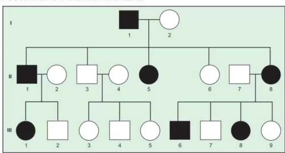
- Identify the pattern of inheritance shown in the pedigree.
- Name 3 conditions with a similar inheritance pattern.
- What is the risk/degree of transmission to offspring of an affected individual?
- Which sex is affected?
Answer
- Inheritance pattern: Autosomal Dominant (AD) (vertical transmission, present in every generation without skipping, equal sex distribution).
- Examples:
- Marfan syndrome
- Achondroplasia
- Neurofibromatosis type 1 (NF-1)
- Tuberous sclerosis
- Hereditary spherocytosis
- Osteogenesis imperfecta
- Transmission risk: 50% (1 in 2) chance of transmission to each offspring in each pregnancy.
- Sex predilection: Both males and females are equally affected and equally capable of transmitting the disease.
MNJ16-003
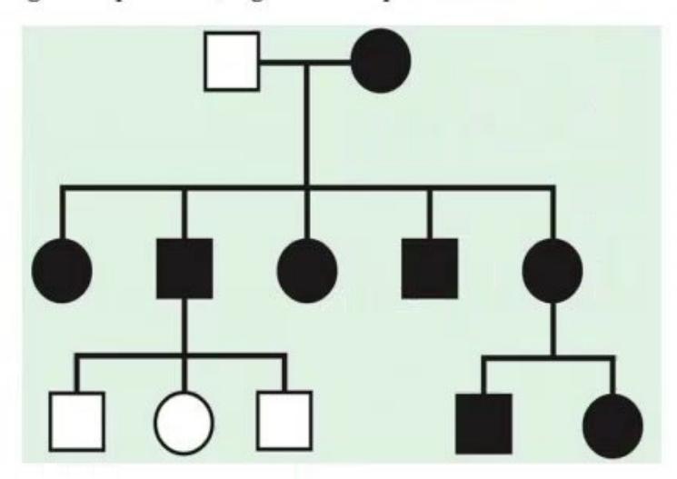
Identify this genetic inheritance pattern and give 4 examples:
Answer
- Inheritance pattern: Mitochondrial (Maternal) Inheritance (affected mother transmits the condition to all of her children [100% of sons and daughters]; affected father transmits to none [0%]).
- Examples:
- MELAS (Mitochondrial Encephalomyopathy, Lactic Acidosis, and Stroke-like episodes)
- MERRF (Myoclonic Epilepsy with Ragged Red Fibers)
- Leber Hereditary Optic Neuropathy (LHON)
- Kearns-Sayre Syndrome (KSS)
- Pearson syndrome
- NARP (Neuropathy, Ataxia, Retinitis Pigmentosa)
MNJ16-004
A 39-year-old female Ritu, 10 weeks pregnant, is concerned about Down syndrome:
- Which invasive antenatal diagnostic tests will you advise, along with their gestational timing?
- Name the serum screening markers used in antenatal screening.
- If the mother is a Robertsonian translocation carrier
, what is the recurrence risk to the offspring? - Name the chromosomes most commonly involved in Robertsonian translocations in Down syndrome.
- The recurrence risk is highest (
) with which translocation?
Answer
- Invasive diagnostic tests:
- Chorionic Villus Sampling (CVS): 10–12 weeks of gestation
- Amniocentesis: 15–20 weeks of gestation
- Cordocentesis (Percutaneous Umbilical Blood Sampling):
- Screening markers:
- First Trimester Combined Screen: Free
(elevated), PAPP-A (low), plus Fetal USG Nuchal Translucency (NT ). - Second Trimester Quadruple Screen: Maternal serum AFP (low), unconjugated estriol (
- low), (high), and Inhibin-A (high). - Cell-free fetal DNA (NIPT): High-accuracy non-invasive screening after 10 weeks.
- First Trimester Combined Screen: Free
- Recurrence risk for maternal
carrier: 10–15% (risk is only ~1–2% if father is carrier). - Chromosomes involved: Chromosomes 14, 21, 13, 15, 22 (acrocentric chromosomes).
- Highest risk translocation:
/ 21q21q isochromosome translocation ( recurrence risk to all viable offspring).
MNJ16-005
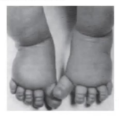
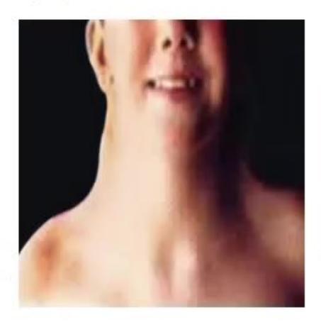

Images of a female child Pooja at birth (congenital pedal lymphedema) and at 6 years of age (webbed neck, short stature):
- Identify the syndrome and state the chromosomal anomaly.
- What is the phenotypic sex in this syndrome?
- Mention 6 cardinal clinical features of this syndrome.
- Name an easy outpatient bedside screening procedure for diagnosis.
- Which hormone therapies will you offer during childhood and adolescence?
- What is the phenotypically similar condition seen in males and females with normal karyotypes?
Answer
- Syndrome & Karyotype: Turner Syndrome, 45,X (monosomy X or mosaicism 45,X/46,XX).
- Phenotypic sex: Phenotypic Female.
- Cardinal clinical features:
- Proportionate short stature
- Webbed neck (pterygium colli) and low posterior hairline
- Broad "shield" chest with widely spaced hypoplastic nipples
- Congenital lymphedema of hands and feet in neonates
- Cubitus valgus and short 4th metacarpals
- Ovarian dysgenesis / streak gonads (primary amenorrhea, hypergonadotropic hypogonadism, infertility)
- Cardiovascular anomalies: Bicuspid aortic valve (most common), Coarctation of the aorta
- Renal: Horseshoe kidney
- Bedside screening test: Buccal smear examination for Barr body (absent / negative Barr body in 45,X).
- Hormone therapies:
- Recombinant Growth Hormone (rhGH): Initiated early to optimize final adult height.
- Estrogen replacement therapy: Initiated at 11–12 years for pubertal induction and secondary sexual characteristics.
- Progesterone: Added after 2 years of estrogen or upon breakthrough bleeding to cycle endometrium.
- Thyroxine: If associated autoimmune Hashimoto thyroiditis is present.
- Phenotypic analogue: Noonan Syndrome (autosomal dominant, normal 46,XX or 46,XY karyotype, commonly associated with pulmonic stenosis).
MNJ16-006
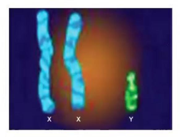
- Which syndrome is represented by this 47,XXY karyotype?
- What is the phenotypic sex?
- State the salient clinical features.
- What investigations will you perform?
- What is the main medical treatment?
Answer
- Syndrome: Klinefelter Syndrome (
due to meiotic non-disjunction). - Phenotypic sex: Phenotypic Male.
- Salient clinical features:
- Tall eunuchoid stature with long limbs (increased lower segment:upper segment ratio)
- Small, firm testes (
) and micropenis / hypogonadism - Gynecomastia in adolescence
- Sparse facial and body hair
- Azoospermia and primary infertility
- Learning difficulties, delayed speech/reading skills, behavioral and executive function issues
- Investigations:
- Chromosomal karyotype: Confirms
- Endocrine profile: Elevated serum FSH and LH (hypergonadotropic) with low/subnormal testosterone after mid-puberty
- Semen analysis: Azoospermia
- Testicular biopsy: Hyalinization and fibrosis of seminiferous tubules with Leydig cell hyperplasia
- Chromosomal karyotype: Confirms
- Treatment:
- Intramuscular Testosterone replacement therapy (e.g., testosterone enanthate/cypionate depot) initiated around 11–12 years of age to promote virilization, muscle mass, bone mineral density, and prevent osteoporosis.
- Educational/psychological support and screening for metabolic syndrome and breast malignancy.
MNJ16-007
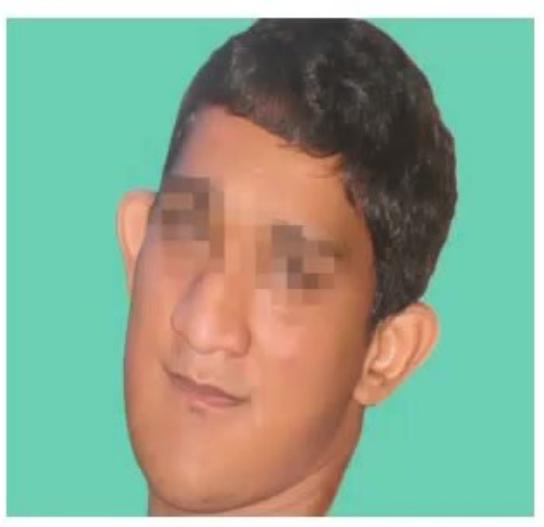
A male child presents with developmental delay, long narrow face, large prominent ears, and intellectual disability:
- Identify the syndrome and explain the structural cytogenetic term "fragile site".
- What is the exact gene locus involved?
- What is the molecular mechanism of mutation / inheritance?
- What are the clinical, cognitive, and behavioral manifestations?
- Mention 4 other triplet repeat expansion disorders with their respective trinucleotide repeats.
Answer
- Syndrome & Fragile Site:
- Fragile X Syndrome (Martin-Bell syndrome).
- A fragile site is a specific, non-staining gap or constriction on a chromosome that fails to condense properly during mitosis and exhibits a propensity to break under folate-deficient culture conditions.
- Gene Locus: Xq27.3 (FMR1 gene - Fragile X Messenger Ribonucleoprotein 1).
- Molecular mechanism: Trinucleotide (CGG) repeat expansion in the 5' untranslated region of FMR1:
- Normal: 5–44 repeats
- Premutation: 55–200 repeats
- Full mutation:
hypermethylation and transcriptional silencing of FMR1.
- Manifestations:
- Physical: Elongated narrow facies, prominent forehead, large prominent/protruding ears, high-arched palate, macroorchidism (post-pubertal enlarged testes
), joint hypermobility, flat feet, mitral valve prolapse. - Cognitive / Behavioral: Moderate intellectual disability, severe speech/language delay, autistic spectrum features (hand-flapping, poor eye contact, tactile defensiveness), ADHD, hyperactivity.
- Physical: Elongated narrow facies, prominent forehead, large prominent/protruding ears, high-arched palate, macroorchidism (post-pubertal enlarged testes
- Other Triplet Repeat Disorders:
- Huntington Disease: CAG expansion (Chromosome 4p)
- Myotonic Dystrophy (DM1): CTG expansion (Chromosome 19q)
- Friedreich Ataxia: GAA expansion (Chromosome 9q)
- Spinocerebellar Ataxias (SCAs): CAG expansion
MNJ16-008
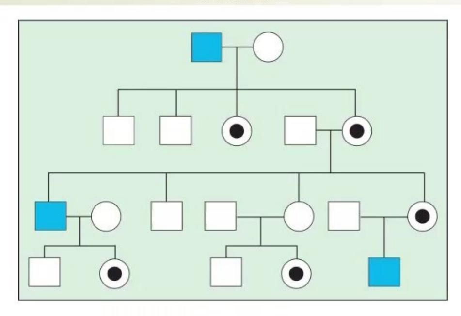
- Identify the pattern of inheritance shown in this pedigree.
- Name 4 conditions with this inheritance pattern.
- Which sex is predominantly affected? What is the risk of transmission to offspring of a carrier mother?
Answer
- Inheritance pattern: X-Linked Recessive (XLR) (males affected; carrier females transmit the condition; no male-to-male transmission).
- Examples:
- Hemophilia A and Hemophilia B
- Duchenne and Becker Muscular Dystrophy (DMD/BMD)
- Glucose-6-Phosphate Dehydrogenase (G6PD) deficiency
- Wiskott-Aldrich Syndrome (WAS)
- Menkes kinky hair disease
- Hunter syndrome (MPS II)
- Chronic Granulomatous Disease (CGD - X-linked form)
- Transmission risks for a carrier mother (
): - Sons: 50% risk of being affected (
). - Daughters: 50% risk of being asymptomatic carriers (
); 0% risk of full disease.
- Sons: 50% risk of being affected (
MNJ16-009
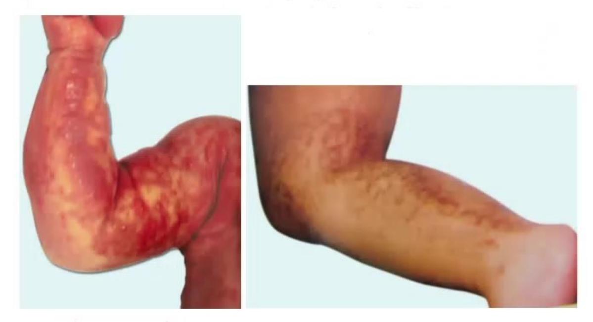
- Identify the syndrome shown with whorled cutaneous hyperpigmentation and state its pattern of inheritance.
- What are the 4 dermatological stages and which gene is affected?
- Give 3 other examples of disorders with a similar mode of inheritance.
- What is the risk of transmission to the offspring of an affected male?
Answer
- Syndrome & Inheritance: Incontinentia Pigmenti (Bloch-Sulzberger syndrome); X-linked dominant (XLD) (typically male lethal in utero).
- Stages & Gene:
- Gene: IKBKG / NEMO gene on chromosome Xq28.
- 4 Cutaneous Stages:
- Stage 1 (Vesicular): Linear vesicles and pustules on extremities at birth/neonatal period.
- Stage 2 (Verrucous): Warty, hyperkeratotic papules and plaques (weeks to months).
- Stage 3 (Hyperpigmented): Swirled, marble-cake "lines of Blaschko" hyperpigmentation on trunk and thighs (infancy to childhood).
- Stage 4 (Atrophic / Hypopigmented): Pale, hairless, atrophic streaks on lower legs (adolescence/adulthood).
- Other XLD examples:
- Hypophosphatemic (X-linked) Rickets
- Ornithine Transcarbamylase (OTC) deficiency
- Rett syndrome
- Alport syndrome (XLD form)
- Transmission from an affected male (
): - 100% of daughters will inherit the mutant X chromosome and be affected.
- 0% of sons will inherit the disease (receive the Y chromosome; male-to-male transmission never occurs in X-linked inheritance).
MNJ16-010

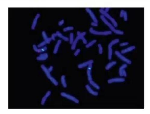
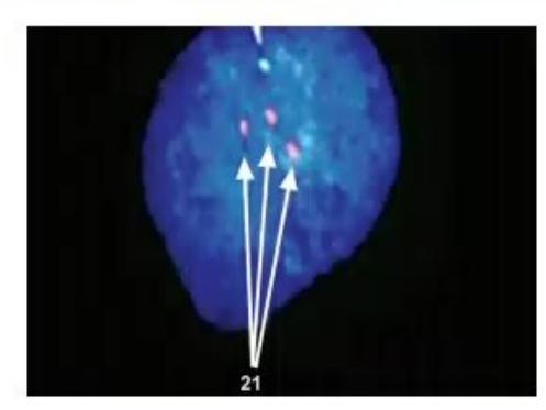
- Identify the diagnostic cytogenetic technique and state the diagnosis shown in Slide B (showing 3 fluorescent signals for chromosome 21).
- Name 4 microdeletion syndromes in which this technique is used for diagnosis.
Answer
- Technique & Slide B Diagnosis:
- Technique: Fluorescent In Situ Hybridization (FISH) (using locus-specific fluorochrome-labeled DNA probes).
- Slide B Diagnosis: Trisomy 21 (Down Syndrome) (interphase FISH showing 3 specific fluorescent hybridization signals for the chromosome 21 probe).
- Microdeletion syndromes diagnosed by FISH / CMA:
- 22q11.2 deletion syndrome (DiGeorge / Velocardiofacial syndrome)
- Prader-Willi syndrome / Angelman syndrome (15q11-q13 deletion)
- Williams syndrome (7q11.23 deletion / elastin gene)
- Miller-Dieker lissencephaly syndrome (17p13.3 deletion)
- Cri du chat syndrome (5p15 deletion)
- Wolf-Hirschhorn syndrome (4p16.3 deletion)
MNJ16-011
- State three invasive procedures for antenatal genetic diagnosis, along with the earliest gestational age at which each can be performed.
- What is the procedure-related risk of fetal loss for each?
- Name 4 common diseases for which antenatal diagnosis is readily available.
- Name 3 viral/non-viral vectors used in gene therapy, and 3 candidate single-gene disorders for gene therapy.
Answer
-
& 2. Antenatal Procedures & Fetal Loss Risk:
- Chorionic Villus Sampling (CVS): 10–12 weeks; Fetal loss risk
. - Amniocentesis: 15–18 weeks; Fetal loss risk
. - Percutaneous Umbilical Blood Sampling (Cordocentesis):
; Fetal loss risk .
- Chorionic Villus Sampling (CVS): 10–12 weeks; Fetal loss risk
-
Conditions for antenatal diagnosis:
- Chromosomal aneuploidies (Trisomy 21, 18, 13, Turner syndrome)
- Hemoglobinopathies (
-Thalassemia major, Sickle cell anemia) - Muscular dystrophies (Duchenne/Becker muscular dystrophy)
- Spinal Muscular Atrophy (SMA)
- Cystic Fibrosis
- Inborn errors of metabolism (Gaucher disease, MPS, CAH)
-
Gene Therapy Vectors & Candidate Disorders:
- Vectors: Adeno-Associated Virus (AAV), Lentivirus / Retrovirus, Adenovirus, Lipid nanoparticles (LNPs / Naked plasmid DNA).
- Candidate Disorders: Spinal Muscular Atrophy (SMN1), Severe Combined Immunodeficiency (ADA-SCID, X-SCID),
-Thalassemia, Hemophilia A/B, Duchenne Muscular Dystrophy, Leber Congenital Amaurosis (RPE65).
MNJ16-012
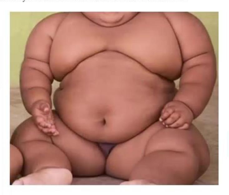
A 7-year-old male child presents with marked hyperphagia, truncal obesity, small hands and feet, developmental delay, neonatal hypotonia, and hypogonadism:
- Identify the syndrome and the specific chromosomal region involved.
- Name the genetic mechanisms of inheritance responsible for this disorder.
- Which other neurodevelopmental syndrome is mapped to the same chromosomal region due to maternal vs paternal genomic imprinting?
Answer
- Syndrome & Region: Prader-Willi Syndrome (PWS); Chromosome 15q11.2–q13.
- Genetic mechanisms: Loss of expression of paternally inherited genes on 15q11-q13:
- Paternal 15q11-q13 deletion (~70% of cases)
- Maternal Uniparental Disomy (UPD 15) (~25–30% of cases)
- Imprinting center defect / mutation (~1–3%)
- Imprinting counterpart: Angelman Syndrome ("Happy Puppet" syndrome), resulting from loss of maternal UBE3A expression on chromosome 15q11-q13 (characterized by severe speech impairment, ataxia, paroxysms of inappropriate laughter, seizures, and microcephaly).
MNJ16-013
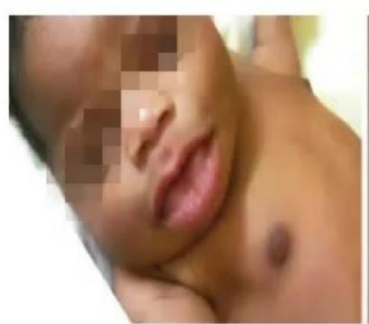
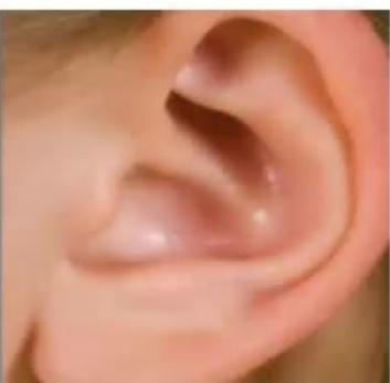
- Identify the overgrowth syndrome shown (showing macroglossia, anterior abdominal wall defect, ear lobe creases).
- Which metabolic complication must be screened and managed in the neonatal period?
- Give 3 salient clinical features.
- Mention the chromosomal region involved and the underlying imprinting defect.
Answer
- Syndrome: Beckwith-Wiedemann Syndrome (BWS).
- Metabolic complication: Severe Neonatal Hypoglycemia (due to pancreatic islet cell hyperplasia and hyperinsulinism).
- Salient features:
- Macrosomia / Large for gestational age (LGA)
- Macroglossia (large protruding tongue)
- Abdominal wall defects (Omphalocele, umbilical hernia, diastasis recti)
- Anterior earlobe creases and posterior helical ear pits
- Hemi-hypertrophy / lateralized overgrowth
- Organomegaly (nephromegaly, hepatomegaly)
- Predisposition to embryonal tumors (Wilms tumor, hepatoblastoma; requiring 3-monthly abdominal USG and serum AFP screening until 8 years of age)
- Genetics: Chromosome 11p15.5 imprinting region (loss of methylation at IC2/KCNQ1OT1, gain of methylation at IC1/H19, or paternal uniparental disomy 11p15).
MNJ16-014
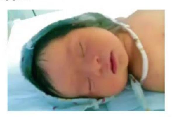
A newborn is suspected of having Down syndrome:
- Name 3 organ systems commonly involved and state 2 specific conditions in each.
- Name an essential screening test that must be performed prior to hospital discharge.
- What are key radiological features of Down syndrome?
- Outline a structured long-term multidisciplinary follow-up plan.
Answer
- Systems & Associated Anomalies:
- Cardiovascular: Endocardial cushion defect (AV canal / AVSD - 40%), VSD, ASD, PDA.
- Gastrointestinal: Duodenal atresia ("double bubble" sign), annular pancreas, tracheoesophageal fistula, Hirschsprung disease, celiac disease.
- Ocular: Brushfield spots, congenital cataract, strabismus, severe refractive errors, nystagmus.
- Endocrine: Congenital and acquired hypothyroidism, Type 1 diabetes mellitus.
- Essential screening before discharge:
- Echocardiogram (screening for congenital heart defects)
- Newborn thyroid screen (TSH / Free
) - Universal Newborn Hearing Screening (OAE / BERA)
- Radiological features:
- Pelvic X-ray: Decreased acetabular and iliac angles, flaring of iliac wings.
- Chest/Neck X-ray: 11 pairs of ribs, atlantoaxial subluxation/instability (increased atlantodens interval
on flexion-extension views). - Abdominal X-ray: "Double bubble" gas pattern in duodenal atresia.
- Hand X-ray: Hypoplastic middle phalanx of the 5th digit with clinodactyly.
- Follow-up plan:
- Annual thyroid screening (TSH)
- Regular audiometry (6–12 monthly) and ophthalmological evaluation
- Celiac disease serology screening at 2–3 years
- Screening for hematological malignancy (CBC for transient myeloproliferative disorder / AML / ALL)
- Cervical spine radiographs for atlantoaxial instability prior to sports/anesthesia
- Early intervention, physiotherapy, and speech therapy.
MNJ16-015
- Diagnose the condition characterized by facial angiofibromas (adenoma sebaceum), ash-leaf hypopigmented macules, subependymal giant cell astrocytomas (SEGA), and renal angiomyolipomas.
- What is the mode of inheritance?
- What is the recurrence risk for the next offspring if one parent is affected?
- Which sex is involved?
- Which genes and gene products are implicated in this condition?
Answer
- Diagnosis: Tuberous Sclerosis Complex (Bourneville disease).
- Inheritance: Autosomal Dominant with high penetrance and variable expressivity (~65–70% represent de novo mutations).
- Recurrence risk: 50% (1 in 2) if a parent is affected.
- Sex: Equal in males and females (
). - Genes & Proteins:
gene on chromosome 9q34 encodes Hamartin. gene on chromosome 16p13.3 encodes Tuberin.
(The hamartin-tuberin complex acts as a GTPase-activating protein to inhibit the mTOR signaling pathway).
MNJ16-016
- Name 4 teratogenic drugs contraindicated in pregnancy.
- Name 3 drug-induced teratogenic syndromes with one characteristic anomaly for each.
Answer
-
Teratogenic drugs:
- Thalidomide
- Warfarin
- Valproic acid / Sodium valproate
- Phenytoin
- Isotretinoin (Retinoic acid)
- Methotrexate
- ACE inhibitors / ARBs
- Lithium
-
Teratogenic Syndromes & Characteristic Anomalies:
- Fetal Alcohol Spectrum Disorder (FASD): Smooth philtrum, thin upper vermilion border, short palpebral fissures, microcephaly, and CNS dysfunction.
- Fetal Hydantoin (Phenytoin) Syndrome: Distal digital hypoplasia / nail hypoplasia, cleft lip/palate, hypertelorism, growth deficiency.
- Fetal Valproate Syndrome: Neural tube defects (spina bifida aperta), characteristic facial dysmorphism (broad nasal bridge, shallow philtrum), congenital heart defects.
- Fetal Warfarin Syndrome (Conradi-Hünermann phenocopy): Nasal hypoplasia (depressed nasal bridge), stippled epiphyses (chondrodysplasia punctata), hypoplastic distal phalanges.
- Thalidomide Embryopathy: Phocomelia (severe limb reduction defects), amelia, anotia/microtia.
MNJ16-017
Write the pattern of inheritance (AD, AR, XLR, or XLD) for each condition listed below:
- Achondroplasia
- Congenital adrenal hyperplasia (CAH)
- Cystic fibrosis
- Hemophilia A
- Duchenne muscular dystrophy
- Huntington's disease
- Neurofibromatosis type 1
- Marfan syndrome
- Phenylketonuria (PKU)
- Sickle cell disease
- Glucose-6-phosphate dehydrogenase (G6PD) deficiency
- Hemophilia B
Answer
- Achondroplasia
Autosomal Dominant (AD) - Congenital adrenal hyperplasia
Autosomal Recessive (AR) - Cystic fibrosis
Autosomal Recessive (AR) - Hemophilia A
X-Linked Recessive (XLR) - Duchenne muscular dystrophy
X-Linked Recessive (XLR) - Huntington's disease
Autosomal Dominant (AD) - Neurofibromatosis type 1
Autosomal Dominant (AD) - Marfan syndrome
Autosomal Dominant (AD) - Phenylketonuria (PKU)
Autosomal Recessive (AR) - Sickle cell disease
Autosomal Recessive (AR) - G6PD deficiency
X-Linked Recessive (XLR) - Hemophilia B
X-Linked Recessive (XLR)
MNJ16-018
Define the following terms used in dysmorphology and give examples of each:
- Association
- Sequence
- Developmental Field Defect
- Syndrome
Answer
- Association: A non-random occurrence in individuals of multiple congenital anomalies not explained by chance, but without a known unifying genetic etiology or specific cascade.
- Examples: VACTERL association (Vertebral, Anal, Cardiac, Tracheoesophageal fistula, Renal, Limb), CHARGE association.
- Sequence: A pattern of multiple secondary defects that result cascades from a single primary structural defect or mechanical disruption during development.
- Examples: Potter sequence (renal agenesis
oligohydramnios pulmonary hypoplasia, compressed facial features, clubbed feet); Pierre Robin sequence (micrognathia glossoptosis cleft palate).
- Examples: Potter sequence (renal agenesis
- Developmental Field Defect: A pattern of anomalies resulting from the aberrant development of an entire morphogenetic unit or contiguous embryonic region during morphogenesis.
- Examples: Holoprosencephaly field defect, Poland sequence/anomaly, Moebius syndrome.
- Syndrome: A recognizable, clinically consistent constellation of multiple congenital anomalies affecting multiple organ systems, known or presumed to have a single unifying etiology (e.g., chromosomal, single-gene mutation, or environmental teratogen).
- Examples: Down syndrome (Trisomy 21), Turner syndrome (45,X), Marfan syndrome (FBN1).
MNJ16-019
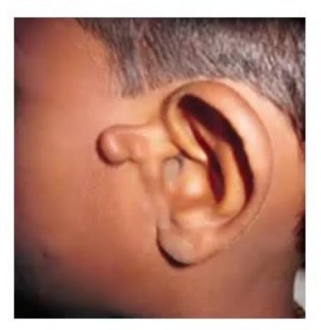
- Spot the diagnosis and give the alternate name of this condition.
- What is the embryological defect causing this condition?
- What is the overall prognosis and cognitive outcome?
Answer
- Diagnosis: Goldenhar Syndrome (also known as Oculo-Auriculo-Vertebral [OAV] Spectrum / First and Second Branchial Arch Syndrome).
- Embryological defect: Vascular disruption / abnormal morphogenesis of structures derived from the first and second branchial arches and clefts (typically unilateral, resulting in preauricular tags, microtia, hemifacial microsomia, epibulbar dermoids, and vertebral anomalies like hemivertebrae).
- Prognosis: Generally favorable; normal lifespan and normal intelligence in
of patients (mild intellectual disability in ~10–15%). Managed with hearing aids, reconstructive craniofacial surgery, and spine monitoring.
MNJ16-020
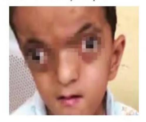
A child presents with a tall forehead, craniosynostosis (turribrachycephaly), midface hypoplasia, and symmetric complex osseous and cutaneous syndactyly of the hands and feet ("mitten hands" and "sock feet"):
- Spot the diagnosis of this child and name the causative gene.
- What is the timing and role of surgical intervention for facial dysmorphism?
Answer
- Diagnosis & Gene: Apert Syndrome (Acrocephalosyndactyly Type I); caused by gain-of-function mutations in the
(Fibroblast Growth Factor Receptor 2) gene on chromosome 10q26. - Surgical interventions:
- Infancy (3–12 months): Early cranial vault decompression and remodeling (fronto-orbital advancement) to relieve elevated intracranial pressure.
- Hand surgery (1–3 years): Staged syndactyly release to create functional digits and an opposable thumb.
- Midface advancement: LeFort III osteotomy / monobloc distraction osteogenesis performed in mid-childhood (6–10 years) or pre-adolescence after substantial facial growth to correct severe midface hypoplasia, proptosis, and obstructive sleep apnea.
MNJ16-021
A 6-year-old male child Panna is brought with concerns of a flat facial profile, low IQ, delayed milestones, hypotonia, and recurrent respiratory infections. On examination, he has epicanthal folds, short broad hands, brachycephaly, and a Grade 4 pansystolic murmur over the lower left sternal border:
- Keeping the diagnosis in mind, which specific joint articulation must you evaluate carefully and why?
- When evaluating for treatable/reversible causes of intellectual and developmental delay in this child, which endocrine test will you perform?
- On regular long-term follow-up, which hematological surveillance test should be done?
- Elaborate the cytogenetic nomenclature:
.
Answer
- Joint of concern: Atlantoaxial joint (C1-C2 articulation) for Atlantoaxial instability (AAI) (due to transverse odontoid ligament laxity). Subluxation can cause spinal cord compression, quadriparesis, gait changes, or sudden death.
- Endocrine test: Serum TSH and Free
(screening for congenital/acquired primary hypothyroidism, which occurs in up to 15–30% of children with Down syndrome and represents a reversible cause of cognitive and growth deterioration). - Hematological monitoring: Complete Blood Count (CBC) with peripheral smear annually to screen for acute leukemia (10- to 20-fold increased risk of ALL and Acute Megakaryoblastic Leukemia [AML-M7]).
- Cytogenetic explanation:
- 46: Total number of chromosomes in the cell.
- XY: Male chromosomal complement.
- t: Translocation (Robertsonian/reciprocal translocation).
- (14;21): Involves chromosomes 14 and 21.
- (q11;q22): Breakpoint is at band q11 on the long arm of chromosome 14, and band q22 on the long arm of chromosome 21.
MNJ16-022
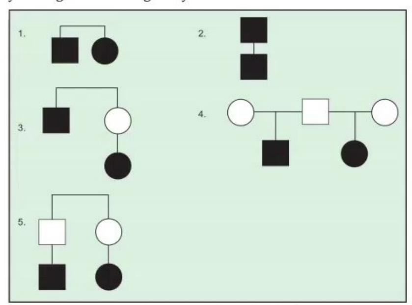
Define and classify the degrees of consanguinity and genetic relationship between relatives:
Answer
- First-degree relatives (Share 50% /
of genes): - Parents and children
- Full brothers and sisters (siblings)
- Second-degree relatives (Share 25% /
of genes): - Grandparent and grandchild
- Uncle / Aunt and Nephew / Niece
- Half-siblings
- Double first cousins
- Third-degree relatives (Share 12.5% /
of genes): - First cousins (children of siblings — most common consanguineous marriage worldwide)
- Great-grandparent and great-grandchild
MNJ16-023
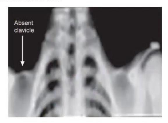
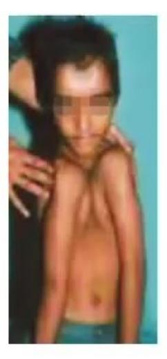
- Identify the skeletal dysplasia shown in the clinical picture and chest radiograph.
- State the mode of inheritance and the gene implicated.
- What characteristic abnormalities are seen on the hand radiograph and skull?
- What classic clinical maneuver / sign demonstrates this condition on physical examination?
Answer
- Diagnosis: Cleidocranial Dysostosis (Cleidocranial Dysplasia) (radiograph showing complete absence / aplasia or hypoplasia of clavicles and bell-shaped thorax).
- Inheritance & Gene: Autosomal Dominant with complete penetrance and variable expressivity; caused by heterozygous mutations in the
(CBFA1) gene on chromosome 6p21. - Radiological features:
- Clavicles: Total or partial absence of clavicles.
- Skull: Widely patent cranial fontanelles and delayed suture closure, multiple Wormian bones, frontal/parietal bossing, hypertelorism.
- Hand X-ray: Long second metacarpals, hypoplastic tapering distal phalanges, cone-shaped epiphyses, accessory ossification centers.
- Dentition: Delayed eruption of secondary teeth, supernumerary impacted teeth.
- Clinical maneuver: Hypermobility of the shoulders — the patient can easily approximate both shoulders in the anterior midline of the chest.
MNJ16-024
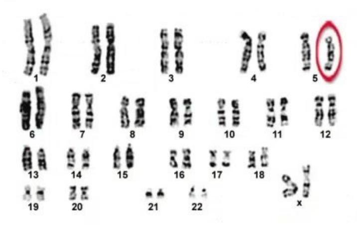
- Identify the karyotype anomaly shown (showing terminal deletion of the short arm of chromosome 5).
- What is the specific cytogenetic defect?
- List 4 cardinal clinical features of this syndrome.
Answer
- Karyotype & Syndrome:
or — Cri du Chat (Cat Cry / 5p-minus) Syndrome. - Genetic defect: Partial deletion of the short arm of chromosome 5 (5p15.2–p15.3 critical region).
- Clinical features:
- High-pitched, monochromatic monotonous cat-like cry in infancy (due to abnormal laryngeal development).
- Severe intellectual disability and developmental delay.
- Microcephaly with round "moon" face and hypertelorism.
- Low-set, dysplastic ears, epicanthal folds, downward-slanting palpebral fissures.
- Micrognathia, broad nasal bridge, hypotonia in infancy, and single palmar (simian) crease.
MNJ16-025
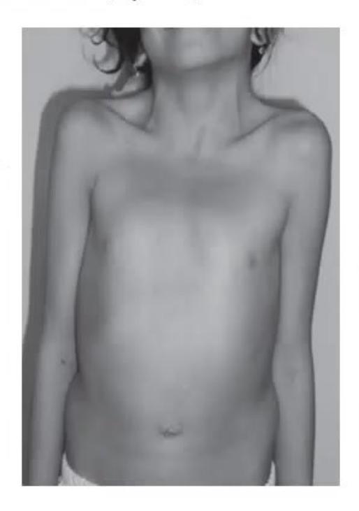
A 10-year-old male child Raman presents with short stature, webbed neck, low posterior hairline, chest deformity (pectus excavatum/carinatum), hypertelorism, and cryptorchidism:
- What is the diagnosis of this condition?
- What chromosomal karyotype is typically expected in this condition?
- What are 3 cardiac manifestations, and which one is the most common?
Answer
- Diagnosis: Noonan Syndrome (autosomal dominant, RASopathy; PTPN11, SOS1, RAF1, RIT1 mutations).
- Karyotype: Normal
(or in females; normal chromosomal analysis distinguishes Noonan syndrome from Turner syndrome). - Cardiac manifestations:
- Dysplastic Pulmonic Valve Stenosis (PVS) — Most common (~50–60%).
- Hypertrophic Cardiomyopathy (HCM) (asymmetric septal hypertrophy — ~20–30%).
- Atrial Septal Defect (ASD - ostium secundum) and Ventricular Septal Defect (VSD).