MNJ28-001
Peripheral smear:
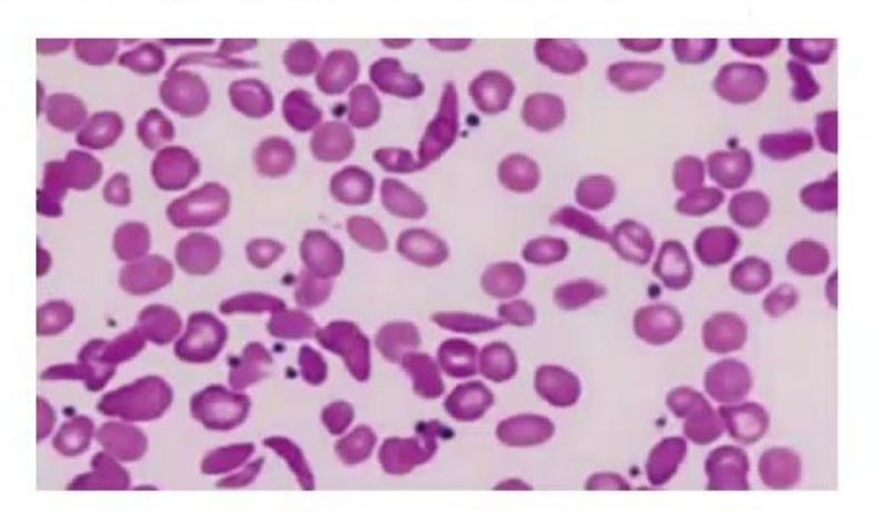
- What is the diagnosis of this PBF?
- What is the drug of choice for the prevention of painful episodes in this condition?
- What are measures for the primary prevention of stroke in these children?
- What are the antibiotics of choice in acute chest syndrome?
- What type of renal malignancy is common in this disorder?
Answer
- Sickle cell anemia (showing sickle/drepanocytic RBCs and target cells).
- Hydroxyurea (15–20 mg/kg/day).
- Primary prevention of stroke:
- Annual Transcranial Doppler (TCD) screening from 2 to 16 years of age (monitoring time-averaged mean maximum velocity [TAMMV] in distal internal carotid/middle cerebral artery).
- If TCD velocity
, initiate chronic transfusion therapy to maintain .
- Third-generation cephalosporin (e.g., Ceftriaxone/Cefotaxime) PLUS a macrolide (e.g., Azithromycin) to cover atypical pathogens (Mycoplasma pneumoniae, Chlamydophila pneumoniae).
- Renal medullary carcinoma (strongly associated with sickle cell trait and disease).
MNJ28-002
A 6-month-old boy Hritik presents with pallor, tri-phalangeal thumbs and mild hepatosplenomegaly. Hb: 7 g/dL, MCV: 100 fL, reticulocyte count: <1%, peripheral smear: normocytic to macrocytic RBCs, normal WBCs and platelets. Vitamin
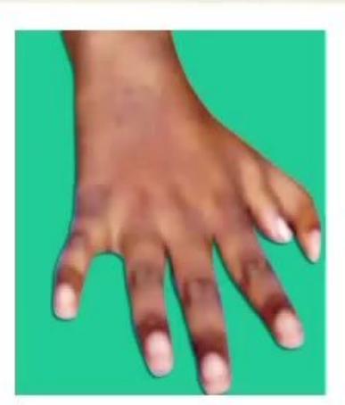
- What is the diagnosis?
- What is the underlying defect?
- What is the closest differential diagnosis?
- Give two points to differentiate them.
- Name at least one malignancy it can predispose to.
Answer
- Diamond-Blackfan anemia (DBA / Congenital pure red cell aplasia).
- Ribosomopathy (mutations in ribosomal protein genes, e.g., RPS19, RPL5) causing intrinsic deficiency and apoptosis of erythroid progenitor cells (CFU-E / BFU-E) with insensitivity to erythropoietin.
- Transient Erythroblastopenia of Childhood (TEC).
- Differentiating points between DBA and TEC:
- Age of onset: DBA presents
months (often at birth), TEC occurs after 6 months (1–3 years). - Congenital anomalies: Present in DBA (40–50%, e.g., triphalangeal thumb, craniofacial defects); absent in TEC.
- RBC indices & markers: MCV is elevated (macrocytic), HbF elevated, and erythrocyte adenosine deaminase (eADA) elevated in DBA; normal in TEC.
- Preceding viral illness: Absent in DBA; common in TEC.
- Age of onset: DBA presents
- Acute myeloid leukemia (AML), myelodysplastic syndrome (MDS), osteosarcoma.
MNJ28-003
An 8-year-old boy Tarun with fever and cough since 2 months, breathlessness since 14 days is being treated with anti-tuberculous drugs since the last 1.5 months without relief. Give the differential diagnosis of mediastinal widening in anterior, middle and posterior mediastinum:
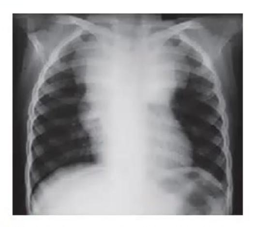
Answer
Anterior Mediastinum (4 T's + Lymphoma):
- Lymphoma (Hodgkin lymphoma, Non-Hodgkin lymphoma / T-ALL)
- Thymoma / Thymic hyperplasia / Thymic cyst
- Teratoma / Germ cell tumors
- Thyroid masses / substernal goiter
- Vascular: Aneurysm, hemangioma, lipoma, cystic hygroma
Middle Mediastinum:
- Lymphadenopathy (Tuberculosis, Sarcoidosis, Lymphoma, Histoplasmosis, fungal infections)
- Bronchogenic cyst, pericardial cyst, enteric duplication cyst
- Vascular lesions: Great vessel anomalies, pulmonary artery enlargement
- Diaphragmatic hernia (Morgagni hernia)
Posterior Mediastinum:
- Neurogenic tumors: Neuroblastoma, ganglioneuroma, ganglioneuroblastoma, schwannoma, neurofibroma
- Ewing sarcoma / Askin tumor of chest wall
- Thoracic meningocele / Neurenteric cyst / Enterogenous cyst
- Posterior mediastinal lymphoma / rhabdomyosarcoma
MNJ28-004
A 10-year-old male child Dinesh presents with pallor, fatigue and hepatosplenomegaly for one month. His CBC shows pancytopenia:
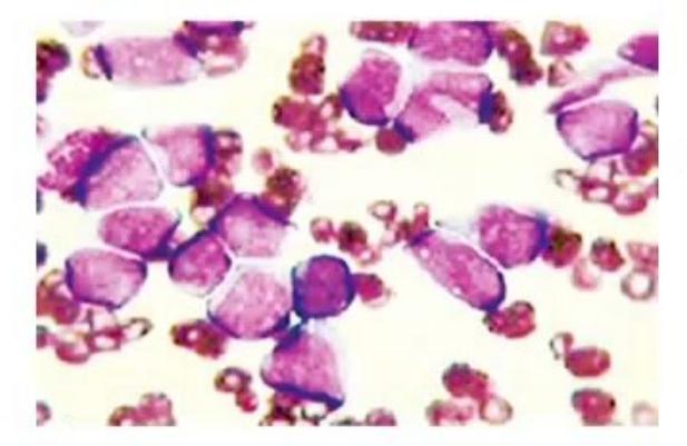

- What is the bone marrow diagnosis?
- Give five poor prognostic factors associated with this condition.
Answer
- Acute Myeloid Leukemia (AML) (showing myeloblasts with Auer rods / monoblasts).
- Poor prognostic factors in AML:
- High initial WBC count (
) - Adverse cytogenetics: Monosomy 7, monosomy 5, del(5q), 11q23 (MLL) rearrangements, complex karyotype (
abnormalities) - Molecular mutations: FLT3-ITD mutation (high allelic ratio)
- Secondary AML (therapy-related or evolving from prior MDS/aplastic anemia)
- Poor response to induction / Persistent minimal residual disease (MRD) after induction
- High initial WBC count (
MNJ28-005
A 3.5-month-old infant Rakesh, exclusively breastfed, presents with loose motions since the early neonatal period. On examination, he is pale and is failing to thrive. His CBC shows a decrease in all cell lines. His blood sugar is 280 mg/dL:
- What is the most likely diagnosis?
- Which 2 investigations will clinch the diagnosis?
- What is the treatment of this condition?
Answer
- Pearson marrow-pancreas syndrome (a mitochondrial deletion disorder).
- Diagnostic investigations:
- Bone marrow aspiration: Shows macrocytic anemia, ringed sideroblasts (Perls' Prussian blue stain), and vacuolization of erythroid and myeloid precursor cells.
- Mitochondrial DNA (mtDNA) analysis: Shows large single mitochondrial DNA deletion/duplication.
- Treatment:
- Supportive: Packed RBC and platelet transfusions, G-CSF for severe neutropenia
- Pancreatic enzyme replacement therapy (for exocrine pancreatic insufficiency)
- Insulin therapy (for neonatal/infantile diabetes mellitus)
- Management of metabolic/lactic acidosis and vitamin/carnitine supplementation
MNJ28-006
An 11-year-old boy Lalu, a known case of hemophilia A, is admitted with pain and swelling in the right knee joint after a mild injury while playing. Examination reveals acute hemarthrosis:
- What is the baseline level of factor VIII in severe, moderate, and mild hemophilia?
- Factor VIII replacement for acute hemarthrosis:
i. Target level of factor VIII to be achieved
ii. Dose of factor VIII - If the patient has to undergo major surgery:
i. Target level of factor VIII to be achieved
ii. Dose and maintenance of factor VIII - What are the other precautions?
- What is the role of DDAVP (Desmopressin) in hemophilia A?
Answer
- Classification by Factor VIII level:
- Severe:
( ) - Moderate:
( ) - Mild:
( )
- Severe:
- Factor VIII for hemarthrosis:
i. Target level:( )
ii. Dose:IV (1 unit/kg raises factor VIII by 2%), repeated every 12–24 hours until symptoms resolve. - For major surgery:
i. Target level:( ) pre-op, maintained for 7–10 days post-op.
ii. Dose: Loading dose ofIV, followed by continuous infusion ( ) or every 8–12 hours. - Other precautions:
- Avoid intramuscular (IM) injections and NSAIDs / Aspirin / antiplatelet agents.
- Screen for inhibitor antibodies (Bethesda assay) and transfusion-transmitted infections (Hepatitis B, C, HIV).
- Early joint rehabilitation / physiotherapy after acute bleed resolves.
- Role of DDAVP (Desmopressin): Stimulates endogenous release of Factor VIII and vWF from endothelial Weibel-Palade bodies; effective in mild Hemophilia A (baseline
) for minor bleeds and dental procedures.
MNJ28-007
A 10-year-old boy Nithish is tested for sickle cell disease preoperatively. His father is known to have sickle cell trait. Results: Hb 9.1 g/dL, RBC:
- What hemoglobinopathy does the boy have?
- What hemoglobinopathy do you expect on testing the mother?
Answer
- Sickle
-thalassemia (compound heterozygosity for HbS and -thalassemia mutation). - Beta-thalassemia trait / minor (
-thalassemia carrier) (since father has sickle cell trait, the -thalassemia gene was inherited from the mother).
Reference ranges on Hb electrophoresis/HPLC:
- Sickle Cell Anemia (HbSS):
, , , . - Sickle Cell Trait (HbAS):
, , . : , , , . : , , , .
MNJ28-008
A 4-year-old girl Ritu with a solid tumor around eye has developed periorbital hemorrhage shown below:
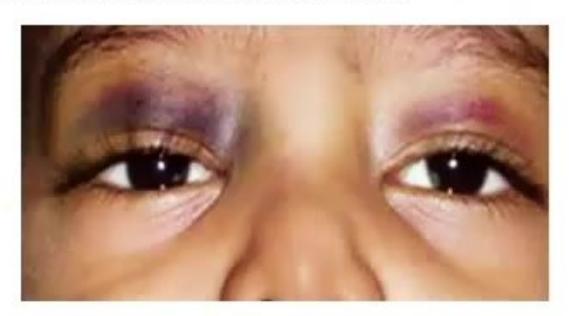
- Name the tumor and the eye finding.
- Name any two conditions associated with this tumor.
Answer
- Neuroblastoma; Periorbital ecchymosis / "raccoon eyes" (due to retrobulbar metastatic infiltration and venous obstruction).
- Associated conditions / syndromes:
- Neurofibromatosis type 1 (NF-1)
- Hirschsprung disease (congenital aganglionic megacolon)
- Beckwith-Wiedemann syndrome
- Central hypoventilation syndrome (Ondine curse)
- Fetal hydantoin syndrome / Fetal alcohol syndrome
- Opsoclonus-myoclonus-ataxia syndrome (paraneoplastic)
MNJ28-009
A 6-day-old neonate with birth asphyxia was referred to the hematology services for a very high WBC count in the range of 78,000/µL, the platelet count being 6.7 lakhs/µL. Sepsis in the newborn was ruled out. The baby was cyanosed on examination but all other examination is normal:
- What is the most likely diagnosis?
- Is the condition primary or secondary?
- Name tests to be performed to prove your diagnosis.
Answer
- Polycythemia / Leukemoid reaction secondary to perinatal hypoxia/asphyxia (Secondary neonatal polycythemia).
- Secondary polycythemia (hypoxia-driven increased erythropoietin production).
- Diagnostic workup:
- Venous hematocrit / packed cell volume (PCV
defines polycythemia) - Arterial Blood Gas (ABG) to assess arterial oxygen saturation (
) - Serum Erythropoietin (EPO) level (elevated in secondary polycythemia, low in primary)
- Carboxyhemoglobin level, blood glucose, and serum calcium
- Abdominal and renal USG (to rule out renal tumors/cystic lesions or hydronephrosis producing EPO)
- Leukocyte Alkaline Phosphatase (LAP) score
- JAK2 V617F mutation analysis (negative in secondary)
- Venous hematocrit / packed cell volume (PCV
MNJ28-010
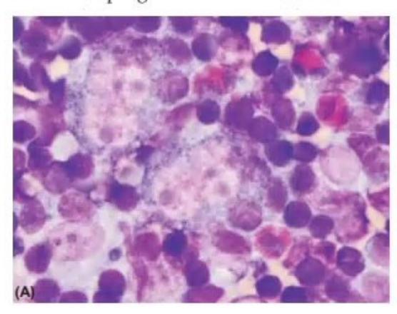
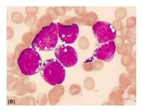
- Diagnosis of slide A and slide B.
- What are "B symptoms" in the disease of slide A?
- What is the prognosis for the disease in slide B?
Answer
- Slide A: Reed-Sternberg cell (classic "owl-eye" appearance)
Hodgkin Lymphoma. Slide B: L3 type lymphoblasts with deep basophilic cytoplasm and lipid vacuoles Burkitt Lymphoma / Mature B-cell ALL. - "B symptoms" of Hodgkin Lymphoma:
- Unexplained fever
(including Pel-Ebstein fever) - Drenching night sweats
- Unexplained weight loss
of baseline body weight within 6 months
- Unexplained fever
- Prognosis: Burkitt lymphoma carries a good prognosis with modern intensive chemotherapy protocols (
cure rate); however, if left untreated or with delayed therapy, it is one of the fastest-growing tumors with high risk of tumor lysis syndrome.

MNJ28-011
A male infant is brought to the hospital on day 7 of life with bleeding in stool, hematuria, and epistaxis. All other examination of this child appears normal. Child was apparently normal and taking breastfeeds till today morning. No fever, distress, or altered sensorium. Baby was born by NVD, cried immediately, and is on exclusive breastfeeds from birth. After collecting appropriate samples, FFP was given. On investigations: Hb: 7.2 g/dL, WBC: 10,300/µL (N: 44, L: 55), platelets: 2,00,000/µL, PT: prolonged, INR: prolonged, aPTT: prolonged, FDP: normal, fibrinogen level: normal:
- What is the most likely cause?
- What further treatment option with formulations and dose?
- Enumerate factors of "extrinsic and common pathway" of the coagulation cascade.
- Which coagulation factor numbers do not exist today?
Answer
- Vitamin K Deficiency Bleeding (VKDB) / Classic Hemorrhagic Disease of the Newborn (HDN).
- Treatment:
- Vitamin
(Phytomenadione): 1 mg IV slowly (or SC). - Fresh Frozen Plasma (FFP): 10–15 mL/kg IV for acute active bleeding or severe hemorrhage.
- Packed RBC transfusion if severe anemia / hemodynamic instability.
- Vitamin
- Coagulation cascade pathways:
- Extrinsic pathway: Factor VII, Tissue Factor (Factor III).
- Common pathway: Factor X, Factor V, Factor II (Prothrombin), Factor I (Fibrinogen), Factor XIII.
- Coagulation factor numbers no longer in use:
- Factor III (Tissue thromboplastin / Tissue factor - not a single plasma protein)
- Factor IV (Calcium ions - not a protein)
- Factor VI (Accelerin - proven to be the activated form of Factor V, i.e., Va).
MNJ28-012
A 6-year-old healthy girl Meenu presented with a sudden onset of a petechial rash all over the body, gradually increasing over the last two days. She also developed epistaxis from the left nostril and bleeding from gums while brushing this morning. She is active and playful. She has no history of fever, taking medications, or ingestion. She has been normal till now with no significant past history. On examination: no pallor, lymphadenopathy, or hepatosplenomegaly. Hb: 10.2 g/dL, WBC: 6,400/µL (N: 34, L: 55, M: 7), platelet count: 16,000/µL, no abnormal cells on peripheral smear. PT and aPTT: normal:
- What is the diagnosis of this condition?
- What immediate treatment will you start if financial constraint is not an issue?
- Is there a role for antibiotics?
- Name a cause of thrombocytopenia with small platelet size.
- Name causes of functional disorders of platelets (thrombasthenia).
Answer
- Immune Thrombocytopenic Purpura (ITP / Primary Immune Thrombocytopenia).
- First-line therapy:
- Intravenous Immunoglobulin (IVIg):
for 1–2 days (or for 5 days), OR - Intravenous Anti-D immunoglobulin (
in Rh-positive, non-splenectomized patients), OR - Oral Prednisolone (
for 1–2 weeks).
- Intravenous Immunoglobulin (IVIg):
- No, antibiotics have no role in acute ITP.
- Small-sized platelets with thrombocytopenia: Wiskott-Aldrich syndrome (WAS / WASP gene mutation).
- Platelet functional disorders:
- Glanzmann thrombasthenia (deficiency of GPIIb/IIIa receptor)
- Bernard-Soulier syndrome (deficiency of GPIb/IX/V receptor - large platelets)
- Gray platelet syndrome / Storage pool deficiencies
MNJ28-013
A 7-year-old male child Prashant presents with orbital swelling as shown in picture along with fever, loss of weight, anemia, hepatosplenomegaly, and rash over the body for the last 10 days. On examination his energy level is very low and he is feeling tired:
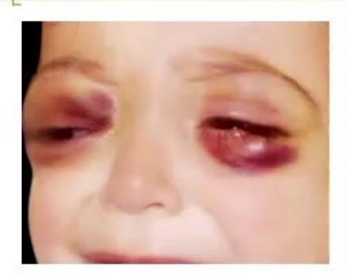
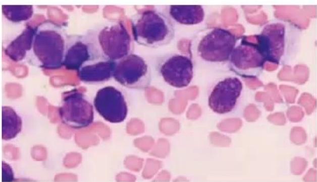
- What is the probable diagnosis?
- What is the specific finding in the PBF shown?
- What is the skin / orbital mass condition associated with this disorder?
- What chromosomal translocation is characteristically seen in this?
Answer
- Acute Myeloid Leukemia (AML) (specifically AML with granulocytic sarcoma / chloroma).
- Myeloblasts with intracellular Auer rods (fused azurophilic granules).
- Chloroma / Granulocytic sarcoma (Myeloid sarcoma / leukemic infiltration of orbit/skin: leukemia cutis).
- Translocation
( ) or .
MNJ28-014
Match the anemia classification with its corresponding cause:
| Anemia Type | Clinical Condition |
|---|---|
| 1. Microcytic with low reticulocyte count | a. Chronic renal failure |
| 2. Microcytic with high reticulocyte count | b. Congenital dyserythropoietic anemia (CDA) |
| 3. Macrocytic with low reticulocyte count | c. Lead poisoning / Iron deficiency anemia |
| 4. Macrocytic with high reticulocyte count | d. Hemolytic Uremic Syndrome (HUS) |
| 5. Normocytic with low reticulocyte count | e. Hypothyroidism / Vitamin |
| 6. Normocytic with high reticulocyte count | f. Hereditary pyropoikilocytosis / Hemoglobin H disease |
Answer
-
- Microcytic with low reticulocyte
c. Lead poisoning / Iron deficiency anemia
- Microcytic with low reticulocyte
-
- Microcytic with high reticulocyte
f. Hereditary pyropoikilocytosis / Hemoglobin H disease
- Microcytic with high reticulocyte
-
- Macrocytic with low reticulocyte
e. Hypothyroidism / Vitamin deficiency
- Macrocytic with low reticulocyte
-
- Macrocytic with high reticulocyte
b. Congenital dyserythropoietic anemia (or active hemolysis with reticulocytosis)
- Macrocytic with high reticulocyte
-
- Normocytic with low reticulocyte
a. Chronic renal failure / Aplastic anemia
- Normocytic with low reticulocyte
-
- Normocytic with high reticulocyte
d. Hemolytic Uremic Syndrome (HUS) / Immune hemolytic anemia
- Normocytic with high reticulocyte
(Match: 1-c, 2-f, 3-e, 4-b, 5-a, 6-d)
MNJ28-015
A 1.5-month-old male child Raju presents with complaints of severe pallor, snub nose, hypertelorism, triphalangeal thumb and absent radial pulse:
- What is the probable diagnosis?
- What is the status of HbF in this patient?
- What are important differential diagnoses?
- What is the treatment?
Answer
- Diamond-Blackfan Anemia (DBA / Congenital pure red cell aplasia).
- Elevated HbF levels (and elevated erythrocyte adenosine deaminase [eADA]).
- Differential diagnoses:
- Fanconi anemia
- Transient erythroblastopenia of childhood (TEC)
- Congenital dyserythropoietic anemia (CDA)
- Parvovirus B19 induced red cell aplasia
- Treatment:
- Corticosteroids (Oral Prednisolone 2 mg/kg/day; effective in ~80%)
- Chronic packed RBC transfusions with iron chelation therapy (for steroid non-responders)
- Hematopoietic stem cell transplantation (HSCT - only curative modality)
MNJ28-016
State True or False:
- Physiological anemia of infancy does not need treatment in term baby.
- Upregulation of EPO is present in this condition.
- In preterm babies, it presents earlier than in term babies.
- Premature infants may need EPO, protein, and vitamin E as treatment.
Answer
- True (Term infants experience a benign physiological nadir at 8–12 weeks, Hb 9–11 g/dL, requiring no therapy).
- False (There is a physiologic down-regulation / reduced erythropoietin production in response to increased tissue oxygenation after birth).
- True (In preterm infants, anemia of prematurity occurs earlier at 4–8 weeks and is more severe, Hb 7–9 g/dL).
- True (Recombinant EPO, iron, protein, and vitamin E supplementation are used in anemia of prematurity).
(Answers: 1-True, 2-False, 3-True, 4-True)
MNJ28-017
State True or False regarding Iron Deficiency Anemia:
- Pallor is visible only at Hb level < 9 g/dL.
- WBC and platelet counts are consistently elevated.
- Erythrocyte zinc protoporphyrin (ZPP) level > 80 µmol/mol is diagnostic for field surveys.
- Serum ferritin level < 12 µg/L is the most useful test and is not affected by liver disease.
- Subjective improvement with iron treatment occurs only after 3 days.
- Adding vitamins to iron syrups is generally unnecessary/useless.
- Elliptocytes and pencil/cigar-shaped cells are present in PBF.
Answer
- False (Clinical pallor becomes apparent at
, especially at conjunctiva and palmar creases). - False (Platelets may be elevated in mild-moderate IDA due to reactive thrombocytosis, but WBC count is typically normal).
- True (ZPP
heme indicates iron-deficient erythropoiesis and is useful for screening). - False (Serum ferritin is an acute-phase reactant; it is elevated in liver disease, inflammation, and infections even in the presence of iron deficiency).
- False (Subjective improvement in appetite, mood, and alertness occurs within 24–48 hours due to replenishment of intracellular iron-containing enzymes).
- True (Elemental iron alone is sufficient; routine vitamin combinations add cost without proven therapeutic advantage).
- True (Pencil cells / cigar cells and elliptocytes are characteristic findings in severe microcytic hypochromic IDA).
(Answers: 1-False, 2-False, 3-True, 4-False, 5-False, 6-True, 7-True)
MNJ28-018
A 7-year-old male child Radhay presents with complaints of fatigue and exercise intolerance for the last one month. He had a history of exchange transfusion in the neonatal period and gallstone removal last year. On examination: pallor and splenomegaly are present. Blood tests show low Hb, normal MCV, MCHC: 37 g/dL:
- What is the diagnosis of this condition and its mode of inheritance?
- For which infection and acute complication is this child susceptible?
- What are possible long-term complications in this disease?
- What cell will you find in PBF and what are other causes of this cell in children?
- What is the diagnostic test?
- What are recent guidelines for splenectomy in this disease?
Answer
- Hereditary Spherocytosis (HS); Autosomal dominant in 75% (mutations in ankyrin, band 3,
-spectrin). - Infection: Parvovirus B19 infection; Complication: Aplastic crisis (and hyperhemolytic / megaloblastic crisis).
- Long-term complications: Pigment gallstones (cholelithiasis), splenomegaly with hypersplenism, leg ulcers, gout, growth retardation.
- PBF: Microspherocytes (dense, round RBCs lacking central pallor). Other causes of spherocytes:
- ABO incompatibility / autoimmune hemolytic anemia (AIHA)
- Thermal injury / severe burns
- Clostridial sepsis / snake bite envenomation
- Hypophosphatemia / Microangiopathic hemolytic anemia
- Diagnostic tests:
- Eosin-5-maleimide (EMA) binding test by flow cytometry (gold standard, high sensitivity & specificity)
- Osmotic fragility test (acidified glycerol lysis test)
- Splenectomy guidelines:
- Treatment of choice for moderate-to-severe HS, but delayed until
years of age to minimize post-splenectomy sepsis risk. - Not indicated in mild HS (
, reticulocytes ). - Administer pneumococcal, meningococcal, and H. influenzae type b vaccinations at least 2–4 weeks prior, with lifelong prophylactic penicillin.
- Treatment of choice for moderate-to-severe HS, but delayed until
MNJ28-019
A 6-year-old male child Gopal presents with fever for 7 days. He is a known case of sickle cell disease. On examination: temperature = 102°F, BP: 88/44 mmHg. Blood test shows WBC:
- Does this child need hospital admission or daycare management?
- What scale will you use to determine pain intensity in children with sickle cell disease?
- What are recent advances to reduce pain in these children?
- What are treatment modalities for priapism in SCD?
- What is the modality to diagnose SCD in newborns?
Answer
- Emergency inpatient hospital admission (due to functional asplenia, fever with leukocytosis and hypotension indicates life-threatening encapsulated bacterial sepsis / pneumococcal bacteremia or acute splenic sequestration).
- Wong-Baker FACES Pain Rating Scale (for younger children) or Numerical Rating Scale (NRS) / Visual Analog Scale (VAS) for older children.
- Advances in pain reduction / disease modification:
- Hydroxyurea (increases HbF, reduces vaso-occlusive crises)
- Crizanlizumab (anti-P-selectin monoclonal antibody)
- Voxelotor (HbS polymerization inhibitor)
- L-glutamine (reduces oxidative stress)
- Treatment of priapism:
- Supportive: Hydration, analgesia, warm sitz bath, urination
- If prolonged
hours: Aspiration of blood from corpus cavernosum and irrigation with dilute alpha-adrenergic agonist (e.g., phenylephrine or adrenaline / epinephrine) - Exchange transfusion / surgical shunt if refractory
- Newborn screening: High-Performance Liquid Chromatography (HPLC) or Isoelectric Focusing (IEF) from dried blood spots (Guthrie card).
MNJ28-020
What are false statements about hereditary methemoglobinemia?
- Methemoglobinemia is due to deficiency of NADH-cytochrome b5 reductase.
- Cyanosis is visible when level
. - Daily oral ascorbic acid (200–500 mg/day) is the treatment of choice for long-term therapy.
- Regular use of vitamin C is associated with high chance of renal stone formation.
- Another treatment option is methylene blue, and that can be used in patients with G6PD deficiency.
Answer
- Statement 1: True (Deficiency of NADH-cytochrome b5 reductase [CYB5R3] is the primary enzymatic cause).
- Statement 2: False (Clinical cyanosis becomes apparent when methemoglobin level exceeds
or of total hemoglobin; normal reduced Hb produces cyanosis at ). - Statement 3: True (Oral ascorbic acid 200–500 mg/day or riboflavin is effective for chronic maintenance).
- Statement 4: False (Ascorbic acid at recommended maintenance doses does not commonly induce nephrolithiasis in children).
- Statement 5: False (Methylene blue is CONTRAINDICATED in G6PD deficiency because it requires NADPH generated by G6PD to reduce to leukomethylene blue; in G6PD deficiency it can precipitate severe acute hemolysis).
(False statements: 2, 4, 5)
MNJ28-021
State True or False about Thalassemia syndromes:
- Even when there is severe anemia, reticulocytopenia is present.
- Before starting transfusion, RBC extended phenotyping should be obtained.
- Plasma non-transferrin bound iron (NTBI) is most likely responsible for serious tissue iron toxicity.
- Quantitative iron by liver biopsy is standard, but T2* MRI is widely useful nowadays.
- Congestive heart failure and arrhythmias are the leading causes of death in poorly chelated patients.
Answer
- True (Ineffective erythropoiesis and intramedullary destruction of erythroblasts result in an inappropriately low reticulocyte count relative to the severe degree of anemia).
- True (Extended RBC phenotyping before first transfusion prevents alloimmunization against minor blood group antigens).
- True (NTBI and labile plasma iron [LPI] generate reactive oxygen species causing parenchymal organ damage).
- True (T2* MRI of heart and liver has replaced liver biopsy as the non-invasive gold standard for monitoring tissue iron overload).
- True (Myocardial iron overload leading to cardiomyopathy, heart failure, and fatal arrhythmias is the leading cause of mortality).
(All statements are True)
MNJ28-022
A 6-year-old previously asymptomatic boy Vedprakash is admitted to the hospital with complaints of sudden onset of jaundice and red-colored urine for 3 days. On examination, he is pale and icteric. He gave a history of cough and cold 4 days back for which he took aspirin:
- What is the possible diagnosis?
- What are other drugs that can precipitate this?
- What is the inheritance of this disease? Can this present in female children also?
- What is the diagnostic test and what is the diagnostic value?
- What findings will you see in the PBF?
Answer
- Glucose-6-Phosphate Dehydrogenase (G6PD) deficiency with acute intravascular hemolytic crisis.
- Precipitating drugs / agents:
- Antimalarials: Primaquine, pamaquine, chloroquine
- Antibacterials: Sulfonamides, co-trimoxazole, dapsone, nitrofurantoin, nalidixic acid
- Analgesics: Aspirin (high doses), phenacetin
- Chemicals: Naphthalene balls (mothballs), Methylene blue
- Foods: Fava beans (favism)
- X-linked recessive. Yes, females can be affected if homozygous, compound heterozygous, or heterozygous with skewed X-inactivation (Lyonization).
- Diagnostic test: Quantitative G6PD spectrophotometric assay in red blood cells (
of normal activity is diagnostic). (Note: Test during acute hemolytic episode may be falsely normal due to higher enzyme levels in surviving young reticulocytes; repeat after 2–3 months). - PBF findings: "Bite cells" (degmacytes), "blister cells", polychromasia, and Heinz bodies on supravital (methyl violet / crystal violet) staining.
MNJ28-023
A 5-year-old male child Hritik presents with complaints of short stature. On examination, he has an absent radius, 4 café-au-lait spots, epicanthic folds, and severe pallor. Blood tests show a decrease in all cell lines. Bone marrow examination shows a hypocellular bone marrow:
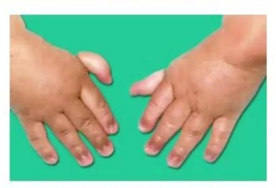
- What is the possible diagnosis?
- What is the specific diagnostic test for this disease?
- What are causes of pancytopenia with a cellular bone marrow?
- What are future neoplastic complications of this disease?
- What treatment will you advise?
Answer
- Fanconi Anemia (FA).
- Chromosomal breakage study / Diepoxybutane (DEB) and Mitomycin C (MMC) test on peripheral blood lymphocytes (demonstrating increased chromosome breaks, radial forms, and gaps).
- Causes of pancytopenia with hypercellular / normocellular bone marrow:
- Acute leukemia (ALL, AML)
- Myelodysplastic syndromes (MDS)
- Megaloblastic anemia (Vitamin
/ folate deficiency) - Hypersplenism
- Hemophagocytic Lymphohistiocytosis (HLH)
- Visceral leishmaniasis (Kala-azar)
- Gaucher disease / Niemann-Pick disease
- Future malignancies: Acute Myeloid Leukemia (AML), MDS, squamous cell carcinoma of head and neck, esophagus, and anogenital tract.
- Treatment:
- Allogeneic Hematopoietic Stem Cell Transplantation (HSCT - only curative therapy)
- Androgens (e.g., Oxymetholone) and G-CSF for hematologic support
- Supportive transfusions (leukoreduced, irradiated blood products)
- Surveillance for secondary solid tumors.
MNJ28-024
The mother of an 11-year-old female child Radhika complains of abnormal pigmentation at the neck that started 5 months back, followed by abnormal nail changes:
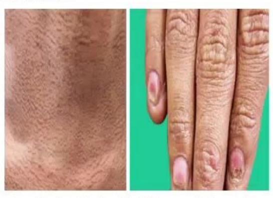
- What is the diagnosis of this condition?
- What is the characteristic diagnostic triad?
- Which is the most common finding on clinical examination?
- What are possible complications?
- What treatment options are available?
Answer
- Dyskeratosis Congenita (Zinsser-Cole-Engman syndrome, a telomere biology disorder).
- Classic triad:
- Reticulated / lacy hyperpigmentation of neck and upper chest
- Nail dystrophy (ridging, splitting, pterygium)
- Oral mucosal leukoplakia
- Reticulated skin pigmentation (present in
of patients). - Complications:
- Progressive bone marrow failure / aplastic anemia
- Pulmonary fibrosis
- Liver cirrhosis
- Malignancy: Myelodysplastic syndrome (MDS), AML, squamous cell carcinoma of mouth/esophagus
- Treatment:
- Allogeneic Hematopoietic Stem Cell Transplantation (HSCT)
- Androgen therapy (Danazol / Oxymetholone to elongate telomeres and improve cytopenias)
- Supportive hematologic care and cancer surveillance
MNJ28-025
A 6-year-old farmer's son Raju is admitted with complaints of fever, ecchymosis over the body and fatigue for the last 15 days. His father states that he spends 5–6 hours per day at the farm, with a history of pica. On examination: lymph nodes palpable, 2 cm hepatomegaly. Blood test shows pancytopenia (Hb: 5 g/dL, ANC:
- What is the diagnosis and what is the severity grade?
- What viruses can cause this disease?
- What are flow cytometry findings in the differential diagnosis of PNH?
- What treatment options are available?
- What are future complications in terms of clonal evolution?
Answer
- Severe Aplastic Anemia (SAA) (likely pesticide-induced/acquired). Criteria for SAA met: Bone marrow cellularity
plus at least 2 of: , , (or ). - Viral etiologies: Hepatitis viruses (Non-A, Non-B, Non-C post-hepatitis aplastic anemia), Epstein-Barr Virus (EBV), Cytomegalovirus (CMV), Parvovirus B19, HIV.
- Flow cytometry in Paroxysmal Nocturnal Hemoglobinuria (PNH): Deficiency / absence of GPI-anchored proteins: CD55 (DAF) and CD59 (MIRL) on granulocytes and red blood cells (using FLAER).
- Treatment options:
- Matched sibling donor (MSD) Hematopoietic Stem Cell Transplantation (treatment of choice for young patients)
- Immunosuppressive therapy (IST): Antithymocyte globulin (ATG) + Cyclosporine + Eltrombopag + short-course steroids
- Clonal evolution / future neoplastic risks: Myelodysplastic syndrome (MDS), Acute Myeloid Leukemia (AML), and Paroxysmal Nocturnal Hemoglobinuria (PNH).
MNJ28-026
A 14-year-old girl presents with a history of excessive menorrhagia. She has a history of excessive postoperative bleeding at 9 years of age when an incision was made for boil removal. Her blood test shows prolonged Bleeding Time (BT) and prolonged aPTT. Other tests are normal:
- What is the diagnosis of this condition?
- Is it important to know the blood group of the child in this disease and why?
- What 2 important tests will you order for definitive diagnosis?
- What treatments are available?
Answer
- von Willebrand Disease (vWD).
- Yes, because plasma vWF and Factor VIII levels vary with ABO blood group (individuals with blood group O have
lower baseline vWF levels compared to non-O blood groups: ). - Definitive diagnostic tests:
- vWF Antigen (vWF:Ag) quantitative assay
- Ristocetin cofactor activity (vWF:RCo) / platelet-binding activity
- Factor VIII coagulant activity (FVIII:C)
- vWF multimer analysis (for subtyping)
- Treatment modalities:
- Desmopressin (DDAVP) (effective in Type 1 vWD)
- vWF-containing Factor VIII concentrates (e.g., Humate-P, Wilate) or recombinant vWF
- Antifibrinolytics: Tranexamic acid, Epsilon-aminocaproic acid (EACA)
- Combined oral contraceptives / Levonorgestrel IUD for menorrhagia
MNJ28-027
A 4-year-old male Rohit presents with a 2-week history of abdominal discomfort, poor appetite, and weight loss. No significant past or family history. No urinary symptoms, fever, or other complaints. Development and school performance are normal. On examination you palpate a 4 cm radius lump on the left side of the abdomen. Other systemic examination is normal:
- What are three most helpful investigations to reach the diagnosis with 2 possible differential diagnoses?
- What is the role of doing urine routine/microscopy if this patient has a possible renal tumor?
- What are 4 prognostic factors for Wilms' tumor?
- What is a nephrogenic rest and what is its relationship with Wilms' tumor?
Answer
- Investigations:
- Contrast-enhanced CT (CECT) / MRI abdomen and pelvis
- USG abdomen with Doppler
- 24-hour urinary catecholamines: VMA and HVA (to differentiate from neuroblastoma)
- Chest CT / X-ray (to assess pulmonary metastasis)
Differential diagnoses: Wilms' tumor (Nephroblastoma) and Neuroblastoma.
- Role of Urine R/M: Detects microscopic hematuria (present in up to 30–50% of Wilms' tumors due to renal pelvic/parenchymal invasion) and rules out UTI or proteinuria.
- Prognostic factors in Wilms' tumor:
- Histopathology: Favorable histology vs. Unfavorable / Anaplastic histology (most critical factor)
- Stage of the disease at diagnosis (Stage I–V)
- Age at diagnosis (better prognosis if
years) - Tumor weight / size (
favorable) - Molecular markers (loss of heterozygosity at 1p and 16q, 1q gain).
- Nephrogenic rests: Foci of abnormally retained, persistent embryonal metanephric blastema beyond 36 weeks of gestation; they are premalignant precursors found in
of unilateral Wilms' tumors and nearly of bilateral Wilms' tumors.
MNJ28-028
A 5-year-old child Raju is on chemotherapy for ALL receiving: Methotrexate, Cyclophosphamide, Doxorubicin, L-asparaginase, Ondansetron, Domperidone, Ganciclovir, Amphotericin B, Vancomycin, and Ceftriaxone.
Match each side effect with the responsible medication:
| Clinical Side Effect | Responsible Medication |
|---|---|
| 1. Hemorrhagic cystitis | a. Doxorubicin |
| 2. Acute dystonic reaction | b. Cyclophosphamide |
| 3. Deranged clotting profile / Coagulopathy | c. L-asparaginase |
| 4. Hypokalemia | d. Domperidone / Metoclopramide |
| 5. "Red man syndrome" / Flushing | e. Amphotericin B |
| 6. Megaloblastic anemia / Mucositis | f. Vancomycin |
| 7. Cardiomyopathy | g. Methotrexate |
Answer
-
- Hemorrhagic cystitis
Cyclophosphamide (due to metabolite acrolein; prevented with Mesna)
- Hemorrhagic cystitis
-
- Acute dystonic reaction
Domperidone / Metoclopramide (dopamine D2 receptor antagonism)
- Acute dystonic reaction
-
- Deranged clotting profile / Coagulopathy
L-asparaginase (inhibits hepatic synthesis of antithrombin III, protein C/S, and fibrinogen)
- Deranged clotting profile / Coagulopathy
-
- Hypokalemia
Amphotericin B (tubular leak)
- Hypokalemia
-
- "Red man syndrome" / Flushing
Vancomycin (histamine release with rapid infusion)
- "Red man syndrome" / Flushing
-
- Megaloblastic anemia / Mucositis
Methotrexate (dihydrofolate reductase inhibition; rescued with folinic acid/leucovorin)
- Megaloblastic anemia / Mucositis
-
- Cardiomyopathy
Doxorubicin (free radical mediated cumulative cardiotoxicity)
- Cardiomyopathy
MNJ28-029
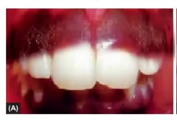
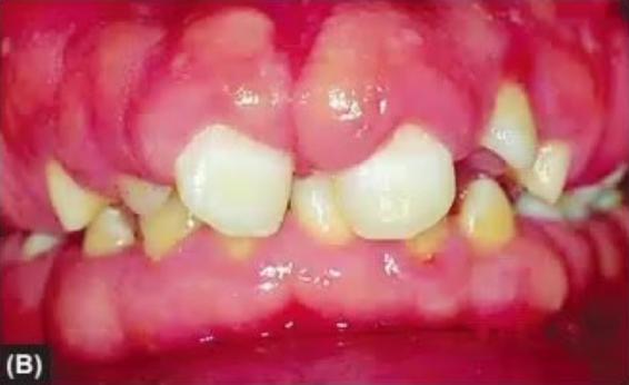
- What are the clinical findings in images A and B?
- What are the causes of each (write two causes for each)?
Answer
- Clinical findings:
- Image A: Gingival hypertrophy / hyperplasia.
- Image B: Mucocutaneous / oral hyperpigmentation (melanoplakia).
- Causes:
- Gingival hypertrophy (Image A):
- Phenytoin toxicity
- Cyclosporine therapy
- Calcium channel blockers (e.g., Amlodipine, Nifedipine)
- Acute Monocytic Leukemia (AML-M4 / M5)
- Oral hyperpigmentation (Image B):
- Addison's disease (Primary adrenal insufficiency / high ACTH)
- Peutz-Jeghers syndrome
- Heavy metal toxicity (Lead line / Bismuth)
- Drugs (e.g., Hydroxyurea, Cyclophosphamide, Clofazimine)
- Gingival hypertrophy (Image A):
MNJ28-030
Read the following clinical scenarios and identify the causative drug from the list: (Heparin, Prednisolone, Penicillin, Aspirin, Co-trimoxazole, Hydroxyurea, Ketoconazole, Phenytoin, Naproxen):

- A 13-year-old girl Reeta is on an anticonvulsant for 1 year. On examination she has pallor. CBC showed MCV 110 fL.
- A 7-year-old boy Ram with sickle cell disease was started on a disease-modifying drug. CBC on follow-up showed MCV of 118 fL.
- A 10-year-old boy Raj came to OPD 6 days after cardiac valve repair surgery. You notice petechiae on the neck and order CBC, which shows a platelet count of 60,000/µL.
- A 3-year-old girl Rani is on VUR antibiotic prophylaxis since 6 months of age. Presented with URI; blood tests show Hb 8 g/dL, TLC
, Platelets .
Answer
- Phenytoin (causes macrocytic anemia by interfering with folate absorption and metabolism).
- Hydroxyurea (induces macrocytosis / elevated MCV, which serves as a marker of drug adherence and efficacy).
- Heparin (Heparin-Induced Thrombocytopenia [HIT type II], an immune-mediated reaction occurring 5–10 days post-exposure).
- Co-trimoxazole (causes drug-induced marrow suppression / pancytopenia via antifolate activity).
MNJ28-031
A 7-year-old boy Reftar is admitted to the hospital with complaints of dark/black urine for 1 day. On examination he is pale. His father gave him an aspirin tablet for headache yesterday. Investigations show acute intravascular hemolysis and low G6PD level:
- What will be the level of haptoglobin and methemalbumin / methemoglobin in this child?
- What will you find in the peripheral blood film?
- What is the characteristic peripheral blood film finding after splenectomy?
- What is the immediate management of a hemolytic crisis in a G6PD-deficient patient?
Answer
- Serum haptoglobin is markedly decreased / undetectable (depleted by binding free Hb); plasma methemalbumin and urinary hemosiderin/free hemoglobin are increased.
- PBF findings: "Bite cells" (degmacytes), "blister/hemi-ghost cells", polychromasia, and Heinz bodies on supravital staining.
- Post-splenectomy PBF findings: Howell-Jolly bodies (nuclear remnants), Pappenheimer bodies, Heinz bodies, target cells, and acanthocytes.
- Management of hemolytic crisis:
- Immediately discontinue offending drug/agent (Aspirin)
- Maintain vigorous hydration and urine alkalinization to prevent renal tubular obstruction by hemoglobin casts
- Packed RBC transfusion if severe anemia or cardiovascular compromise
- Phototherapy / exchange transfusion in severe neonatal jaundice.
MNJ28-032
A 3-year-old boy Gaurav Garg is brought with a history of easy bruising followed by excessive bleeding. He had a history of bleeding from the umbilical cord. Both parents are healthy. You notice bruises over the shin. Blood tests show normal platelet count and normal BT, but a prolonged activated partial thromboplastin time (aPTT):
- What is the probable diagnosis?
- How will you classify the severity of this disease?
- In the history you find that mother's sister's son has the same problem, but maternal uncle's son is healthy. Explain the genetic basis.
- What is the use of AMICAR (
-aminocaproic acid / tranexamic acid) in hemophilic patients?
Answer
- Hemophilia A (Factor VIII deficiency) or Hemophilia B (Factor IX deficiency). (Factor XIII deficiency can also present with umbilical cord bleeding, but has normal aPTT).
- Severity classification by clotting factor level:
- Severe:
( ) spontaneous bleeds, frequent hemarthroses - Moderate:
( ) bleeding with minor trauma - Mild:
( ) bleeding only with major trauma / surgery
- Severe:
- Genetic basis: Hemophilia is an X-linked recessive disorder. The mother is an obligate or potential carrier (
) who inherited the mutant allele from a common ancestor. Her sister also inherited the mutated X chromosome and transmitted it to her son ( ). The maternal uncle passes his Y chromosome to his son (who receives an X from his unrelated mother) and therefore the maternal uncle's son cannot inherit the X-linked disease. - Use of AMICAR (Epsilon-aminocaproic acid) / Tranexamic acid: Antifibrinolytic agent that competitively inhibits plasminogen activation and prevents clot lysis. It is particularly useful for mucosal bleeding (dental extractions, oral/nasal bleeds) by blocking salivary/mucosal fibrinolytic enzymes.
MNJ28-033
Match the iron profile with the type of anemia:
| Iron Profile | Condition |
|---|---|
| 1. Serum iron low, TIBC high, Transferrin saturation 10%, Ferritin low | a. Anemia of chronic disease |
| 2. Serum iron normal/low, TIBC low, Transferrin saturation 30%, Ferritin high/normal | b. Iron deficiency anemia |
| 3. Serum iron normal/high, TIBC normal, Transferrin saturation 30–50%, Ferritin normal/high | c. Beta-thalassemia minor |
| 4. Serum iron high, TIBC low/normal, Transferrin saturation 100%, Ferritin high | d. Sideroblastic anemia |
Answer
| Parameter | Iron Deficiency Anemia | Anemia of Chronic Disease | Sideroblastic Anemia | |
|---|---|---|---|---|
| Serum Iron | Low | Low | Normal / High | High |
| TIBC | High | Low | Normal | Low / Normal |
| Transferrin Saturation | Low ( |
Normal / Low ( |
Normal ( |
High ( |
| Serum Ferritin | Low ( |
High / Normal | Normal / High | Very High |
- 1
b. Iron deficiency anemia - 2
a. Anemia of chronic disease - 3
c. Beta-thalassemia trait - 4
d. Sideroblastic anemia
MNJ28-034
A 4-year-old boy Samay presented when his mother noticed swelling in the abdomen during a bath. He has no other symptoms and no significant past history. On examination: no anemia, jaundice, or lymphadenopathy. Height and weight are appropriate for age. Abdomen reveals a 12 cm diameter smooth, firm mass on the right side. His BP is 116/76 mmHg. Urine examination shows microscopic RBCs, and USG abdomen shows an intrarenal mass without calcification:
- What is the commonest malignant and commonest non-malignant abdominal mass in children?
- What are two differential diagnoses, and what is the most probable diagnosis in this child?
- What cardiovascular/renal complication is present in this child and what is the underlying mechanism?
- Name 3 syndromes associated with this diagnosis.
- What is the prognosis in this case?
Answer
- Commonest malignant abdominal mass in children: Neuroblastoma / Wilms' tumor (Wilms is commonest renal malignant mass). Commonest non-malignant mass: Fecal impaction / Hydronephrosis / Multicystic dysplastic kidney.
- Differential diagnoses: Wilms' tumor (Nephroblastoma) and Neuroblastoma. Most probable diagnosis: Wilms' tumor (smooth, intrarenal, does not cross midline, hematuria, no calcification).
- Hypertension (elevated BP) due to renal ischemia and hyperreninism caused by compression of the renal vasculature by the tumor mass.
- Associated syndromes:
- WAGR syndrome (Wilms, Aniridia, Genitourinary anomalies, Retardation)
- Beckwith-Wiedemann syndrome (BWS - hemihypertrophy, macroglossia, omphalocele)
- Denys-Drash syndrome (Wilms, pseudohermaphroditism, progressive glomerulopathy)
- Prognosis: Excellent; overall 5-year survival rate for favorable histology Wilms' tumor exceeds
.
MNJ28-035
Monika, a 9-year-old child, presents with a lump on the left side of her neck for one month. She has had no fever, night sweats, or weight loss. On examination: BCG scar is present, and a 3 cm diameter mobile lymph node is palpable on the left side of the neck. She had a cold lasting 2 days one month back without fever. Investigations: Hb 10.7 g/dL, normal TLC and platelets, CRP 12 mg/L, ESR 25 mm/hr, Paul-Bunnell test negative, chest X-ray normal:
- What are 5 differential diagnoses for chronic cervical lymphadenopathy?
- What treatment will you advise at the first visit?
- What is the most likely diagnosis?
- What will you do if after 10–14 days of treatment the lump size remains the same?
- What level of protection does the BCG vaccine provide against tuberculous and non-tuberculous mycobacteria?
Answer
- Differential diagnoses:
- Reactive / infective lymphadenitis (bacterial, e.g., Staphylococcus, Streptococcus)
- Tubercular lymphadenitis (Scrofula)
- Non-tuberculous mycobacterial (NTM) lymphadenitis
- Cat scratch disease (Bartonella henselae)
- Infectious mononucleosis (EBV / CMV)
- Lymphoma (Hodgkin / Non-Hodgkin)
- First-line management: Oral course of broad-spectrum antibiotic covering Staphylococcus aureus and Streptococcus pyogenes (e.g., Amoxicillin-Clavulanate or Cephalexin for 10–14 days) and observation.
- Reactive infective lymphadenitis (post-viral/upper respiratory tract infection).
- If no regression after 10–14 days: Perform Fine Needle Aspiration Cytology (FNAC) or Excisional Lymph Node Biopsy for histopathology, GeneXpert (CBNAAT), and mycobacterial/fungal culture.
- BCG protection: Provides
protection against severe childhood tuberculosis (TB meningitis and miliary TB), against pulmonary TB, and minimal to variable ( ) protection against non-tuberculous mycobacteria.
MNJ28-036
Amita, a 6-year-old girl with leukemia, completed her 2nd course of chemotherapy 8 days back and returned home. Her family members are suffering from a viral illness. In emergency, she is febrile and her central venous catheter (port/line) exit site is inflamed. Other systemic examination is normal:
- What is the single most important immediate investigation and why?
- What is the probable diagnosis and is this an emergency?
- What are risk factors for infection in a child following intensive chemotherapy?
- How will you manage this child?
Answer
- Complete Blood Count (CBC) with absolute neutrophil count (ANC) and paired blood cultures (from central line and peripheral vein) to diagnose febrile neutropenia and identify bacteremia.
- Febrile neutropenia with central line-associated bloodstream infection (CLABSI). YES, this is a medical emergency requiring immediate hospitalization and empiric broad-spectrum IV antibiotics within 1 hour.
- Risk factors for infection:
- Severe neutropenia (
or expected nadir ) - Mucositis / breakdown of mucosal barriers
- Indwelling central venous catheter
- Immunosuppression from chemotherapy and corticosteroids
- Malnutrition and prolonged hospital stay
- Severe neutropenia (
- Management:
- Immediate inpatient admission
- Obtain blood cultures from all lumens of central line and peripheral vein, urine culture, and swab of catheter exit site
- Prompt empirical IV monotherapy with anti-pseudomonal
-lactam (e.g., Piperacillin-Tazobactam, Cefepime, or Meropenem) Vancomycin (indicated due to central line site inflammation) - IV fluid hydration, strict barrier precautions, and antipyretics (avoid NSAIDs/rectal suppositories).