MNJ33-001
See the X-ray images below:
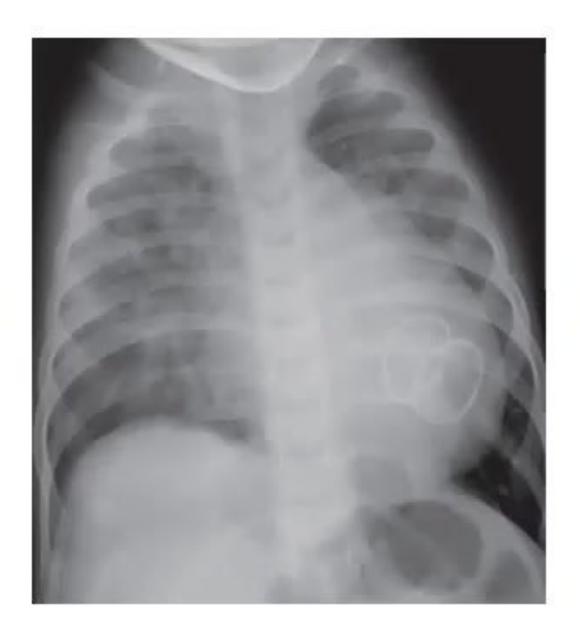
- What was the lesion?
- What was the procedure carried out?
- What are the complications?
Answer
- Lesion: Ventricular Septal Defect (VSD) / Atrial Septal Defect (ASD)
- Procedure: Transcatheter device closure / device implant (e.g., Amplatzer septal occluder)
- Complications:
- Device displacement / embolization
- Thromboembolism formation
- Hemolytic anemia (mechanical hemolysis)
- Arrhythmias / heart block
- Residual shunt
- Vascular access complications
MNJ33-002
See the chest radiograph below:
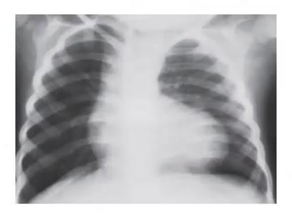
- Give the possible diagnosis based on this X-ray.
- What are the first two steps in the treatment of a hypercyanotic (hypoxic / Tet) spell?
- In a cyanotic newborn, how can you distinguish pulmonary disease from cyanotic congenital heart disease?
- Which cardiac conditions are associated with the following radiographic signs:
- Egg-shaped heart
- Snowman silhouette (figure-of-8)
- Rib notching
Answer
- Diagnosis: Tetralogy of Fallot (TOF) — classic "boot-shaped" heart (coeur en sabot) with normal heart size, upturned cardiac apex, concavity in main pulmonary artery segment, and oligemic lung fields (decreased pulmonary vascular markings).
- First two steps in hypoxic spell management:
- Knee-chest position
- High-flow 100% oxygen supplementation
(Followed by Morphine sulfate, IV Sodium bicarbonate, IV crystalloid bolus, IV Phenylephrine/Propranolol).
- Distinguishing pulmonary vs cyanotic CHD:
- Hyperoxia test: On 100% FiO2 for 10–15 min, PaO2 rises >150–200 mmHg in pulmonary parenchymal disease, but remains <100–150 mmHg in cyanotic CHD (right-to-left shunt).
- Radiographic associations:
- Egg-shaped heart: Transposition of Great Arteries (TGA)
- Snowman silhouette: Supracardiac Total Anomalous Pulmonary Venous Connection (TAPVC)
- Rib notching: Coarctation of Aorta (typically >5–6 years of age)
MNJ33-003
A 2-day-old neonate, birth weight: 3.1 kg. On examination: cyanosis +, soft murmur +, single S2. On investigation: hypoxia and acidosis present, chest X-ray shows normal cardiac size:
- What is the most likely diagnosis?
- What is the emergency medical therapy?
- What is the dosage and what are the potential complications of this drug?
Answer
- Diagnosis: Duct-dependent cyanotic congenital heart disease (e.g., d-TGA with intact IVS, Pulmonary atresia with intact IVS, Critical pulmonary stenosis).
- Emergency therapy: Intravenous Prostaglandin E1 (PGE1 / Alprostadil) infusion to maintain ductal patency.
- Dosage & Complications:
- Dosage: 0.05 to 0.1 µg/kg/min continuous IV infusion, titrated down to effective maintenance dose once stabilized.
- Complications: Apnea (must be prepared for intubation), systemic hypotension, cutaneous flushing, fever, bradycardia, seizures, edema.
MNJ33-004
A term infant suddenly develops cardiac failure in the first week of life. The infant was pink, peripheral pulses were collapsing/bounding, but there is no cardiac murmur. Chest X-ray showed cardiomegaly. Echocardiogram showed a structurally normal heart and great vessels. The PDA was closed:
- What is the likely diagnosis?
- What further investigations are indicated?
Answer
- Diagnosis: Systemic arteriovenous (AV) fistula / malformation (e.g., Vein of Galen malformation, hepatic hemangioendothelioma / AV malformation) causing high-output cardiac failure.
- Investigations:
- Neurovascular imaging: Cranial Doppler ultrasound, Brain MRI / MR Angiography (MRA) or CT Angiography
- Abdominal Doppler ultrasound (to rule out hepatic AV fistula)
- Diagnostic cerebral / visceral digital subtraction angiography (DSA)
MNJ33-005
See the X-ray image below and answer the following questions:
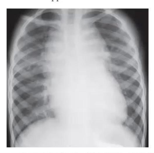
- What is the diagnosis?
- What is this sign called?
- What anatomical structures cause this radiographic appearance?
Answer
- Diagnosis: Total Anomalous Pulmonary Venous Connection (TAPVC) — Supracardiac type
- Sign: "Snowman appearance" / "Figure-of-8 appearance"
- Anatomical structures causing the shadow:
- Dilated vertical vein on the left
- Dilated left innominate (brachiocephalic) vein horizontally
- Dilated superior vena cava (SVC) on the right (forming the upper contour / "head" of the snowman)
- Enlarged right atrium and right ventricle forming the lower contour / "body"
MNJ33-006
A 1-day-old infant appears dusky during feeding. O2 is administered via nasal cannula, and 2 hours later she develops worsening tachypnea. Findings: HR 170/min, RR 80/min, Right arm BP 48/30 mmHg, Right leg BP 52/32 mmHg, SpO2 90% on oxygen:
- If the diagnosis is Hypoplastic Left Heart Syndrome (HLHS), describe physical findings of pulse, murmur, precordium, and S2.
- What was wrong in the initial management in this scenario?
- Mention 5 congenital heart defects that present with duct-dependent circulation in the first week of life.
- What is the emergency management?
Answer
- Physical findings in HLHS:
- Pulses: Weak / thready peripheral pulses with signs of shock/poor systemic perfusion.
- Precordium: Hyperdynamic right ventricular impulse / parasternal heave.
- Heart sounds: Loud, single S2 (due to single anterior pulmonary valve).
- Murmur: Usually no murmur or only a soft, non-specific systolic murmur (of tricuspid regurgitation or PDA).
- Error in management: High-concentration oxygen administration lowers pulmonary vascular resistance (PVR), promoting "pulmonary steal" and precipitously worsening systemic hypoperfusion/shock in duct-dependent systemic flow lesions.
- Duct-dependent CHDs in week 1:
- Duct-dependent systemic blood flow (SBF): HLHS, Interrupted Aortic Arch (IAA), Critical Aortic Stenosis, Critical Coarctation of Aorta.
- Duct-dependent pulmonary blood flow (PBF): Pulmonary Atresia, Critical Pulmonary Stenosis, Tricuspid Atresia.
- Duct-dependent parallel mixing: d-TGA with intact ventricular septum.
- Emergency management: Immediate continuous IV Prostaglandin E1 (PGE1) infusion (0.05–0.1 µg/kg/min) to maintain ductal patency, minimize supplemental FiO2 to avoid pulmonary steal, correct metabolic acidosis, and maintain inotropic support.
MNJ33-007
A 5-day-old infant born at 31 weeks' gestation is on ventilatory support. He has a grade 2 holosystolic/continuous murmur extending past the second heart sound. Pulses are bounding. Precordial palpation is hyperdynamic. Echocardiography reveals a large patent ductus arteriosus (PDA) without other ductal-dependent lesions:
- When would one suspect a hemodynamically significant PDA in a preterm clinically?
- Mention 3 complications of PDA in a preterm neonate.
- Contraindications for indomethacin / ibuprofen therapy.
- Pharmacological closure of PDA — medications and dosages.
Answer
- Clinical suspicion: Tachypnea, tachycardia, increased ventilatory / O2 requirement, failure to wean from ventilator, continuous/systolic murmur, bounding peripheral pulses with wide pulse pressure, active/hyperdynamic precordium, poor weight gain.
- Complications: Bronchopulmonary dysplasia (BPD / CLD), Necrotizing enterocolitis (NEC), Intraventricular hemorrhage (IVH), Pulmonary edema / hemorrhage, Congestive heart failure, Renal impairment.
- Contraindications: Active bleeding (especially GI or IVH), severe thrombocytopenia (<50,000/mm³), coagulation defect, necrotizing enterocolitis, acute renal impairment / oliguria (BUN >30 mg/dL or Cr >1.8 mg/dL), active untreated sepsis.
- Pharmacological regimens:
- Indomethacin (IV): 0.2 mg/kg initially, followed by 2 doses of 0.1–0.25 mg/kg at 12–24 hour intervals based on chronological age.
- Ibuprofen (IV/Oral): 10 mg/kg initial dose, followed by 5 mg/kg at 24 and 48 hours.
- Paracetamol (IV/Oral): 15 mg/kg/dose every 6 hours for 3–7 days.
MNJ33-008
When examining a healthy 3-year-old boy during a health supervision visit, you hear a grade 2 systolic ejection murmur most clearly at the upper left sternal border:
- Differential diagnosis of ejection systolic murmur (ESM) in the second left intercostal space (ICS).
- Classical auscultatory findings in Atrial Septal Defect (ASD).
- Anatomical types of ASD.
- In children with ASD and moderate-to-severe pulmonary hypertension, which associated conditions should be ruled out?
Answer
- Differential diagnoses of ESM at 2nd left ICS:
- Atrial Septal Defect (ASD) — flow murmur across pulmonary valve
- Pulmonary Stenosis (valvular / infundibular)
- Innocent flow murmur (e.g., Pulmonary flow murmur of childhood)
- Increased pulmonary blood flow states (e.g., severe anemia, fever, thyrotoxicosis)
- Partial Anomalous Pulmonary Venous Connection (PAPVC)
- Coarctation of Aorta / Aortic Stenosis
- Auscultatory findings in ASD:
- Normal or split S1
- Wide, fixed splitting of S2
- Grade 2–3/6 ejection systolic murmur at upper left sternal border (pulmonary flow murmur)
- Mid-diastolic low-pitched rumbling murmur at lower left sternal border (due to increased flow across tricuspid valve in large shunts)
- Types of ASD: Ostium secundum (most common, ~70%), Ostium primum (partial AVSD), Sinus venosus ASD (superior/inferior), Coronary sinus ASD.
- Conditions to rule out: Large left-to-right shunt (large ASD/VSD), TAPVC/PAPVC, Cor triatriatum, Mitral stenosis (Lutembacher syndrome), Left ventricular outflow obstruction.
MNJ33-009
Following an uncomplicated delivery, a 3.7 kg term infant develops cyanosis in the first hour of life. Findings at 3 hours of age include: Cyanosis; HR: 140/min; RR: 56/min; no heart murmurs; pulse oximetry on room air: 70% saturation in the right hand and 75% in the foot; on 100% FiO2 via head-hood: saturation increases to 90% in the foot; chest radiography is normal:
- Clinical differentials based on pre- and post-ductal saturation.
- Describe the hyperoxia test.
- Causes of differential cyanosis and reverse differential cyanosis.
Answer
- Clinical differentials: Cyanotic congenital heart disease with duct-dependent circulation, Persistent Pulmonary Hypertension of the Newborn (PPHN), Transposition of Great Arteries (TGA) with Coarctation of Aorta / Interrupted Aortic Arch.
- Hyperoxia test: Measure baseline ABG (PaO2) on room air, then administer 100% FiO2 for 10–15 minutes and repeat ABG:
- PaO2 > 150–200 mmHg: Suggests respiratory / pulmonary parenchymal disease.
- PaO2 < 100–150 mmHg (and little rise with FiO2): Diagnostic of fixed right-to-left intracardiac shunt (cyanotic CHD) or massive PPHN.
- Causes:
- Differential cyanosis (Pre-ductal saturation > Post-ductal saturation):
- Persistent Pulmonary Hypertension of Newborn (PPHN) with R-to-L ductal shunt
- Coarctation of Aorta / Interrupted Aortic Arch (IAA) with PDA
- Critical Aortic Stenosis with PDA
- Reverse differential cyanosis (Post-ductal saturation > Pre-ductal saturation):
- d-TGA with Coarctation of Aorta / Interrupted Aortic Arch
- d-TGA with severe PPHN (suprasystemic pulmonary vascular resistance)
- Differential cyanosis (Pre-ductal saturation > Post-ductal saturation):
MNJ33-010
A 14-year-old girl collapses during a race. She is unconscious, cyanotic, and pulseless, but spontaneously revives within seconds. Both patient and family histories are unremarkable:
- What are the causes of sudden cardiac death (SCD) in children?
- What appropriate diagnostic tests should be ordered?
- What arrhythmias are typically associated with exertional syncope?
Answer
- Causes of sudden cardiac death in children:
- Structural / Functional:
- Hypertrophic Cardiomyopathy (HCM / HOCM)
- Anomalous origin of coronary artery from opposite sinus (e.g., ALCAPA)
- Arrhythmogenic Right Ventricular Cardiomyopathy (ARVC)
- Dilated Cardiomyopathy / Myocarditis
- Aortic dissection / Marfan syndrome
- Valvular aortic stenosis / LVOT obstruction
- Commotio cordis (blunt chest impact)
- Electrical / Channelopathies:
- Long QT syndrome (LQTS) / Short QT syndrome
- Catecholaminergic Polymorphic Ventricular Tachycardia (CPVT)
- Brugada syndrome
- Wolff-Parkinson-White (WPW) syndrome with AF
- Congenital complete heart block
- Structural / Functional:
- Diagnostic tests:
- 12-lead ECG (measure QTc, delta waves, Brugada pattern)
- Echocardiography (assess HCM, LVOT gradient, systolic function, coronary origins)
- 24-hour Holter monitoring / Implantable loop recorder
- Exercise stress testing (graded treadmill test)
- Cardiac MRI (for ARVC, anomalous coronaries, myocardial fibrosis)
- Genetic testing for channelopathies / cardiomyopathies
- Arrhythmias associated with exertional syncope: Ventricular tachycardia (polymorphic VT, CPVT, Torsades de pointes), Ventricular fibrillation, Rapid AF/SVT over accessory pathway in WPW.
MNJ33-011
A healthy 8-month-old boy has had intermittent episodes of intense cyanosis of the hands, lower arms, and feet since birth. The infant appears alert and playful during the episodes. At other times, his arms and legs are pink but have a "mottled" appearance. Physical examination reveals no remarkable findings other than mottling of the extremities:
- Causes of episodic acrocyanosis / peripheral cyanosis.
- Diagnostic tests indicated for this child.
Answer
- Causes of episodic acrocyanosis:
- Benign physiological acrocyanosis / vasomotor instability / cutis marmorata (cold exposure)
- Methemoglobinemia
- Episodic cardiac arrhythmias / paroxysmal tachycardia
- Hypercyanotic (Tet) spells in uncorrected CHD
- Breath-holding spells / seizure disorders
- Raynaud's phenomenon / peripheral vasospasm
- Diagnostic tests:
- 12-lead ECG & Holter monitoring (rule out arrhythmias)
- Arterial blood gas (ABG) with Co-oximetry (measure MetHb levels)
- Echocardiography (rule out structural congenital heart defect)
- Video EEG (if seizure disorder suspected)
MNJ33-012
A 14-year-old boy is followed periodically for bicuspid aortic valve. He presents with a 3-week history of fatigue and intermittent fever up to 101°F:
- What will you look for on physical examination?
- He has elevated WBC and ESR. What other diagnostic tests will you order?
- What organisms are most likely to grow on blood culture?
- What treatment regimen is ideal?
- What risks does this boy face for sudden clinical deterioration?
Answer
- Physical examination findings:
- New or changing heart murmur (especially aortic regurgitation)
- Peripheral stigmata of Infective Endocarditis (IE): Osler nodes, Janeway lesions, Splinter hemorrhages, Roth spots (fundus), Petechiae
- Splenomegaly, clubbing, signs of heart failure
- Diagnostic tests:
- Serial blood cultures (3 sets from separate venipuncture sites before starting antibiotics)
- Transthoracic (TTE) and/or Transesophageal Echocardiogram (TEE) to detect vegetations, abscess, valve perforation, or new regurgitation
- Urine analysis (microscopic hematuria, proteinuria)
- Serum rheumatoid factor, complement levels
- Likely organisms:
- Viridans group streptococci (e.g., Streptococcus mutans, S. mitis)
- Staphylococcus aureus / Coagulase-negative staphylococci (S. epidermidis)
- Enterococcus species, HACEK group organisms
- Treatment regimen:
- Prolonged IV bactericidal antibiotic therapy for 4–6 weeks:
- High-dose IV Ceftriaxone / Ampicillin + Gentamicin (for streptococci/enterococci) or IV Cloxacillin / Vancomycin + Gentamicin (for staphylococci), tailored to MIC.
- Risks for sudden deterioration:
- Acute severe aortic regurgitation with pulmonary edema / cardiogenic shock
- Embolic stroke / systemic embolization (to brain, spleen, renal, coronary arteries)
- Perivalvular abscess with complete heart block / conduction disturbances
- Mycotic aneurysm rupture
MNJ33-013
Fill in the blanks regarding pediatric ECG criteria:
- At what age does the pediatric ECG resemble an adult ECG?
- Tall P waves in lead II (Right Atrial Enlargement) is defined as a height greater than ........ mm.
- Maximum normal P wave duration in children is ........ seconds.
- Maximum normal P wave duration in infants (<12 months) is ........ seconds.
- PR interval — lower normal limit in children less than 3 years is ........ seconds.
Answer
- Age ECG resembles adult: By 3–5 years (approaching adult pattern around 8–12 years)
- Tall P waves (P pulmonale in lead II): > 2.5–3.0 mm
- Max P wave duration in children: 0.10 seconds (2.5 small squares)
- Max P wave duration in infants <12 mo: 0.08 seconds (2 small squares)
- PR interval lower limit (<3 years): 0.08 seconds
MNJ33-014
A 12-year-old boy presented with acute onset pain in the left iliac fossa, fever, and vomiting. A preoperative 12-lead ECG is shown below:
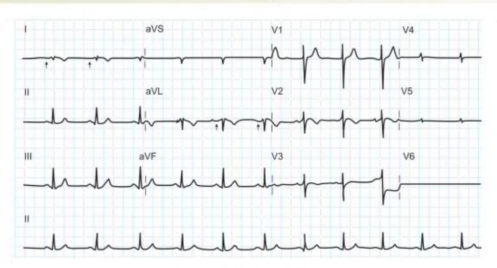
- Analyze the ECG findings.
- What is the diagnosis and what malformations are associated with this condition?
Answer
- ECG Analysis:
- Inverted P waves, QRS complexes, and T waves in Lead I and aVL
- Lead aVR has upright P and QRS waves (resembling normal lead aVL/I)
- Progressively decreasing R-wave voltage across precordial leads V1 to V6 (absence of normal R-wave progression across left chest)
- Diagnosis on ECG: Dextrocardia (Mirror-image dextrocardia / Situs inversus totalis)
- Associated malformations / conditions:
- Situs inversus totalis (organs transposed: stomach/spleen on right, liver/appendix on left — explains acute left iliac fossa appendicitis pain)
- Right-sided aortic arch
- Kartagener syndrome / Primary Ciliary Dyskinesia (triad: Situs inversus, Bronchiectasis, Chronic sinusitis)
- Lung lobe anomalies (right lung: 2 lobes, left lung: 3 lobes)
MNJ33-015
Answer the following questions regarding pulse oximetry:
- At what FiO2 level should a patient be monitored on continuous pulse oximetry?
- What are the causes (external and internal) of inaccurate SpO2 readings?
- A child rescued from a school fire shows 100% SpO2 on pulse oximetry upon admission to the PICU. The intensivist still orders high-flow oxygen and an ABG with co-oximetry. Why?
- What is the oxygen-carrying capacity (arterial oxygen content
) of blood in a child with SpO2 of 97% and Hb of 10 g/dL? - What is the lowest "safe" level of SpO2 saturation in a child with normal Hb, and its corresponding PaO2?
- What should be done when bright ambient light interferes with SpO2 reading?
- When will you suspect "duct-dependent circulation" on day 1 of life using pulse oximetry?
Answer
- FiO2 threshold: Any patient requiring supplemental oxygen (
/ >21–40%) or in respiratory distress. - Causes of inaccurate SpO2:
- External: Bright ambient light / phototherapy, patient motion artifact, nail polish / henna, improper sensor placement / loose probe.
- Internal: Hypothermia, severe vasoconstriction / poor peripheral perfusion / shock, abnormal hemoglobins (carboxyhemoglobin, methemoglobin), intravenous dyes (methylene blue, indocyanine green), severe anemia.
- Fire / smoke inhalation victim: Standard pulse oximeters measure absorbance at 660 nm and 940 nm and cannot differentiate oxyhemoglobin from carboxyhemoglobin (
). Carboxyhemoglobin is read falsely as oxyhemoglobin, displaying a falsely high SpO2 (100%) despite severe tissue hypoxia. Co-oximetry is required to quantify . - Oxygen-carrying capacity calculation:
(or 130 mL/L).
- Lowest safe saturation & PaO2: SpO2 of 90% corresponds to a PaO2 of approximately 60 mmHg on the oxygen-hemoglobin dissociation curve.
- Ambient light interference: Shield or cover the sensor site and patient extremity with an opaque cloth/cover.
- Duct-dependent circulation suspicion on day 1: A difference of
between pre-ductal (right upper limb) and post-ductal (either lower limb) oxygen saturation, or any absolute saturation .
MNJ33-016
Regarding Congestive Cardiac Failure (CCF) in pediatric patients:
- List 4 causes of systolic dysfunction and 4 causes of diastolic dysfunction.
- What are the general principles of management of CCF?
- What are the specific pharmacological agents used in the management of CCF?
Answer
- Causes of dysfunction:
- Systolic dysfunction (impaired contractility / pump failure):
- Dilated cardiomyopathy
- Myocarditis (viral / rheumatic)
- Myocardial ischemia / ALCAPA / infarction
- Acute severe volume overload (e.g., severe AR / MR)
- Sepsis-induced myocardial dysfunction
- Severe chronic anemia
- Diastolic dysfunction (impaired ventricular filling / relaxation):
- Hypertrophic cardiomyopathy (HCM)
- Restrictive cardiomyopathy
- Constrictive pericarditis / Cardiac tamponade
- Severe aortic stenosis / systemic hypertension
- Systolic dysfunction (impaired contractility / pump failure):
- Principles of management:
- Reduce cardiac preload and work (fluid restriction, propped-up position, sedation, oxygen)
- Augment myocardial contractility (inotropic support)
- Reduce cardiac afterload (vasodilators)
- Treat underlying/precipitating causes (correct anemia, control arrhythmias, treat infection/fever, surgical repair of defects)
- Specific drugs used:
- Diuretics: Furosemide, Spironolactone, Chlorothiazide (reduce preload)
- Afterload reducers (ACE inhibitors / ARBs): Captopril, Enalapril
- Inotropes: Digoxin, Milrinone (inodilator), Dobutamine, Dopamine, Epinephrine
- Beta-blockers: Carvedilol, Metoprolol (for chronic compensated failure)
MNJ33-017
See the 12-lead ECG rhythm strip below:
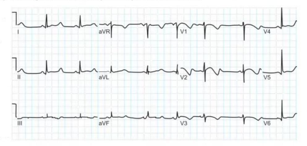
- Identify the abnormality in the ECG.
- What electrolyte abnormality can cause this?
- Why is it important to identify and correct this rhythm disturbance promptly?
Answer
- Abnormality: Prolonged QT interval (Prolonged QTc > 0.44–0.46 s)
- Electrolyte abnormality: Hypocalcemia (also Hypokalemia, Hypomagnesemia)
- Clinical importance: Prolonged QT predisposes to life-threatening polymorphic ventricular tachycardia (Torsades de pointes), ventricular fibrillation, and sudden cardiac death / arrest.
MNJ33-018
State True (T) or False (F) for each of the following statements regarding pediatric ECG:
- Neonates normally have right axis deviation.
- Complete congenital heart block is associated with maternal SLE / anti-Ro/SSA antibodies.
- Right bundle branch block (RBBB) is seen in Coarctation of Aorta, Ebstein's anomaly, and ASD.
- Romano-Ward syndrome is associated with prolonged PR interval.
- A resting heart rate rising to 180 beats/min in a neonate is considered tachycardia.
- Superior QRS axis (left axis deviation) is seen in Tricuspid Atresia and AV canal defect.
Answer
- True (T): Neonates have physiological right ventricular dominance resulting in right axis deviation.
- True (T): Transplacental anti-Ro (SSA) and anti-La (SSB) autoantibodies damage the fetal AV node.
- True (T): RBBB/incomplete RBBB is characteristically seen in ASD, Ebstein's anomaly, and post-cardiac surgery.
- False (F): Romano-Ward syndrome is a congenital Long QT syndrome (autosomal dominant), characterized by prolonged QT interval (normal PR interval and normal hearing).
- True (T): Normal neonatal resting heart rate is 110–160 bpm; sustained heart rate >160–180 bpm represents tachycardia.
- True (T): Superior axis (LAD) is a hallmark of Tricuspid Atresia, AV Canal Defect (AVSD), and Noonan syndrome.
MNJ33-019
State True (T) or False (F) regarding fetal circulation:
- The umbilical vein carries deoxygenated blood to the placenta from the fetus.
- Blood from the superior vena cava preferentially flows across the foramen ovale into the left atrium.
- 45% of right ventricular outflow enters the lungs via the pulmonary arteries in the fetus.
- The descending aorta is connected to the pulmonary artery via the ductus venosus.
Answer
- False (F): The umbilical vein carries oxygenated blood from the placenta to the fetus (umbilical arteries carry deoxygenated blood back to the placenta).
- False (F): Blood from the Inferior Vena Cava (IVC) is preferentially shunted through the foramen ovale into the left atrium; SVC blood flows into the right ventricle.
- False (F): Only ~10–15% of RV outflow enters the high-resistance fetal lungs; ~85–90% is shunted via the ductus arteriosus into the descending aorta.
- False (F): The pulmonary artery connects to the descending aorta via the ductus arteriosus (the ductus venosus shunts oxygenated umbilical vein blood directly to the IVC, bypassing the liver).
MNJ33-020
A 13-year-old girl presents with a 3-month history of palpitations. She is fit and well-grown with no recent illness or medications. She is an excellent student. She feels her heart rate rising and then coming gradually down after 3–4 hours. There is no history of chest pain, presyncope, or collapse. Symptoms occur predominantly during school hours. She plays sports without difficulty. Family history for cardiac disease is negative. Physical and systemic examination and blood pressure are entirely normal:
- What is the most likely diagnosis?
- What test must you perform before sending her home?
- Name 3 causes of potential life-threatening syncope / palpitations in children.
- When should a child with palpitations/syncope be referred to a pediatric cardiologist?
Answer
- Diagnosis: Physiological sinus tachycardia associated with situation-related stress / anxiety.
- Essential test: 12-lead ECG (to rule out pre-excitation/WPW, long QT syndrome, baseline arrhythmias).
- Life-threatening causes:
- Wolff-Parkinson-White (WPW) syndrome
- Long QT syndrome (LQTS) / CPVT / Brugada syndrome
- Hypertrophic Cardiomyopathy (HOCM)
- Arrhythmogenic Right Ventricular Cardiomyopathy (ARVC)
- Anomalous coronary artery origins
- Indications for pediatric cardiology referral:
- Exertional syncope or chest pain during exercise
- Abnormal 12-lead ECG (prolonged QTc, delta wave, LVH/RVH, heart block)
- Family history of sudden unexplained death at young age or inherited channelopathies/cardiomyopathies
- Documented pathologic arrhythmia on ECG/Holter
- Abnormal cardiac physical examination (pathologic murmur, gallop, diminished femoral pulses)
MNJ33-021
From each of the following clinical scenarios, select the most likely diagnosis from the options below:
Options:
- a. Anthracycline-induced cardiomyopathy
- b. Infective endocarditis
- c. Acute rheumatic fever
- d. Systemic Juvenile Idiopathic Arthritis (sJIA)
- e. Systemic Lupus Erythematosus (SLE)
Scenarios:
- A 13-year-old boy treated for rhabdomyosarcoma at 4 years of age with complete remission. Now presents with shortness of breath for 6 months. On examination, he appears fatigued with tachypnea, gallop rhythm, raised JVP, and cardiomegaly on chest X-ray.
- A 15-year-old boy presents with anorexia and low-grade intermittent fever for 1 month. On examination, you find muffled heart sounds, thrombocytopenia, and photosensitive facial rash after outdoor activities.
- A 7-year-old girl presents with a swollen left knee after complete recovery from a swollen right ankle last week. She has fever, an evanescent pink rash over her trunk, and a history of sore throat 1 month ago. ECG demonstrates first-degree heart block.
Answer
- -> a. Anthracycline-induced cardiomyopathy (Doxorubicin/daunorubicin cardiotoxicity requires long-term echocardiographic monitoring).
- -> e. Systemic Lupus Erythematosus (SLE) (Malar photosensitive rash, thrombocytopenia, lupus carditis/pericardial effusion causing muffled heart sounds).
- -> c. Acute rheumatic fever (Jones criteria: migratory polyarthritis, erythema marginatum, carditis / prolonged PR interval [first-degree AV block], antecedent streptococcal pharyngitis).
MNJ33-022
Choose the most appropriate diagnostic cardiac investigation for each given scenario:
Options:
- a. 24-hour Holter ECG monitoring
- b. 12-lead standard ECG
- c. Cardiac catheterization
- d. Echocardiography (ECHO)
- e. MRI thorax
- f. Exercise stress test
Scenarios:
- A 3-year-old boy comes for an upper respiratory infection. On examination, you notice a soft continuous/systolic murmur in the right supraclavicular area that becomes louder on sitting and disappears upon lying down and turning the head (venous hum).
- A 12-year-old boy with recurrent episodes of panic attacks and fast heart rate. On examination, he has an elevated resting heart rate of 120/min with an otherwise normal systemic examination.
- A 6-year-old boy who underwent balloon aortic valvuloplasty for congenital aortic stenosis at 18 months of age presents with shortness of breath on exertion and a harsh systolic murmur in the aortic area.
Answer
- -> b. 12-lead standard ECG (Benign innocent murmur / venous hum; confirm normal baseline ECG and reassure parents).
- -> a. 24-hour Holter ECG monitoring (To evaluate paroxysmal supraventricular tachycardia vs sinus tachycardia).
- -> c. Cardiac catheterization (or d. Echocardiography) (Gold standard to accurately quantify peak-to-peak transvalvular pressure gradient, aortic valve area, and assess restenosis).
MNJ33-023
Choose the most likely diagnosis for each chest pain scenario:
Options:
- a. Aortic stenosis
- b. Pneumonia
- c. Precordial catch syndrome (Texidor's twinge)
- d. Hypertrophic Obstructive Cardiomyopathy (HOCM)
- e. Costochondritis
Scenarios:
- A 12-year-old girl is referred for an 11-month history of recurrent, sharp, localized left-sided chest pain that does not radiate, lasts a few seconds to 1–2 minutes, and resolves spontaneously. She is well-grown and physical examination is completely normal.
- A 15-year-old boy collapses suddenly while playing football. In the emergency department, he describes severe retrosternal chest pain and diaphoresis during maximal exertion. On examination, you find a harsh ejection systolic murmur at the right upper sternal border radiating to the carotids.
- A 10-year-old girl presents with a 3-week history of left-sided chest pain that sharpens on deep breathing. There is no fever, cough, trauma, or palpitation. Cardiac and respiratory exams are normal. On chest wall examination, you elicit reproducible focal tenderness over the 2nd costochondral junction.
Answer
- -> c. Precordial catch syndrome (Texidor's twinge): Benign, sharp, sudden, short-lived pain aggravated by breathing.
- -> a. Aortic stenosis: Exertional syncope/angina with classic systolic ejection murmur at right 2nd ICS radiating to carotids.
- -> e. Costochondritis: Reproducible focal tenderness over costochondral junctions with normal systemic examination.
MNJ33-024
An 8-year-old boy is scheduled for elective minor surgery. The anesthesiologist cannot palpate his femoral pulses. On referral, you find no cyanosis or clubbing. Femoral pulses are absent/markedly delayed with a forceful, heaving apex beat. A grade 3/6 systolic murmur is heard best at the upper left sternal border and left interscapular region. Radiograph and MR angiogram are shown below:
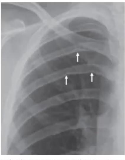
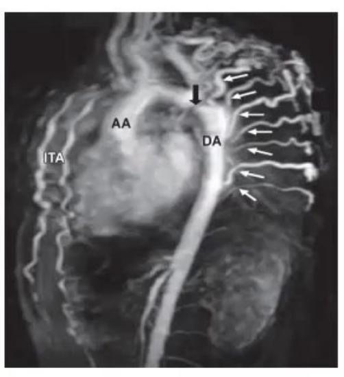
- What are the findings on chest X-ray and MR angiogram?
- What is the diagnosis based on clinical and investigation findings?
- What syndromes are characteristically associated with this condition?
- What is the definitive management plan?
Answer
- Imaging findings:
- Chest X-ray: Inferior rib notching (Roesler's sign on 3rd–8th ribs due to dilated collateral intercostal arteries), "figure-of-3" sign on aortic silhouette.
- MR Angiogram: Severe discrete narrowing / coarctation of the descending thoracic aorta just distal to the origin of the left subclavian artery with extensive collateral arterial circulation.
- Diagnosis: Postductal Coarctation of the Aorta
- Associated syndromes:
- Turner syndrome (45,X — strongly associated with CoA)
- Williams syndrome / PHACES syndrome / Noonan syndrome
- Bicuspid aortic valve (seen in >50–70% of CoA patients)
- Definitive management:
- Refer to pediatric cardiology/cardiothoracic surgical team.
- Transcatheter balloon angioplasty with endovascular stent placement (procedure of choice in children >25 kg / older children) OR surgical resection with end-to-end anastomosis.
- Monitor and manage systemic hypertension post-repair.