THG15-001
A 1-year-old child presents with paroxysms of cough of 2 weeks duration, without fever, with hoarseness of voice, and tachypnea. Respiratory examination is normal.
a. What is the probable diagnosis?
b. How will you confirm the diagnosis?
c. What is the characteristic hematologic abnormality seen?
d. What is the preferred agent of choice in treatment of this condition?
e. What is the desired chemoprophylaxis?
Answer
- a. Pertussis (whooping cough)
- b. Culture (Bordet-Gengou/Regan-Lowe agar) or PCR of nasopharyngeal swab/secretions
- c. Absolute lymphocytosis with normal or elevated total leukocyte count
- d. Macrolides: Azithromycin 10 mg/kg/day once daily for 5 days (preferred in infants <1 month), or Clarithromycin 15 mg/kg/day in 2 divided doses for 7 days, or Erythromycin 40–50 mg/kg/day QID for 14 days
- e. Post-exposure chemoprophylaxis for all household/close contacts: Azithromycin for 5 days or Erythromycin for 14 days (regardless of immunization status)
THG15-002
A 2-year-old child is brought by his mother for tuberculosis screening, as there is a family history of recently diagnosed pulmonary tuberculosis. You are planning to get a tuberculin skin test (TST) with purified protein derivative (PPD).
a. What is the current recommendation of strength of PPD?
b. What is the recommended site for TST administration?
c. How will you know that TST administration technique was proper?
d. How will you store the tuberculin vial after administration?
e. When will you read the TST after administration?
f. How will you read the TST?
g. How will you interpret?
h. How will you approach a patient who reports late for TST reading?
i. What is the impact of live viral vaccine on TST?
Answer
- a. PPD-RT 23 with Tween 80, 2 TU (0.1 mL) is standard. If unavailable, up to 5 TU (PPD-S) can be used.
- b. Volar (ventral) aspect of the forearm, 2–4 inches (5–10 cm) below the elbow joint.
- c. Intradermal injection raising a pale, discrete wheal with distinct margins of 6–10 mm in diameter.
- d. Refrigerated at 2–8°C (protected from light; discard after 28 days of opening if preservative present).
- e. Read between 48 and 72 hours (2–3 days) after administration.
- f. Palpate induration (not erythema) and measure the transverse diameter across the long axis of the forearm in millimeters using a transparent ruler (ballpoint pen method).
- g. Induration ≥10 mm is considered positive in BCG-vaccinated children (≥5 mm in HIV-infected, severely malnourished, or close contact of active TB).
- h. If read between 72 hours and 7 days: a positive reaction (≥10 mm) is valid, but a negative test must be repeated on the other forearm. If after 7 days, repeat the test completely.
- i. Live viral vaccines (e.g., MMR, varicella) given within 6 weeks prior to TST can cause transient suppression of cell-mediated immunity and false-negative results.
THG15-003
This is the X-ray of an adolescent girl who presented with unremitting cough of 3 weeks duration along with weight loss.
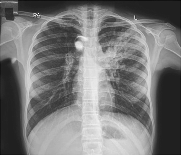
a. How will you proceed based on this X-ray finding?
b. Explain the steps of induced sputum sample collection.
c. What are the criteria for good sputum sample?
d. Which is the preferred container for sample collection?
e. How many samples of sputum are required for molecular tests?
Answer
- a. Chest X-ray reveals left upper zone consolidation with fibro-cavitary changes and paratracheal lymphadenopathy, highly suggestive of pulmonary tuberculosis. Proceed with sputum collection for NTEP-approved rapid molecular testing (CBNAAT / GeneXpert MTB/RIF or TrueNat).
- b. Steps of sputum induction:
-
- Fasting for 2–3 hours prior to procedure
-
- Premedication/priming with inhaled salbutamol (via MDI + spacer or nebulization) to prevent bronchospasm
-
- Nebulization with 5 mL of 3% hypertonic saline for 10–15 minutes
-
- Encourage deep coughing and expectoration into a sterile container
-
- c. Mucoid or mucopurulent expectorate from the lower respiratory tract, volume of 2–5 mL (contains <10 squamous epithelial cells and >25 leukocytes per low power field).
- d. Sterile, leak-proof 50 mL Falcon tube (conical centrifuge tube).
- e. A single good-quality sputum sample is sufficient for rapid molecular tests (CBNAAT/TrueNat).
THG15-004
This toddler, who is on bottle feeds and who has recently completed 5 days course of co-amoxiclav for respiratory infection, has come with this lesion.
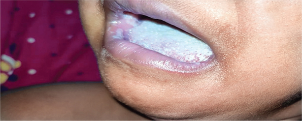
a. Describe the lesion.
b. What is the diagnosis?
c. What is the etiological agent?
d. How will you confirm the diagnosis?
e. What is the recommended treatment?
Answer
- a. White, curdy, adherent plaques on the buccal mucosa, tongue, and gingiva with a "cottage cheese" appearance that reveal erythematous, bleeding base upon scraping.
- b. Oral thrush (pseudomembranous candidiasis)
- c. Candida albicans
- d. Primarily clinical diagnosis. Can be confirmed by 10% KOH mount showing budding yeast cells and pseudohyphae.
- e. Treatment:
- Mild (<50% mucosa involved): Topical clotrimazole 1% mouth paint / drops 5–6 times daily for 7–14 days, or oral nystatin suspension (100,000 U/mL) 0.5–1 mL QID for 7–14 days.
- Severe (>50% mucosa involved / refractory): Oral fluconazole 3–6 mg/kg/day once daily for 7–14 days.
- Sterilize feeding bottles, teats, and pacifiers.
THG15-005
A 6-year-old child, became sick, 5 days after playing in stagnant water after recent heavy rainfall. She developed abrupt onset of high-grade fever, rigors, myalgia (especially in calf), and headache. On examination, she had intense conjunctival hyperemia (suffusion) without discharge, muscle tenderness, and hepatosplenomegaly with lymphadenopathy.
a. What is the most probable diagnosis?
b. What is the causative organism and reservoir?
c. What are the typical hematological findings?
d. How will you confirm the diagnosis?
e. What is the recommended treatment?
f. What reaction is anticipated after parenteral treatment for this disease?
Answer
- a. Leptospirosis (Weil's syndrome if severe with jaundice and renal failure)
- b. Leptospira interrogans (spirochete); Reservoir: Rodents (rats, mice), domestic animals via urine contamination
- c. Neutrophilic leukocytosis, normocytic normochromic anemia, thrombocytopenia, markedly elevated ESR and CRP, elevated CPK, and conjugated hyperbilirubinemia with disproportionate alkaline phosphatase elevation.
- d. Confirmation:
- First week (leptospiremic phase): Blood/urine PCR or blood culture in EMJH medium
- After first week (immune phase): IgM ELISA or Microscopic Agglutination Test (MAT, gold standard: 4-fold rise in titer or single titer ≥1:400)
- e. Treatment:
- Mild disease: Oral doxycycline (2 mg/kg/day BD) or oral amoxicillin (50 mg/kg/day TDS) for 7 days
- Moderate-to-severe: IV crystalline penicillin G (2.5–4 lakh units/kg/day 6 hourly) or IV ceftriaxone (80–100 mg/kg/day OD) for 7 days
- f. Jarisch–Herxheimer reaction (acute fever, rigors, tachycardia, and hypotension within hours of initiating antibiotics due to massive spirochetal lysis).
THG15-006
A 7-year-old child presents with fever, sore throat, and dysphagia of 2 days duration. On third day of illness, child develops papular rash on trunks, axilla, and groin. Later it spreads to extremities sparing palms and soles and circumoral area.
a. What is the probable diagnosis?
b. Name the findings on examination of oral cavity?
c. What is the causative organism?
d. What is the reason for the rash?
e. What is the investigation of choice to confirm the diagnosis?
f. Name two long-term complications of this infection.
g. What is the recommended treatment?
Answer
- a. Scarlet fever (Scarlatina)
- b. Oral cavity findings:
- Hyperemic pharynx and enlarged tonsils with yellowish exudates
- White strawberry tongue (first 1–2 days) progressing to red strawberry tongue (day 4–5)
- Palatal petechiae (Forchheimer spots)
- Circumoral pallor
- c. Group A Beta-Hemolytic Streptococcus (GABHS / Streptococcus pyogenes)
- d. Streptococcal pyrogenic exotoxins (SPE-A, SPE-B, SPE-C / erythrogenic toxins) produced by lysogenized bacteria.
- e. Throat swab culture (gold standard) or Rapid Antigen Detection Test (RADT) for GABHS.
- f. Non-suppurative complications: Acute rheumatic fever, post-streptococcal glomerulonephritis (PSGN), PANDAS, post-streptococcal reactive arthritis.
- g. Oral penicillin V or amoxicillin (50 mg/kg/day in 2–3 divided doses) for a full 10 days (or single dose IM benzathine penicillin G).
THG15-007
Match the antibiotic with its side effect:
| Antibiotic | Side Effect |
|---|---|
| a. Vancomycin | i. Flu-like illness |
| b. Clindamycin | ii. Ototoxicity |
| c. Erythromycin | iii. Red man syndrome |
| d. Ciprofloxacin | iv. Disulfiram-like reaction |
| e. Rifampicin | v. Pseudomembranous colitis |
| f. Gentamicin | vi. Optic neuropathy |
| g. Cefoperazone | vii. Infantile hypertrophic pyloric stenosis |
| h. Ethambutol | viii. Pill esophagitis |
| i. Chloramphenicol | ix. Tendon rupture |
| j. Doxycycline | x. Gray baby syndrome |
Answer
- a - iii (Vancomycin — Red man syndrome)
- b - v (Clindamycin — Pseudomembranous colitis / C. difficile)
- c - vii (Erythromycin — Infantile hypertrophic pyloric stenosis)
- d - ix (Ciprofloxacin — Tendon rupture / tendinopathy)
- e - i (Rifampicin — Flu-like illness / orange body fluids)
- f - ii (Gentamicin — Ototoxicity / nephrotoxicity)
- g - iv (Cefoperazone — Disulfiram-like reaction / bleeding due to MTT ring)
- h - vi (Ethambutol — Optic neuropathy / red-green color blindness)
- i - x (Chloramphenicol — Gray baby syndrome / bone marrow suppression)
- j - viii (Doxycycline — Pill esophagitis / tooth discoloration)
THG15-008
State whether the following statements are True or False:
a. Streptococcus pyogenes can cause necrotizing fasciitis and toxic shock syndrome.
b. All pathogenic species of Staphylococcus are catalase negative.
c. In cystic fibrosis exacerbations, pseudomonal coverage is always needed.
d. Ceftazidime or cefepime can be used in the treatment of melioidosis.
e. All enterococci exhibit high-level resistance to most cephalosporins.
f. There is no “carrier state” in Mycoplasma infection.
g. The primary goal of antibiotic treatment in paroxysmal phase of pertussis is to prevent the complications.
h. Ceftriaxone and cefotaxime are effective agents of chemoprophylaxis in Haemophilus influenzae infection.
i. Moldy odor perspiration is classical sign of brucellosis.
j. If Salmonella isolates are resistant to ampicillin and ceftriaxone, it is referred as multidrug-resistant typhoid.
Answer
- a. True (GABHS is the classic cause of type II monomicrobial necrotizing fasciitis and streptococcal toxic shock syndrome).
- b. False (All staphylococci are catalase positive; streptococci are catalase negative).
- c. True (Pseudomonas aeruginosa is the major chronic respiratory pathogen in CF).
- d. True (IV ceftazidime or meropenem is the intensive-phase drug of choice for melioidosis).
- e. True (Enterococci have intrinsic resistance to cephalosporins due to low-affinity PBPs).
- f. False (Asymptomatic respiratory carriage of M. pneumoniae is well recognized in children).
- g. False (Antibiotics in the paroxysmal phase do not alter the clinical course; primary goal is eliminating nasopharyngeal carriage and interrupting transmission).
- h. False (Rifampicin is the chemoprophylactic agent of choice for H. influenzae type b).
- i. True (Peculiar wet-hay / moldy perspiration odor is described in brucellosis).
- j. False (MDR typhoid is defined as resistance to all three first-line conventional agents: ampicillin, chloramphenicol, and cotrimoxazole).
THG15-009
Q15.9
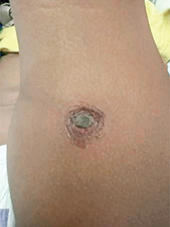
a. Describe the skin lesion?
b. What is the diagnosis?
c. Name two common causative organisms.
d. Name two complications for each organism.
e. What is the recommended treatment?
Answer
- a. Deep ulcerative lesion with an adherent, thick dark-brown/black necrotic crust and an elevated, indurated violaceous erythematous border ("punched-out" ulcer).
- b. Ecthyma (deep / ulcerative impetigo)
- c. Group A Streptococcus (S. pyogenes) and Staphylococcus aureus (including MRSA).
- d. Complications:
- S. pyogenes: Post-streptococcal glomerulonephritis (PSGN), cellulitis/erysipelas, scarlet fever.
- S. aureus: Staphylococcal scalded skin syndrome (SSSS), deep abscess, septic arthritis/osteomyelitis.
- e. Oral antistaphylococcal antibiotic: Cephalexin (50 mg/kg/day QID) or Amoxicillin-clavulanate (or Clindamycin / Cotrimoxazole if MRSA suspected) for 7–10 days along with warm soaks for crust removal.
THG15-010
Find the odd one out from each statement:
a. Gram-positive bacteria:
i. Enterococcus
ii. Neisseria
iii. Corynebacterium
iv. Clostridium
b. Bactericidal antibiotic:
i. Ceftazidime
ii. High-dose azithromycin
iii. Teicoplanin
iv. Aztreonam
c. Pre-extensively drug resistant tuberculosis (pre-XDR TB):
i. Isoniazid resistance
ii. Rifampicin resistance
iii. Levofloxacin resistance
iv. Linezolid resistance
d. Streptococcal pharyngitis clinical feature:
i. Tender anterior cervical adenopathy
ii. Cough
iii. High-grade fever
iv. Age 3–14 years
e. Treatment of Helicobacter pylori infection without antibiotic susceptibility testing:
i. Clarithromycin
ii. Amoxicillin
iii. Metronidazole
iv. Tetracycline
Answer
- a. ii. Neisseria (Gram-negative diplococcus, while others are Gram-positive).
- b. ii. High-dose azithromycin (Bacteriostatic macrolide, while ceftazidime, teicoplanin, and aztreonam are bactericidal).
- c. iv. Linezolid resistance (Pre-XDR TB is MDR/RR-TB + resistance to any fluoroquinolone [levofloxacin/moxifloxacin]; linezolid resistance defines XDR-TB).
- d. ii. Cough (Presence of cough points away from GABHS and towards viral pharyngitis; Centor/McIsaac score gives negative weight to cough).
- e. i. Clarithromycin (Due to high global clarithromycin resistance, empiric clarithromycin triple therapy without DST is no longer recommended).
THG15-011
The following is the complete hemogram and peripheral smear findings of a child who presented with fever of 5 days duration without any respiratory symptoms.
| Parameter | Value |
|---|---|
| White blood cells | 4,350/mm³ |
| Red blood cells | 4.3 million/mm³ |
| Hemoglobin | 12.3 g/dL |
| Hematocrit | 36.9% |
| Mean corpuscular volume (MCV) | 82 µm³ |
| Mean corpuscular hemoglobin (MCH) | 27.46 pg/cell |
| Mean corpuscular hemoglobin concentration (MCHC) | 32% Hb/cell |
| Platelets | 36,000/mm³ |
Peripheral smear: Normal white blood cells without atypical cells or blasts, normocytic normochromic RBCs without abnormal cells, platelets normal in number and shape with clumps seen, and no hemoparasites.
a. What is the abnormality seen in the hemogram?
b. What is the terminology given to this abnormality?
c. What is the most probable diagnosis?
d. What is the causative organism?
e. What is the mode of spread?
f. What is the recommended treatment?
Answer
- a. Automated analyzer shows severe thrombocytopenia (36,000/mm³), but peripheral blood smear reveals normal platelet count with platelet clumping.
- b. Pseudothrombocytopenia (EDTA-dependent platelet clumping/satellitism).
- c. Scrub typhus (or early Rickettsial infection / acute febrile illness)
- d. Orientia tsutsugamushi
- e. Bite of the infected larval stage (chigger) of trombiculid mites (Leptotrombidium deliense).
- f. Oral Doxycycline 2.2 mg/kg/dose BD (max 100 mg BD) for 7–10 days (or oral Azithromycin 10 mg/kg OD for 5 days in young infants/pregnancy).
THG15-012
Match the following:
| Test | Disease / Organism |
|---|---|
| 1. Weil–Felix test | a. Leptospirosis |
| 2. Rapid plasma reagin test (RPR test) | b. Cryptococcus |
| 3. Microscopic agglutination test (MAT) | c. Diphtheria |
| 4. Quantitative buffy coat test | d. Rickettsial infection |
| 5. Indian Ink test | e. Syphilis |
| 6. Paul–Bunnell test | f. Malaria |
| 7. Casoni test | g. Infectious mononucleosis |
| 8. Elek test | h. Staphylococcus |
| 9. D-zone test | i. Tuberculosis |
| 10. Interferon-γ release assay (IGRA) test | j. Hydatid disease |
Answer
- 1 - d (Weil–Felix test — Rickettsial infection)
- 2 - e (RPR test — Syphilis)
- 3 - a (MAT — Leptospirosis)
- 4 - f (Quantitative buffy coat test — Malaria)
- 5 - b (India Ink test — Cryptococcus)
- 6 - g (Paul–Bunnell test / Monospot — Infectious mononucleosis / EBV)
- 7 - j (Casoni test — Hydatid disease / Echinococcus)
- 8 - c (Elek test — Diphtheria toxigenicity)
- 9 - h (D-zone test — Staphylococcus inducible clindamycin resistance)
- 10 - i (IGRA test — Tuberculosis)
THG15-013
An 8-year-old presented to you with nasal discharge and daytime cough of 2 days duration. The child had nasal discharge 1 week back associated with fever for 2 days duration at the onset. The symptoms were gradually improving over past 5–6 days, when the worsening of symptoms started. Child had recurrence of fever with worsening nasal discharge of purulent nature and daytime cough.
a. What is the clinical diagnosis?
b. What are the common organisms implicated?
c. Any laboratory test required for the diagnosis?
d. What is the empiric treatment of choice?
e. Name two complications in this condition?
Answer
- a. Acute bacterial rhinosinusitis (ABRS) with "double sickening" pattern (worsening after initial improvement).
- b. Streptococcus pneumoniae, nontypeable Haemophilus influenzae, and Moraxella catarrhalis.
- c. No. Acute rhinosinusitis is a clinical diagnosis; routine imaging/cultures are not recommended.
- d. Oral Amoxicillin-clavulanate (co-amoxiclav) 45–90 mg/kg/day (amoxicillin component) in 2 divided doses for 10–14 days.
- e. Complications:
- Orbital: Preseptal cellulitis, orbital cellulitis, subperiosteal abscess
- Intracranial: Cavernous sinus thrombosis, epidural/subdural empyema, meningitis, brain abscess
THG15-014
An adolescent hailing from a rural area with agricultural background and cattle farming presents with fever of insidious onset of 2 weeks duration. There is associated malaise, night sweats which has strong, peculiar moldy odor, and arthralgia. On examination, there is hepatosplenomegaly with lymphadenopathy.
a. What is the probable diagnosis?
b. What is the causative organism?
c. What is the most common mode of transmission?
d. How is the diagnosis confirmed?
e. What is the recommended treatment?
Answer
- a. Brucellosis (Malta fever / Undulant fever)
- b. Brucella species (B. melitensis most common and virulent, B. abortus, B. suis, B. canis)
- c. Ingestion of unpasteurized milk, cheese, or dairy products; contact with infected animal tissues/fluids.
- d. Blood/bone marrow culture (Castaneda biphasic media incubated up to 4 weeks), Standard Tube Agglutination Test (STAT titer ≥1:160), or Brucella DNA PCR.
- e. Combination antimicrobial therapy for at least 6 weeks:
- Children ≥8 years: Oral Doxycycline (4 mg/kg/day in 2 divided doses) + oral Rifampicin (15–20 mg/kg/day OD) for 6 weeks (or Doxycycline + Gentamicin/Streptomycin for initial 1–2 weeks).
- Children <8 years: Cotrimoxazole (TMP-SMX) + Rifampicin for 6 weeks.
THG15-015
Fill in the blanks with the name of bacteria:
a. ______ is the most common cause of osteomyelitis in patients with sickle cell anemia
b. Pediatric autoimmune neuropsychiatric disorder associated with ______
c. Albert stain is used in detection of ______
d. Ecthyma gangrenosum is associated with ______
e. Cat-scratch fever is caused by ______
f. Neuromelioidosis is caused by ______
g. ______ is most often seen in conjunctivitis-otitis media syndrome
h. Rusty sputum is seen in pneumonia due to ______
i. Guillain–Barré syndrome is associated with ______
j. Ekiri syndrome is due to infection with ______.
Answer
- a. Salmonella species (e.g., Salmonella enteritidis, S. typhimurium)
- b. Streptococcus pyogenes (Group A Streptococcus)
- c. Corynebacterium diphtheriae (metachromatic volutin granules)
- d. Pseudomonas aeruginosa
- e. Bartonella henselae
- f. Burkholderia pseudomallei
- g. Nontypeable Haemophilus influenzae
- h. Streptococcus pneumoniae
- i. Campylobacter jejuni
- j. Shigella sonnei (or Shigella flexneri)
THG15-016
Match the condition with the antiviral agent of choice:
| Infection | Agent of Choice |
|---|---|
| a. Primary genital herpes simplex virus (HSV) | i. Valganciclovir |
| b. Neonatal herpes | ii. Ganciclovir IV |
| c. Congenital cytomegalovirus (CMV) | iii. Cidofovir |
| d. HSV keratoconjunctivitis | iv. Acyclovir oral |
| e. Chronic hepatitis C <12 years age | v. Vidarabine / Trifluridine |
| f. Chronic hepatitis C >12 years age | vi. Ganciclovir oral |
| g. CMV pneumonitis in immunocompromised | vii. Foscarnet IV |
| h. CMV retinitis in AIDS | viii. Sofosbuvir with ribavirin |
| i. Resistant HSV | ix. Acyclovir IV |
| j. Severe adenoviral infection in immunocompromised | x. Peg-interferon with ribavirin |
Answer
- a - iv (Primary genital HSV — Oral Acyclovir / Valacyclovir)
- b - ix (Neonatal herpes — High-dose IV Acyclovir [60 mg/kg/day])
- c - i or ii (Congenital CMV — Oral Valganciclovir or IV Ganciclovir)
- d - v (HSV keratoconjunctivitis — Topical Trifluridine / Vidarabine / Ganciclovir)
- e - x (Chronic hepatitis C <12 yrs — Peg-interferon with ribavirin)
- f - viii (Chronic hepatitis C >12 yrs — Sofosbuvir with ribavirin / DAA)
- g - ii (CMV pneumonitis — IV Ganciclovir + IVIg)
- h - ii or i (CMV retinitis in AIDS — IV Ganciclovir / Oral Valganciclovir / Foscarnet)
- i - vii (Acyclovir-resistant HSV — IV Foscarnet)
- j - iii (Severe adenoviral infection — IV Cidofovir)
THG15-017
A pregnant woman of 5 weeks gestation is having low-grade fever, rash, and postauricular lymphadenitis. Clinically rubella is suspected.
a. How do you confirm maternal rubella infection?
b. How will you identify if mother is immune?
c. What will be the suggested course of action?
d. How do you interpret the tests result in the mother?
e. What is the suggested action if mother is confirmed to be infected?
Answer
- a. Detection of rubella-specific IgM antibodies in acute maternal serum or a ≥4-fold rise in rubella-specific IgG titer between acute and convalescent sera (paired sera).
- b. Presence of rubella IgG antibodies with absent IgM at the onset of rash/illness confirms pre-existing immunity.
- c. Course of action:
- If initial rubella IgM is negative, draw a second sample after 2–3 weeks and test concurrently with the banked first sample.
- If both are negative, repeat a third sample 6 weeks after exposure.
- d. Interpretation:
- If all serial specimens are negative for IgG and IgM: no infection.
- If initial IgM was negative and subsequent specimen turns IgM positive (or significant IgG seroconversion/low IgG avidity): confirmed acute primary rubella.
- e. Primary rubella infection in the first trimester (<11–12 weeks) carries >80–90% risk of congenital rubella syndrome (CRS) and severe fetal malformations; comprehensive counseling regarding medical termination of pregnancy (MTP) is recommended.
THG15-018
A 12-year-old girl with β-thalassemia major developed low-grade fever, and rhinitis for 4 days followed by erythematous macular rash on cheeks, which spread to trunks and resolved in 10 days. Two weeks later, she had pain and swelling of both hands and wrists and bilateral cervical lymphadenitis which subsided in 3 weeks and progressive pallor. Rheumatoid factor was positive.
a. What is the probable diagnosis?
b. What is the causative virus?
c. Name the diagnostic tests.
d. Name four complications of this virus infection.
e. Name two situations which predispose to development of complications in this viral infection.
f. What is the treatment?
Answer
- a. Erythema infectiosum (Fifth disease / "slapped-cheek" syndrome) with post-viral reactive polyarthritis.
- b. Parvovirus B19 (single-stranded DNA virus targeting erythroid progenitor cells).
- c. Parvovirus B19-specific IgM ELISA and viral DNA detection by PCR in blood.
- d. Complications:
-
- Transient aplastic crisis (in chronic hemolytic anemias)
-
- Pure red cell aplasia / chronic anemia (in immunocompromised)
-
- Hydrops fetalis / intrauterine fetal death (during pregnancy)
-
- Myocarditis and dilated cardiomyopathy
-
- Hemophagocytic lymphohistiocytosis (HLH)
-
- Papular-purpuric gloves and socks syndrome (PPGSS)
-
- e. Underlying chronic hemolytic anemias (e.g., β-thalassemia, sickle cell disease, hereditary spherocytosis), immunodeficiency states, and pregnancy.
- f. Mostly supportive. In severe persistent aplasia or immunocompromised host, trial of intravenous immunoglobulin (IVIg 400 mg/kg/day for 5 days or 1 g/kg/day for 2 days) provides neutralizing antibodies; packed RBC transfusions as needed.
THG15-019
A 10-day-old newborn, delivered normally to an asymptomatic mother, developed fever, irritability, and poor feeding for 1 day. Vesicular skin lesions were seen over face, eyes, and mouth. Child developed respiratory distress and shock.
a. What is the likely diagnosis?
b. What is the mode of transmission?
c. What are the types of clinical presentation?
d. What is the confirmatory test?
e. What is the specific treatment?
Answer
- a. Disseminated Neonatal Herpes Simplex Virus (HSV) infection (HSV-2 > HSV-1).
- b. Intrapartum transmission during delivery through an infected maternal genital tract (primary maternal infection carries >30–50% transmission risk).
- c. Three distinct patterns:
-
- Skin, Eye, and Mouth (SEM) disease (localized, ~45%)
-
- CNS disease / Encephalitis (~30%)
-
- Disseminated disease with multiorgan failure, hepatitis, pneumonitis, and DIC (~25%)
-
- d. HSV DNA PCR from skin vesicle scrapings/swab, blood, and CSF.
- e. Intravenous Acyclovir 60 mg/kg/day divided 8 hourly for 21 days for disseminated/CNS disease (14 days for SEM), followed by oral acyclovir suppressive therapy (300 mg/m²/dose TDS) for 6 months.
THG15-020
An 8-year-old previously normal child is brought with fever, altered sensorium, behavioral changes for 1 day, and focal seizures. He has expressive aphasia. There are no other similar cases reported from that area.
a. What is the most likely diagnosis?
b. Name two features suggesting the etiology?
c. What is the confirmatory laboratory finding?
d. What is the neuroimaging finding you expect?
e. What are the other laboratory test findings?
f. What is the specific treatment?
Answer
- a. Herpes Simplex Encephalitis (HSE, typically HSV-1).
- b. Prominent frontotemporal signs: focal seizures, expressive/fluent aphasia, rapid personality/behavioral changes, and sporadic non-epidemic occurrence.
- c. Detection of HSV DNA in cerebrospinal fluid (CSF) by PCR (sensitivity >95%, specificity >99%).
- d. MRI Brain (T2/FLAIR and DWI): Asymmetric hyperintensities and restricted diffusion involving the temporal lobes, inferior frontal lobes, and insular cortex with characteristic sparing of basal ganglia.
- e.
- CSF analysis: Lymphocytic pleocytosis (10–500 cells/mm³), elevated protein, normal glucose, and presence of RBCs (xanthochromia).
- EEG: Periodic lateralized epileptiform discharges (PLEDs) originating from the frontotemporal regions.
- f. Intravenous Acyclovir 10–15 mg/kg/dose 8 hourly (500 mg/m² 8 hourly) for a full 14–21 days.
THG15-021
A term newborn weighing 2.8 kg was delivered naturally to a mother who developed chickenpox 2 days before delivery. The baby was normal on examination.
a. What is the treatment required for the newborn?
b. What is the rationale of the treatment?
c. Any other alternative treatment can be tried if the primary treatment medicine is not available.
d. For preterm babies of <28 weeks gestation, is the treatment guideline same?
Answer
- a. Varicella Zoster Immunoglobulin (VZIG) 1 vial (125 units) IM administered as soon as possible after birth (preferably within 96 hours, up to 10 days).
- b. Mothers developing varicella from 5 days before to 2 days after delivery transmit high viral load transplacentally without maternal protective anti-VZV IgG antibodies (which take ~5 days to form), predisposing the newborn to severe, fatal progressive varicella (neonatal varicella mortality up to 30%).
- c. Intravenous Immunoglobulin (IVIg 400 mg/kg single dose IV) or IV Acyclovir (10–20 mg/kg 8 hourly).
- d. Preterm infants <28 weeks gestation (or BW <1,000 g) should receive VZIG upon any maternal varicella exposure regardless of timing or maternal history, because transplacental IgG transfer is minimal before 28 weeks.
THG15-022
A term newborn (39 weeks) with birth weight of 1,800 g and head circumference of 32 cm is brought on 8th postnatal day with jaundice and hepatosplenomegaly. Serum bilirubin is 12.8 mg/dL with the direct fraction of 6.8 mg/dL.
a. What is the most likely intrauterine infection?
b. Name two clues in the information provided for the diagnosis.
c. Mention two other features which may be seen in this condition.
d. How will you confirm the diagnosis?
e. How is the child treated?
Answer
- a. Congenital Cytomegalovirus (CMV) infection
- b. Symmetric intrauterine growth restriction / small for gestational age (SGA, 1,800 g at 39 weeks), microcephaly (HC 32 cm), and neonatal conjugated hyperbilirubinemia (cholestatic jaundice) with hepatosplenomegaly.
- c. Sensorineural hearing loss (SNHL), chorioretinitis, periventricular intracranial calcifications, and petechial/purpuric "blueberry muffin" rash.
- d. Detection of CMV DNA by PCR or viral culture from urine or saliva collected within the first 2–3 weeks of life (testing after 3 weeks cannot differentiate congenital from postnatal infection).
- e. Oral Valganciclovir (16 mg/kg/dose BD) for 6 months (or IV Ganciclovir 6 mg/kg/dose 12 hourly for 6 weeks).
THG15-023
An 8-week-old infant develops runny nose followed by cough, low-grade fever with chest retractions, and feeding difficulty. On examination, child is afebrile, respiratory rate is 64 breaths/min with bilateral wheeze a saturation of 92% in room air.
a. What is your clinical diagnosis?
b. What tests are required to diagnose this condition?
c. Name four organisms responsible.
d. Name four high-risk conditions for severe complications associated with the commonest organism
e. How is the causative organism confirmed?
f. What is the treatment?
g. Is there any way serious infection can be prevented in high-risk infants?
Answer
- a. Acute viral bronchiolitis
- b. No routine laboratory tests or radiographs are required; it is a clinical diagnosis.
- c. Respiratory syncytial virus (RSV, >70%), Human metapneumovirus (hMPV), Rhinovirus, Parainfluenza virus, Adenovirus, Influenza virus, and Human bocavirus.
- d. High-risk conditions for severe RSV bronchiolitis:
-
- Prematurity (<35 weeks gestation)
-
- Bronchopulmonary dysplasia (BPD) / chronic lung disease
-
- Hemodynamically significant congenital heart disease (cyanotic or left-to-right shunt)
-
- Congenital or acquired immunodeficiency
-
- Neuromuscular diseases / anatomical airway abnormalities
-
- e. Multiplex RT-PCR from nasopharyngeal aspirate or rapid antigen detection test.
- f. Supportive therapy: Gentle nasal suctioning/clearing of secretions, humidified oxygen (via nasal prongs or HFNC) to maintain SpO2 ≥92%, maintenance of hydration and electrolyte balance (small frequent feeds or IV fluids if RR >60–70). Routine bronchodilators, steroids, and antibiotics are not recommended.
- g. Immunoprophylaxis with Palivizumab (anti-RSV monoclonal antibody, 15 mg/kg IM monthly during RSV season) or single-dose Nirsevimab.
THG15-024
A 4-year-old child brought with fever and headache for 4 days and abdominal pain, vomiting, and gum bleeding for 1 day. Examination reveals a febrile, lethargic child with flushed skin and petechiae on both legs, with heart rate of 130 beats/min, weak peripheral pulses, capillary refill time (CRT) of 3 seconds, and a normal BP.
a. What is the physiologic status?
b. What is the probable diagnosis?
c. How do you classify this infection?
d. Name two tests to confirm the diagnosis.
e. What is the initial treatment?
f. How do you continue if the child responds to initial treatment?
g. How do you monitor the child?
Answer
- a. Compensated shock (tachycardia, prolonged CRT 3 sec, weak pulses, but normal blood pressure maintained).
- b. Severe dengue (Dengue hemorrhagic fever with plasma leakage leading to compensated shock).
- c. WHO (2009) classification:
-
- Dengue without warning signs
-
- Dengue with warning signs (abdominal pain, persistent vomiting, mucosal bleed, lethargy, hepatomegaly >2 cm, fluid accumulation, high hematocrit with rapid platelet drop)
-
- Severe dengue (severe plasma leakage with shock/fluid accumulation, severe bleeding, or severe organ impairment)
-
- d.
- Days 1–4: Dengue NS1 antigen ELISA or Dengue RT-PCR
- Day 5 onwards: Dengue IgM and IgG capture ELISA (or 4-fold rise in IgG titer)
- e. Immediate IV isotonic crystalloid (Ringer's lactate or 0.9% Normal Saline) bolus of 10–20 mL/kg over 1 hour.
- f. Fluid tapering regimen upon improvement:
- 5–7 mL/kg/hr for 1–2 hours
- Then 3–5 mL/kg/hr for 2–4 hours
- Then 2–3 mL/kg/hr for 2–4 hours, and discontinue within 24–48 hours
- g. Frequent monitoring of vital signs (HR, BP, pulse pressure), capillary refill time, serial hematocrit (every 4–6 hours), and hourly urine output (target >1 mL/kg/hr).
THG15-025
A 2-year-old child is brought with history of a stray dog bite in the leg 2 hours ago. There are two bleeding bite marks.
a. How do you categorize animal bites for treatment purpose?
b. What category this bite belongs to?
c. What is the first aid?
d. How do you treat this child?
e. If this child is again bitten by a stray dog after 2 months, what is the treatment recommended?
f. What is pre-exposure prophylaxis?
Answer
- a. WHO / National Rabies Control Program bite categorization:
- Category I: Touching or feeding animals, licks on intact skin (No risk)
- Category II: Minor scratches or abrasions without bleeding, nibbling of uncovered skin (Moderate risk)
- Category III: Single or multiple transdermal bites or scratches with bleeding, licks on broken skin, contamination of mucous membranes with saliva, or any bat exposure (High risk)
- b. Category III bite.
- c. Immediate vigorous washing of all bite wounds with soap and running water for at least 15 minutes, followed by application of virucidal antiseptic (povidone-iodine / 70% alcohol). Do not suture immediately.
- d. Full Post-Exposure Prophylaxis (PEP):
-
- Rabies Immunoglobulin (RIG): Infiltrate full calculated dose into and around the wound (Human RIG: 20 IU/kg, or Equine RIG: 40 IU/kg, or Rabies mAb: 3.33 IU/kg)
-
- Anti-Rabies Vaccine (ARV): Modern cell culture vaccine via intramuscular Essen regimen (days 0, 3, 7, 14, 28) or updated Thai Red Cross intradermal regimen (2 sites on days 0, 3, 7, 28)
-
- Tetanus prophylaxis and antibiotic cover.
-
- e. If re-exposed within 3 months of complete PEP: Only local wound washing with soap and water is required (no vaccine or RIG needed).
- f. Pre-exposure prophylaxis (PrEP): ARV given on days 0, 7, and 21 (or 28) for high-risk individuals (vets, animal handlers, travelers).
THG15-026
State True/False regarding Entamoeba histolytica (EH) infection in children:
a. Nearly 30% of infected individuals develop amebic colitis—T/F.
b. Presence of EH cysts in the stool examination can be associated with colitis—T/F.
c. Stool microscopy is the most sensitive and specific test for amebic colitis—T/F.
d. Entamoeba dispar closely mimics EH in stool microscopy—T/F.
e. Cyst passers need not be treated—T/F.
f. Serology [immunoglobulin M enzyme-linked immunosorbent assay (IgM ELISA)] has the highest sensitivity in extraintestinal amebiasis—T/F.
g. Invasive amebiasis can produce splenic abscess—T/F.
h. Only symptomatic children with EH need to be treated—T/F.
i. Metronidazole as a single drug is enough to treat amebic colitis—T/F.
j. Young age and immunocompromised state are the risk factors for severe EH infection—T/F.
Answer
- a. False (Only ~10% of infected individuals develop invasive amebic colitis; 90% remain asymptomatic).
- b. False (Motile hematophagous trophozoites with ingested RBCs indicate acute colitis; cysts indicate non-invasive luminal carriage).
- c. False (Stool microscopy has poor sensitivity [<60%] and cannot distinguish E. histolytica from non-pathogenic E. dispar; stool antigen ELISA or PCR has >95% sensitivity and specificity).
- d. True (E. dispar is morphologically identical to E. histolytica on microscopy).
- e. False (Asymptomatic cyst passers must be treated with a luminal amebicide to prevent transmission and invasive disease).
- f. True (Serum amebic antibodies are positive in >95% of patients with extraintestinal disease like amebic liver abscess).
- g. True (Rare extraintestinal hematogenous dissemination can cause splenic, brain, or pulmonary abscesses).
- h. False (Both symptomatic patients and asymptomatic cyst passers require treatment).
- i. False (Tissue amebicides like metronidazole/tinidazole eliminate invasive trophozoites but do not clear luminal cysts; must be followed by a luminal amebicide such as diloxanide furoate or paromomycin).
- j. True (Young infants, malnutrition, corticosteroid therapy, and immunocompromise increase risk of fulminant necrotizing colitis).
THG15-027
In influenza infection:
a. What are the common organisms causing secondary bacterial infection?
b. How did 2009 H1N1 differ from the previous pandemics?
c. Name two common complications.
d. What are the benefits of oseltamivir treatment in children?
e. What are the advantages of a pregnant woman taking the vaccine?
f. What is the efficacy of available influenza vaccines?
Answer
- a. Streptococcus pneumoniae, Staphylococcus aureus (including MRSA), and Group A Streptococcus.
- b. 2009 H1N1 pandemic features:
- Disproportionate morbidity and mortality in children, pregnant women, and young adults without prior underlying chronic disease
- Higher frequency of gastrointestinal symptoms (vomiting, diarrhea)
- Higher rate of acute respiratory distress syndrome (ARDS) and neurological complications (encephalopathy, seizures)
- c. Acute otitis media, secondary bacterial pneumonia, acute sinusitis, myositis/rhabdomyolysis, and acute encephalitis.
- d. Benefits of Oseltamivir:
- Shortens duration of illness by 1–1.5 days if started within 48 hours
- Reduces symptom severity and duration of viral shedding
- Reduces risk of secondary bacterial pneumonia and otitis media
- Decreases hospitalizations and ICU admissions in high-risk patients
- e. Protects the pregnant mother against severe influenza/pneumonia, reduces risk of prematurity/miscarriage, and confers passive protective maternal antibodies to the infant for the first 6 months of life.
- f. Approximately 60–70% efficacy when vaccine strains are well-matched to circulating strains.
THG15-028
A 20-month-old child brought for influenza vaccine who previously received one dose at 6 months.
a. What is the recommendation for influenza vaccine now?
b. What are the two types of influenza vaccine?
c. What is the efficacy of influenza vaccines?
d. What are the constituents of current influenza vaccines?
e. It was noticed after 2 weeks that second dose of MMR (measles-mumps-rubella) has been missed. Can it be given now, 1 week after the influenza injection? What is the rationale?
Answer
- a. Child requires 2 doses of the current season's quadrivalent influenza vaccine administered 4 weeks apart (children <9 years who have not received ≥2 prior doses in previous seasons require a 2-dose primary series).
- b. Inactivated Influenza Vaccine (IIV, administered IM) and Live Attenuated Influenza Vaccine (LAIV, administered intranasally).
- c. Approximately 60–70% in well-matched seasons.
- d. Quadrivalent formulation containing 2 Influenza A strains (A/H1N1 and A/H3N2) and 2 Influenza B lineages (Victoria and Yamagata).
- e. Yes. Since the injected influenza vaccine is an inactivated (killed) vaccine, live vaccines (like MMR) can be administered simultaneously or at any interval before or after it without interference.
THG15-029
A 3-year-old child with moderate wasting has abdominal pain frequently. He passed two long worms looking-like earth worms.
a. What is the diagnosis?
b. Name two common symptoms of this condition.
c. Name two complications of this condition.
d. Name the pulmonary complication and clinical features.
e. What is the treatment of choice?
f. What is the preventive strategy recommended by World Health Organization (WHO)?
Answer
- a. Intestinal ascariasis (Ascaris lumbricoides roundworm infestation).
- b. Recurrent colicky abdominal pain, abdominal distension, anorexia, vomiting, and passage of worms in stool or vomitus.
- c. Mechanical intestinal obstruction (bolus of worms at ileocecal junction), biliary colic / acute cholangitis, acute pancreatitis, appendicitis, and intussusception.
- d. Loeffler's syndrome (pulmonary larval migration): dry cough, dyspnea, wheezing, low-grade fever, fleeting pulmonary infiltrates on chest X-ray, and marked peripheral blood eosinophilia.
- e. Oral Albendazole 400 mg single dose (or Mebendazole 100 mg BD for 3 days or 500 mg single dose).
- f. Periodic preventive chemotherapy (deworming) with single-dose albendazole (400 mg for >2 years, 200 mg for 1–2 years) once or twice yearly for all preschool and school-age children in endemic communities.
THG15-030
An 8-year-old boy had high-grade fever with chills for past 3 days. For past 1 day, he has altered sensorium and a generalized seizure. He has pallor and hepatosplenomegaly. Rapid diagnostic test (RDT) for malaria is positive.
a. What is the diagnosis?
b. What is the specific treatment for this child?
c. What is the sensitivity and specificity of RDT in this condition?
d. What is the principle in RDT? What are the antigens employed in the RDT?
e. What are the limitations of the RDT?
f. Child is suspected to have a relapse after 2 months. Can RDT be used for diagnosis?
g. What is the definition of hyperparasitemia?
Answer
- a. Cerebral malaria (Severe Plasmodium falciparum malaria).
- b. Intravenous Artesunate 2.4 mg/kg IV at 0, 12, and 24 hours, then once daily for minimum 24 hours. Once conscious and tolerating orally, complete a full 3-day course of oral Artemisinin-based Combination Therapy (ACT: Artemether-Lumefantrine or Artesunate-Amodiaquine) + single-dose primaquine (0.25 mg/kg).
- c. Sensitivity and specificity >95% at parasite densities >200 parasites/µL.
- d. Immunochromatographic capture assay using dye-labeled monoclonal antibodies against target parasite antigens:
-
- Histidine-Rich Protein II (HRP-2): specific for P. falciparum
-
- Plasmodium Lactate Dehydrogenase (pLDH): species-specific (Pf, Pv) or pan-malarial
-
- Plasmodium aldolase: pan-malarial
-
- e. Limitations of RDT:
- HRP-2 can persist in blood for 2–4 weeks after parasite clearance, causing false-positive tests
- Gene deletions of pfhrp2/3 cause false-negative results
- Prozone effect at extremely high parasitemia
- Poor sensitivity at very low parasitemia (<100 parasites/µL)
- Susceptible to heat and humidity degradation
- f. No. Relapse/re-infection should be confirmed by light microscopy (Giemsa-stained thick and thin smears) or PCR, because HRP-2 antigens from the prior treated episode can persist and yield a false positive.
- g. Hyperparasitemia: Parasitemia ≥5% (low transmission areas) or ≥10% (high transmission areas) of circulating red blood cells.