MNJ23-001
- Site for primary focus in congenital tuberculosis.
- Management plan for a child born to a mother diagnosed with TB in the 3rd trimester as per IAP.
Answer
-
Primary focus in congenital tuberculosis:
- Liver (primary complex in the liver with periportal lymphadenopathy, acquired via transumbilical route) or Lungs (acquired via aspiration of infected amniotic fluid).
-
Management plan as per IAP:
- Continue breastfeeding (safe if mother wears a surgical mask and practices hand hygiene; breast milk does not transmit TB).
- Administer BCG vaccine at birth.
- Perform Chest X-ray (CXR) and clinical evaluation of the neonate:
- If asymptomatic with normal CXR: Give Isoniazid (INH) chemoprophylaxis (
) for 6 months (6 HR/INH prophylaxis), followed by Mantoux testing. - If symptomatic or abnormal CXR / suspected congenital TB: Perform gastric aspirate for CBNAAT (GeneXpert) and initiate full Anti-Tubercular Therapy (ATT): 2 months intensive phase (
) + 4–7 months continuation phase ( ).
- If asymptomatic with normal CXR: Give Isoniazid (INH) chemoprophylaxis (
MNJ23-002
A 6-year-old child Ravi, resident of Bihar, is admitted with fever for 10 days, loss of appetite for 10 days, and hepatosplenomegaly. His peripheral blood smear / bone marrow aspirate is shown below:
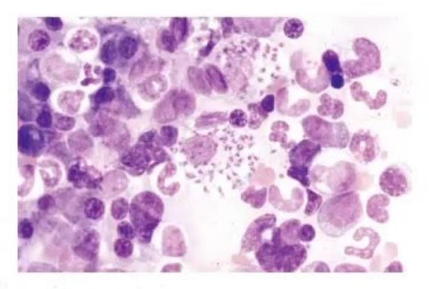
- See the smear and give the finding with form of parasite.
- Name the culture media used for diagnosis.
- Drugs used to treat resistant cases of this disease.
Answer
-
Smear finding & parasite form:
- Leishman-Donovan (LD) bodies — Amastigote form of Leishmania donovani (intracellular non-flagellated round-to-oval bodies with a nucleus and rod-shaped kinetoplast within reticuloendothelial macrophages).
-
Culture media:
- NNN medium (Novy-MacNeal-Nicolle medium) — shows flagellated promastigote forms.
-
Drugs used to treat resistant cases / Kala-azar:
- Liposomal Amphotericin B (Drug of choice)
- Miltefosine (Oral)
- Paromomycin (Aminosidine)
- Pentamidine isethionate
- Recombinant IFN-
as an adjunct - Adjunct splenectomy in severe refractory hypersplenism
MNJ23-003
Name direct and indirect evidences for laboratory diagnosis of Kala-azar (2 each).
Answer
-
Direct evidences (demonstration of parasite):
- Microscopic visualization of LD bodies (amastigotes) in bone marrow, splenic, liver, or lymph node aspirates.
- Culture of promastigotes on Novy-MacNeal-Nicolle (NNN) medium.
- Leishmania DNA detection by Polymerase Chain Reaction (PCR).
-
Indirect evidences (serology / non-specific tests):
- rK39 antigen-based rapid immunochromatographic dipstick test / ELISA
- Aldehyde test of Napier (formol-gel test; positive after 3 months of illness due to hypergammaglobulinemia) and Chopra's antimony test
- Peripheral blood examination showing pancytopenia (anemia, leukopenia, thrombocytopenia) with reversal of A:G ratio.
MNJ23-004
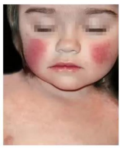
Study this picture of a 7-month-old infant who developed a rash during the declining phase of fever starting with the cheeks:
- What is the most probable diagnosis?
- What is the causative organism?
- Name two situations where infection with this organism may be life-threatening.
Answer
-
Most probable diagnosis:
- Erythema infectiosum (Fifth disease / "Slapped cheek" syndrome)
-
Causative organism:
- Parvovirus B19 (Single-stranded DNA virus)
-
Two life-threatening situations:
- Transient Aplastic Crisis (TAC) in patients with underlying chronic hemolytic anemias (e.g., Sickle Cell Disease, Hereditary Spherocytosis, Thalassemia, Autoimmune Hemolytic Anemia).
- Non-immune Hydrops Fetalis / intrauterine fetal demise during maternal infection in pregnancy (due to severe fetal anemia and myocardial failure).
- Pure Red Cell Aplasia / chronic marrow failure in immunocompromised individuals (HIV, chemotherapy, SCID).
MNJ23-005
- Define TB contact and give its preventive treatment.
- Define treatment failure as per RNTCP (NTEP).
- Treatment for BCG adenitis.
Answer
-
TB Contact & Treatment:
- Definition: Any child living in a household with an adult or family member who has active pulmonary TB or was treated with ATT in the past 2 years.
- Preventive Treatment (TPT): After ruling out active TB (normal CXR and no symptoms):
- Isoniazid (INH) at
daily for 6 months (6H), OR - 3 months of daily Isoniazid + Rifampicin (3HR), OR
- 3 months of weekly Rifapentine + Isoniazid (3HP).
- Isoniazid (INH) at
-
Treatment failure (RNTCP / NTEP definition):
- A tuberculosis patient whose biological specimen is smear- or culture-positive at the end of the intensive phase / at 12 weeks of treatment or at the end of treatment despite full adherence, or who becomes smear/culture-positive again during continuation phase.
-
Treatment for BCG adenitis:
- Non-suppurative BCG adenitis: Conservative management / reassurance; no active intervention or ATT is required as most resolve spontaneously within weeks to months.
- Suppurative BCG adenitis: Needle aspiration (avoids sinus formation); avoid surgical incision and drainage.
MNJ23-006
- Define malarial treatment failure / drug resistance.
- What is premunition?
Answer
-
Malarial treatment failure / Drug resistance (WHO Classification):
- Early Treatment Failure (ETF):
- Development of danger signs or severe malaria on Days 1, 2, or 3 with parasitemia;
- Parasitemia on Day 2 higher than on Day 0, irrespective of temperature;
- Parasitemia on Day 3 with axillary temp
; - Parasitemia on Day 3
of Day 0 count.
- Late Clinical Failure (LCF):
- Development of danger signs / severe malaria in presence of parasitemia on any day between Day 4 and Day 28 (or Day 42) in patients not meeting ETF criteria;
- Parasitemia on any day between Day 4 and Day 28 (or Day 42) with axillary temp
(or history of fever).
- Late Parasitological Failure (LPF):
- Presence of parasitemia on any day between Day 7 and Day 28 (or Day 42) with axillary temp
in patients not meeting ETF or LCF criteria.
- Presence of parasitemia on any day between Day 7 and Day 28 (or Day 42) with axillary temp
- Adequate Clinical and Parasitological Response (ACPR):
- Absence of parasitemia on Day 28 (or Day 42) without previously meeting criteria for ETF, LCF, or LPF.
- Early Treatment Failure (ETF):
-
Premunition:
- A state of acquired partial non-sterilizing immunity seen in individuals living in malaria-endemic areas. Low-level asymptomatic parasitemia persists, suppressing inflammatory cytokine storm and protecting against severe life-threatening disease and death upon subsequent reinfection.
MNJ23-007
- Name the drugs used for chronic Hepatitis B infection.
- Favorable predictors for treatment response in chronic HBV.
- What are the end points for therapy?
Answer
-
Drugs used for chronic Hepatitis B:
- Nucleos(t)ide Analogues: Entecavir (ETV), Tenofovir Disoproxil Fumarate (TDF), Tenofovir Alafenamide (TAF), Lamivudine, Adefovir.
- Immunomodulators: Pegylated Interferon alfa-2a (PEG-IFN-
) / Interferon alfa-2b.
-
Favorable predictors of treatment response:
- High baseline serum ALT/SGPT levels (
) - Low baseline serum HBV DNA levels (
) - Higher histological activity score on liver biopsy
- Older age / acquired later in childhood
- HBV Genotypes A and B (respond better to interferon than C and D)
- High baseline serum ALT/SGPT levels (
-
Endpoints of therapy:
- Ideal/Definitive endpoint: Loss of HBsAg with or without development of anti-HBs seroconversion.
- Satisfactory intermediate endpoints:
- Undetectable serum HBV DNA by sensitive PCR
- In HBeAg-positive patients: Loss of HBeAg with seroconversion to anti-HBe
- Normalization of serum ALT levels
- Histological improvement (reduction of necroinflammation and fibrosis)
MNJ23-008
- Management plan for acute HBV contact (intimate contacts / percutaneous exposure) in children.
- Management plan for Hepatitis B prophylaxis in a patient with nephrotic syndrome on hemodialysis.
Answer
-
Management of acute HBV contact (unvaccinated/non-immune):
- Administer Hepatitis B Immune Globulin (HBIG) at
IM immediately (ideally within 24 hours, maximum 7 days). - Simultaneously initiate the complete Hepatitis B vaccine series (
/ pediatric dose IM) at 0, 1, and 6 months at a different anatomical site.
- Administer Hepatitis B Immune Globulin (HBIG) at
-
Hepatitis B prophylaxis in hemodialysis / immunocompromised patients:
- Administer double the standard dose (
or high-dose formulation) on a 4-dose schedule at 0, 1, 2, and 6 months. - No HBIG required unless acute parenteral exposure.
- Check annual anti-HBs titers; administer booster if anti-HBs falls
.
- Administer double the standard dose (
MNJ23-009
- Give criteria to define polio eradication in an area.
- What is the incidence of Vaccine-Associated Paralytic Poliomyelitis (VAPP) after the 1st dose?
- What is the effectiveness of IPV vaccine after 3 doses (8 weeks apart)?
- What is the minimal age after which IPV can be given and why?
Answer
-
Criteria to define polio eradication:
- Zero cases of paralytic poliomyelitis caused by indigenous wild poliovirus for at least 3 consecutive years.
- Routine immunization coverage maintained at
. - High-quality, certified acute flaccid paralysis (AFP surveillance) system meeting WHO benchmarks (non-polio AFP rate
per 100,000 children years and adequate stool specimen collection ). - Poliovirus laboratory containment.
-
Incidence of VAPP after 1st dose:
- Approximately 1 case per 1.4 to 2.8 million first doses of OPV (highest risk with the first dose).
-
Effectiveness of IPV after 3 doses:
- Near 100% (99–100%) protective seroconversion against all three poliovirus serotypes (Types 1, 2, and 3).
-
Minimal age for IPV and rationale:
- 6 to 8 weeks of age (routinely given at 6 and 14 weeks as fractional IPV / fIPV or 6, 10, 14 weeks as full-dose IPV).
- Rationale: High levels of transplacentally acquired maternal anti-polio antibodies in the first few weeks can interfere with the infant's active immune response to injectable IPV; giving it after 6–8 weeks allows maternal antibodies to decline sufficiently.
MNJ23-010
- Give the WHO definition of a confirmed case of Japanese encephalitis.
- Name the vector that transmits the disease in India (MC agent/agents).
- Name the sign which if present on MRI clinches the diagnosis.
Answer
-
WHO definition of a confirmed case of Japanese Encephalitis (JE):
- A suspected case of Acute Encephalitis Syndrome (AES) with laboratory confirmation by ANY of:
- Presence of JE-specific IgM antibodies in CSF (or serum) by IgM capture ELISA;
- A
-fold rise in JE-specific antibody titers in paired acute and convalescent sera; - Detection of JE virus genome in serum, CSF, or brain tissue by RT-PCR;
- Isolation of JE virus from CSF, serum, or brain tissue.
- A suspected case of Acute Encephalitis Syndrome (AES) with laboratory confirmation by ANY of:
-
Vector transmitting JE in India:
- Culex tritaeniorhynchus and Culex vishnui group mosquitoes (breed in irrigated rice paddies).
-
MRI findings:
- Bilateral symmetric hyperintensity on T2/FLAIR in the thalami (classic), basal ganglia, and midbrain substantia nigra ("Eye of the Tiger" / "Double Panda" sign in midbrain/pons).
MNJ23-011
A 12-year-old male child presented with complaints of worsening school performance, altered behavior, and sleep pattern with some brief abnormal movements. Physical examination is normal. No past history of any medical illness except a febrile rash at the age of 7–8 months:
- What is the most probable diagnosis?
- Give its clinical stages.
- Diagnostic modalities.
- Available specific treatment, if any.
Answer
-
Most probable diagnosis:
- Subacute Sclerosing Panencephalitis (SSPE) (slow virus encephalitis caused by mutant, defective Measles virus).
-
Clinical stages of Jabbour:
- Stage I (Behavioral & Cognitive): Subtle changes in personality, declining school performance, mood changes, irritability, lethargy.
- Stage II (Myoclonic stage): Stereotypic massive myoclonic jerks/spasms, drop attacks, choreoathetosis, visual deficits, dementia.
- Stage III (Extrapyramidal & Pyramidal stage): Rigidity, spasticity, decerebrate/decorticate posturing, autonomic instability, unresponsiveness.
- Stage IV (Terminal/Vegetative stage): Akinetic mutism, loss of cortical function, coma, flaccidity, autonomic failure, death.
-
Diagnostic modalities:
- CSF and Serum anti-measles IgG antibody titers: Markedly elevated (with oligoclonal bands and high CSF:serum ratio
). - Electroencephalography (EEG): Pathognomonic periodic, high-amplitude, slow-wave complexes (Radermecker complexes) occurring synchronously every 4–10 seconds against a suppressed background.
- Neuroimaging (MRI/CT): Progressive multifocal white matter T2/FLAIR hyperintensities, diffuse cortical atrophy, and ventricular dilatation.
- CSF and Serum anti-measles IgG antibody titers: Markedly elevated (with oligoclonal bands and high CSF:serum ratio
-
Available treatment:
- No curative therapy; treatment is largely palliative and slows progression:
- Oral Isoprinosine (Inosiplex) (
) - Intraventricular / Intrathecal Interferon-alpha (IFN-
) via Ommaya reservoir - Antiepileptics (Valproate, Clonazepam) for myoclonus
- Intrathecal/oral Ribavirin, IVIG, supportive nursing care.
- Oral Isoprinosine (Inosiplex) (
- No curative therapy; treatment is largely palliative and slows progression:
MNJ23-012
A 2-year-old male child presented to emergency department with complaints of low-grade fever for the last 2–3 weeks, headache and vomiting for the last 2 weeks, spillage of milk while drinking for the last 1 week, and altered sensorium with focal seizures of left half of body at the time of admission. The only past history that is found is a follow-up case of cyanotic CHD (operated at 3 months of age), rest everything is normal:
- What is the most probable diagnosis?
- What is the etiological agent expected?
- Diagnostic modalities.
- Initial medical treatment.
- Indications for surgical interventions.
Answer
-
Most probable diagnosis:
- Brain Abscess (Right cerebral hemisphere, causing left-sided focal seizures / left upper motor neuron facial palsy).
-
Expected etiological agents:
- Streptococcus viridans, Streptococcus milleri / anginosus group, Staphylococcus aureus, anaerobic streptococci, Bacteroides species (polymicrobial in cyanotic CHD due to right-to-left shunt bypassing pulmonary phagocytic filtration).
-
Diagnostic modalities:
- Contrast-Enhanced CT (CECT) Head / MRI Brain with DWI: Shows a ring-enhancing lesion with surrounding vasogenic edema and restricted central diffusion on DWI.
- Blood cultures.
- Lumbar puncture is CONTRAINDICATED due to high risk of uncal/transtentorial herniation.
-
Initial medical treatment:
- Empirical IV Antibiotics (for 4–6 weeks):
- Third-generation Cephalosporin (Ceftriaxone / Cefotaxime) + Metronidazole + Vancomycin.
- Antiepileptics: IV Phenytoin or Levetiracetam for seizure control.
- Anti-edema measures: IV Mannitol / 3% Hypertonic Saline, Dexamethasone if severe mass effect with impending herniation.
- Empirical IV Antibiotics (for 4–6 weeks):
-
Indications for surgical intervention (Burr hole aspiration / excision):
- Abscess diameter
- Significant mass effect, midline shift, or impending herniation
- Threatened rupture into ventricles
- Gas-containing abscess or fungal etiology
- Failure to improve / expanding lesion after 2–3 weeks of targeted medical therapy
- Readily accessible superficial unilocular lesion.
- Abscess diameter
MNJ23-013
A pregnant woman at 26 weeks' gestation does not know her varicella immunization status. Her 5-year-old son has been diagnosed with varicella a day prior. What will you advise the mother?
Answer
-
Immediate Serological Testing:
- Immediately test maternal serum for Varicella-Zoster Virus (VZV) IgG antibodies.
- If results are available within 72–96 hours:
- If VZV IgG positive: Mother is immune; reassure her, no further prophylaxis is needed.
- If VZV IgG negative (non-immune) or results unavailable within 96 hours:
- Administer Varicella-Zoster Immune Globulin (VZIG) at a dose of
IM (minimum 125 units, maximum 625 units) as soon as possible, ideally within 96 hours (acceptable up to 10 days post-exposure).
- Administer Varicella-Zoster Immune Globulin (VZIG) at a dose of
-
Oral Antiviral Therapy:
- If VZIG is not available or if the pregnant woman subsequently develops fever/rash: Initiate oral Acyclovir (
5 times daily for 7 days) or IV Acyclovir ( 8-hourly for varicella pneumonia/severe disease). - Note: Live attenuated Varicella vaccine is strictly contraindicated during pregnancy.
- If VZIG is not available or if the pregnant woman subsequently develops fever/rash: Initiate oral Acyclovir (
MNJ23-014
Plan antiretroviral therapy regimen for a 3-year-old child who has recently been diagnosed with HIV infection with complaints of failure to thrive, hepatomegaly, thrombocytopenia, CD4 <25%, and viral load
- Write the drug regimen and dosages.
- What are the tests to be ordered prior to initiation and after 15–30 days of treatment?
Answer
-
Antiretroviral Therapy (ART) Regimen:
- Preferred First-line Regimen (WHO / NACO Guidelines):
- ABC + 3TC + DTG (Abacavir + Lamivudine + Dolutegravir)
- Alternative / Protease Inhibitor Regimen: AZT + 3TC + LPV/r (Zidovudine + Lamivudine + Lopinavir/ritonavir)
- Dosages:
- Zidovudine (AZT):
PO BID (or BID) - Lamivudine (3TC):
PO BID - Abacavir (ABC):
PO BID - Dolutegravir (DTG):
PO once daily (weight-band based) - Lopinavir/ritonavir (LPV/r):
PO BID - Also initiate Cotrimoxazole Preventive Therapy (CPT).
- Zidovudine (AZT):
- Preferred First-line Regimen (WHO / NACO Guidelines):
-
Laboratory tests to order:
- Prior to initiation (Baseline):
- Complete Blood Count (Hb, TLC, DLC, Platelet count) — especially check for anemia/thrombocytopenia before ZDV
- Baseline CD4 count / CD4 percentage and plasma HIV viral load
- Renal function tests (BUN, Serum Creatinine, electrolytes)
- Liver function tests (SGOT/AST, SGPT/ALT, Bilirubin)
- Fasting lipid profile & blood glucose (for PI regimens)
- Screening for active TB, Hepatitis B, and Hepatitis C
- HLA-B*5701 testing (if initiating Abacavir)
- At 15–30 days post-initiation:
- Repeat CBC (monitor for ZDV-induced bone marrow suppression/anemia)
- Repeat LFT and RFT (monitor for drug-induced hepatotoxicity / nephrotoxicity)
- Clinical assessment for drug adherence, adverse effects, and Immune Reconstitution Inflammatory Syndrome (IRIS).
- Prior to initiation (Baseline):
MNJ23-015
Mark True/False regarding osteomyelitis statements:
- Staph aureus is the most common organism.
- The ESR and CRP are usually elevated.
- Blood cultures are positive in over 75% of all cases of osteomyelitis.
- Plain films of bone may be normal in the first 1–2 weeks of the course.
- Treatment usually requires a total time of antibiotics (IV and PO) of 4–6 weeks.
Answer
- True (Staphylococcus aureus is responsible for 70–80% of pediatric acute hematogenous osteomyelitis).
- True (CRP rises within 6–8 hours and ESR peaks later, serving as sensitive markers of disease activity and treatment response).
- False (Blood cultures are positive in only 30–50% of acute osteomyelitis cases, not >75%).
- True (Early radiographic signs like periosteal reaction and lytic bone destruction take 10–14 days to become visible on plain X-rays).
- True (Standard treatment consists of 4–6 weeks of targeted antimicrobial therapy, with transition to high-dose oral therapy once clinically improved and CRP declines).
MNJ23-016
A 15-year-old female presents with fever for past 24 hours. Fever is high grade associated with sweating. For past 2 hours patient is complaining of ghabrahat, chakkar, headache, vomiting. On examination: toxic looking, drowsy with Glasgow Coma Scale of 10. Blood pressure systolic: 70 mmHg, pulse rate: 140/min, capillary refill time: 4 seconds, RR: 30/min. There is rash (petechial/purpuric) over abdomen and limbs:
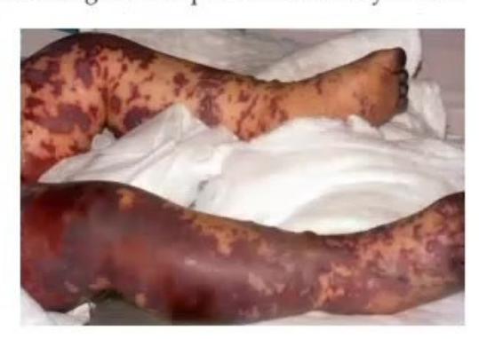
- Probable diagnosis of this given condition.
- First step in management other than airway and breathing.
- What class of antibiotic you would like to give to this patient?
- What drug you would like to give in dopamine-refractory shock?
Answer
-
Probable diagnosis:
- Meningococcemia (Fulminant Neisseria meningitidis sepsis / Purpura fulminans / Waterhouse-Friderichsen syndrome).
-
First step in management (Circulation):
- Aggressive fluid resuscitation: Establish rapid IV/IO access and infuse isotonic crystalloid boluses (
over 10–15 minutes, repeated up to ) while monitoring for signs of fluid overload.
- Aggressive fluid resuscitation: Establish rapid IV/IO access and infuse isotonic crystalloid boluses (
-
Class of antibiotics:
- Third-generation Cephalosporins: IV Ceftriaxone (
) or IV Cefotaxime.
- Third-generation Cephalosporins: IV Ceftriaxone (
-
Drug for dopamine-refractory shock:
- Norepinephrine (for warm shock/vasodilatory shock) or Epinephrine (for cold shock).
- IV Hydrocortisone (for suspected absolute/relative adrenal insufficiency in catecholamine-resistant septic shock / Waterhouse-Friderichsen syndrome).
MNJ23-017
A 9-day-old male child Raju was brought to your clinic with complaints of difficulty in feeding, crying excessively, and seizures/spasms. Parents of baby were laborers by occupation. Mother had not received any ANC care. Baby was born at home, had moderate cry, and was well till day 8. AF: at level, sutures: normal. Tone: increased in all 4 limbs with neck retraction, intermittent spasms worsened with touch/stimulus:
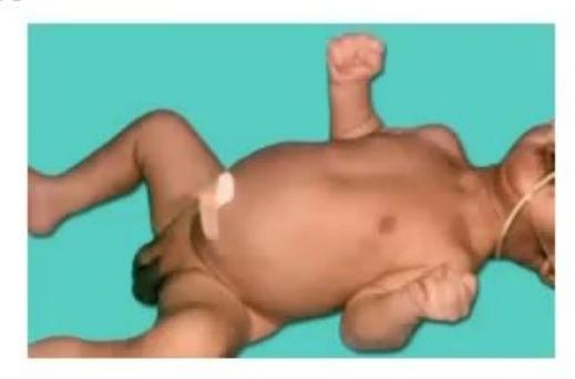
- What is the diagnosis of this baby?
- What is the treatment?
- What is the usual cause of death in this condition?
- What are the various clinical types of this disease?
Answer
-
Diagnosis:
- Neonatal Tetanus (Tetanus neonatorum / Clostridium tetani infection).
-
Treatment:
- Neutralization of toxin: Human Tetanus Immunoglobulin (HTIG)
IM single dose (or Equine Tetanus Antitoxin ). - Eradicate vegetative bacteria: IV/oral Metronidazole (
divided 6-hourly) for 10–14 days (preferred over penicillin). - Control muscle spasms: IV Diazepam (
), Midazolam infusion, Baclofen, and neuromuscular blockade with mechanical ventilation if refractory. - Supportive care: Minimal handling in a dark, quiet environment, umbilical wound debridement, tube feeding / TPN, airway protection.
- Active immunization: Administer DTwP/Td vaccine at recovery (tetanus infection does NOT confer natural immunity).
- Neutralization of toxin: Human Tetanus Immunoglobulin (HTIG)
-
Usual cause of death:
- Respiratory failure / Apnea secondary to severe laryngospasm, chest wall muscle rigidity, aspiration pneumonia, and autonomic nervous system dysfunction (cardiovascular collapse).
-
Clinical types of tetanus:
-
- Generalized tetanus
-
- Neonatal tetanus
-
- Localized tetanus
-
- Cephalic tetanus
-
MNJ23-018
A 10-year-old child Rohan came to you with complaints of fever for 5 days, bodyache, and decreased oral intake. Mother noticed small water-filled lesions on trunk and back:
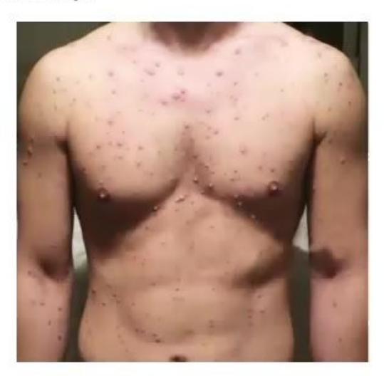
- What is the diagnosis of this condition?
- Name three complications that can occur in this child.
- What is the period of infectivity?
Answer
-
Diagnosis:
- Varicella (Chickenpox) — classic "dewdrops on a rose petal" pleomorphic vesicular lesions.
-
Three complications:
- Secondary bacterial skin and soft tissue infection (Staphylococcus aureus, Group A Streptococcus)
- Acute cerebellar ataxia / Varicella encephalitis
- Varicella pneumonia
- Acute Disseminated Encephalomyelitis (ADEM) / Reye's syndrome
- Purpura fulminans / hemorrhagic varicella
-
Period of infectivity:
- From 1 to 2 days before the onset of rash until all lesions have crusted over (usually 5–7 days after the appearance of the rash).
MNJ23-019
A 2-year-old girl Neha presents with severe pruritus, especially at night and after taking a bath with warm water. Pruritus is most significant in web spaces of hands and toes. Her family members have similar complaints:
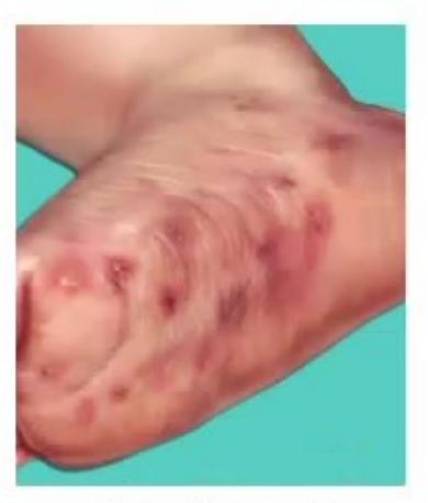
- Diagnosis of this condition.
- What is the causative organism?
- What is the topical treatment?
- What is the oral treatment?
Answer
-
Diagnosis:
- Scabies (Infestation characterized by intensely pruritic burrows, papules, and vesicles).
-
Causative organism:
- Sarcoptes scabiei var. hominis (Itch mite).
-
Topical treatment:
- 5% Permethrin dermal cream: Applied from neck to toe (including scalp, face, and neck in infants), left on for 8–14 hours, then washed off. Repeat after 7–14 days.
- Alternatives: 1% Gamma benzene hexachloride (Lindane - avoid in infants), 10% Crotamiton cream, 5–10% precipitated sulfur ointment in petrolatum (safe in neonates).
-
Oral treatment:
- Oral Ivermectin:
single dose with food; repeat dose after 1–2 weeks (indicated for crusted/Norwegian scabies, institutional outbreaks, or failure of topicals; approved for children ). - Note: All household members and close contacts must be treated simultaneously.
- Oral Ivermectin:
MNJ23-020
A healthcare worker is reported with percutaneous scratch with a solid needle contaminated with blood of a patient with primary HIV infection and a low CD4 count:
- What is the recommended regimen of Post-Exposure Prophylaxis (PEP)? Include drugs, dose, and duration of treatment.
- What is the testing schedule for HIV counseling and testing following exposure?
Answer
-
Post-Exposure Prophylaxis (PEP) Regimen:
- Recommended 3-drug regimen:
- Tenofovir Disoproxil Fumarate (TDF)
+ Lamivudine (3TC) (or Emtricitabine ) + Dolutegravir (DTG) once daily. - Alternative: TDF + 3TC + Lopinavir/ritonavir (
BID) or Atazanavir/ritonavir. - Timing: Initiate as soon as possible, ideally within 2 hours (maximum within 72 hours).
- Duration: Complete 28 days (4 weeks) of therapy.
- Tenofovir Disoproxil Fumarate (TDF)
- Recommended 3-drug regimen:
-
HIV Testing Schedule:
- Baseline: At the time of exposure (to rule out pre-existing HIV infection)
- 6 weeks post-exposure
- 12 weeks (3 months) post-exposure
- 6 months post-exposure (especially if exposed to co-infected HCV patient).
MNJ23-021
A 4-year-old boy Amir came with fever for 10 days, cough, lower back pain, and a skin lesion in the armpit area shown below:
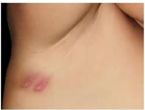
- Describe the lesion given in this image.
- What is the diagnosis of this condition?
- What is the causative organism?
- This disease is transmitted by which vector?
- Write down any 3 complications.
- Write down two treatments available.
Answer
-
Description of lesion:
- Eschar — a painless, round-to-oval ulcer with a central black necrotic crust surrounded by an erythematous indurated halo and regional lymphadenopathy (resembling a cigarette burn).
-
Diagnosis:
- Scrub Typhus (Tsutsugamushi disease).
-
Causative organism:
- Orientia tsutsugamushi (obligate intracellular bacterium).
-
Transmission vector:
- Bite of the larval stage (chigger) of trombiculid mites (Leptotrombidium deliense).
-
Three complications:
- Meningoencephalitis / acute cerebellitis
- Acute Respiratory Distress Syndrome (ARDS) / pneumonitis
- Acute Kidney Injury (AKI)
- Myocarditis / circulatory collapse / shock
- Disseminated Intravascular Coagulation (DIC)
-
Two available treatments:
- Doxycycline:
PO divided BID (or BID) for 7–10 days. - Azithromycin:
PO once daily for 5 days (drug of choice in young children and pregnant women).
- Doxycycline:
MNJ23-022
A 10-year-old boy Karzan Khan presents with fatigue, fever, and malaise for 8 days. On examination, he has generalized lymphadenopathy, splenomegaly, and hepatomegaly. His throat examination and peripheral blood smear are shown below:
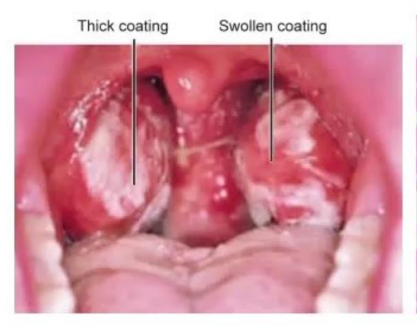
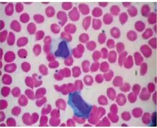
- Describe the main features in each image.
- What is the diagnosis of this condition?
- Write 3 complications that may occur.
- What medication is contraindicated in this disease?
- What is the treatment available?
Answer
-
Description of images:
- Image 1 (Throat): Exudative tonsillopharyngitis with marked tonsillar enlargement, pharyngeal erythema, palatal petechiae, and thick grayish-white pseudomembrane.
- Image 2 (Peripheral smear): Atypical reactive lymphocytes (Downey cells) — large pleomorphic lymphocytes with abundant basophilic cytoplasm scalloping around adjacent red blood cells.
-
Diagnosis:
- Infectious Mononucleosis ("Glandular fever" caused by Epstein-Barr Virus / EBV).
-
Three complications:
- Splenic rupture (spontaneous or traumatic)
- Upper airway obstruction due to massive tonsillar hypertrophy
- Autoimmune hemolytic anemia (AIHA) / severe thrombocytopenia
- Neurological complications: Guillain-Barré syndrome, meningoencephalitis, Bell's palsy
- Hepatitis / fulminant hepatic necrosis.
-
Contraindicated medication:
- Ampicillin / Amoxicillin (and aminopenicillins), which trigger a characteristic pruritic maculopapular rash in
of patients.
- Ampicillin / Amoxicillin (and aminopenicillins), which trigger a characteristic pruritic maculopapular rash in
-
Treatment:
- Supportive: Rest, hydration, antipyretics/analgesics.
- Strict avoidance of contact sports / strenuous exercise for at least 3–4 weeks to prevent splenic rupture.
- Systemic corticosteroids (Prednisolone): Indicated for impending airway obstruction, severe thrombocytopenia, or autoimmune hemolytic anemia.
MNJ23-023
A 12-year-old boy Dharmender developed throat pain two days back. He returned with his elder sister from his boarding school in Tripura to Kolkata. His temperature was 36°C. Heart rate and blood pressure were normal. He had also developed a petechial/purpuric rash, and his blood/CSF grew the following:
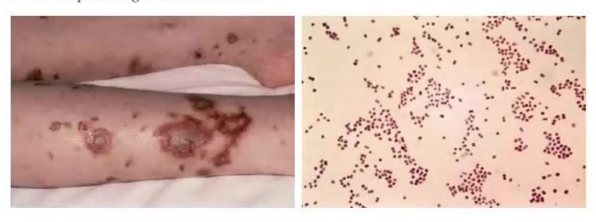
- Diagnosis of this condition.
- Drug treatment and dose.
- What treatment will you advise for his sister?
- How to protect against this condition?
Answer
-
Diagnosis:
- Meningococcal Infection / Meningococcemia (Neisseria meningitidis — Gram-negative diplococci with adjacent flat surfaces / bean-shaped).
-
Drug treatment and dose:
- IV Ceftriaxone:
IV once daily (or in 2 divided doses, max ) for 7 days. - Alternative: IV Cefotaxime (
in 4 divided doses) or IV Crystalline Penicillin G ( divided 4-hourly).
- IV Ceftriaxone:
-
Chemoprophylaxis for close contact (sister):
- Rifampicin:
(max ) PO BID for 2 days (4 doses), OR - Ceftriaxone:
IM single dose ( if years), OR - Ciprofloxacin:
PO single dose (in adults/older adolescents).
- Rifampicin:
-
Protection / Prevention:
- Meningococcal Conjugate Vaccine (MenACWY / MenB): Quadrivalent conjugate vaccine containing serogroups A, C, W-135, and Y (and Serogroup B recombinant vaccine).
MNJ23-024
Study the image of a 10-year-old child:
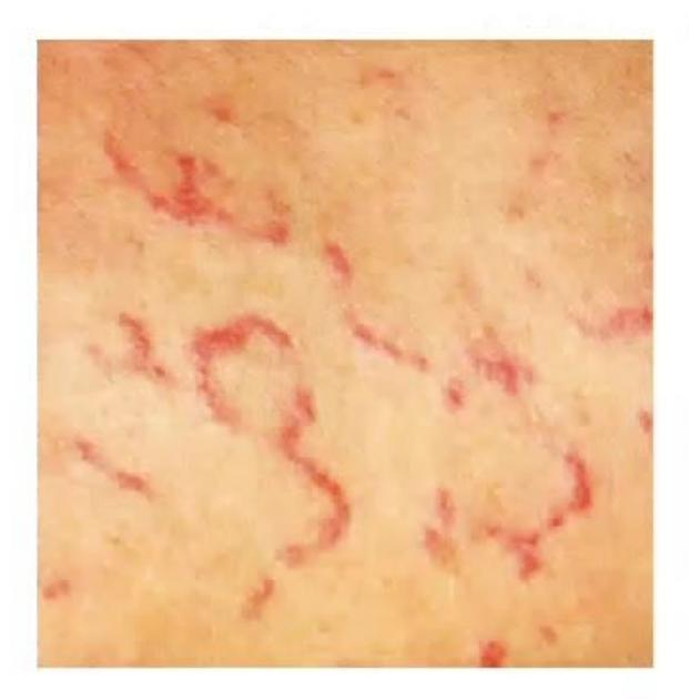
- Identify the rash seen in the image.
- Name the condition in which it is seen.
Answer
-
Identification of rash:
- Erythema Marginatum — an evanescent, non-pruritic, erythematous maculopapular rash with sharp, raised outer borders and central clearing (serpiginous / polycyclic appearance), commonly distributed on the trunk and proximal limbs, sparing the face.
-
Condition:
- Acute Rheumatic Fever (ARF) (one of the Major Jones criteria).
MNJ23-025
Two children A and B came to your clinic with complaints of rash over the diaper/perianal area. Child A is 20 days old and always wears diapers, but the mother of Child B uses diapers only at night. Give the diagnosis and distinguishing features of A and B:
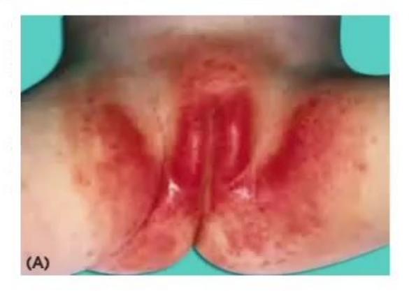
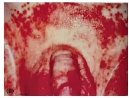
Answer
-
Child A (Image A):
- Diagnosis: Irritant Diaper Dermatitis (Contact / Ammoniacal diaper dermatitis).
- Characteristic features: Involves convex surfaces of the buttocks, lower abdomen, and perineum; characteristically spares the intertriginous skin folds/creases.
- Management: Frequent diaper changes, barrier creams containing Zinc Oxide / petroleum jelly, aeration.
-
Child B (Image B):
- Diagnosis: Candidal Diaper Dermatitis (Candida albicans infection).
- Characteristic features: Beefy-red confluent erythema with elevated margins, involvement of deep intertriginous skin folds/creases, and distinct pinpoint satellite papules and pustules.
- Management: Topical antifungal cream (Clotrimazole, Miconazole, or Nystatin) applied 2–3 times daily for 7–10 days.
MNJ23-026
A 9-year-old girl Kareena Sharma on long-term steroids and mycophenolate mofetil presents with acute onset cough, breathlessness, and tachypnea. Evaluation reveals normal temperature,
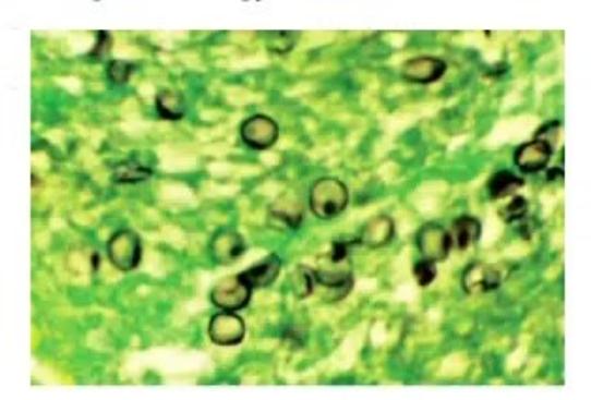
- What is the most likely diagnosis?
- What is the likely finding on chest X-ray?
- Which diagnostic modality is best for confirmation?
- Name 2 stains for confirming the etiology.
Answer
-
Most likely diagnosis:
- Pneumocystis jirovecii Pneumonia (PJP / PCP) (opportunistic fungal infection in an immunocompromised host).
-
Chest X-ray findings:
- Bilateral diffuse perihilar symmetrical interstitial/alveolar ground-glass infiltrates with classic apical sparing.
-
Best diagnostic modality:
- Bronchoalveolar Lavage (BAL) with microscopic staining or PCR (Induced sputum is an alternative).
-
Stains for confirmation:
- Grocott-Gomori Methenamine Silver (GMS) stain (stains cyst walls black/brown)
- Toluidine Blue O stain
- Direct Fluorescent Monoclonal Antibody (DFA) stain
- Giemsa / Calcofluor white stain.
MNJ23-027
A 9-year-old child Akshay came to you with lesions over the face, eyes, and lips that progressed over the last 3 days. Before this, he was on antibiotic/anticonvulsant treatment for cough/cold and suddenly developed these lesions:
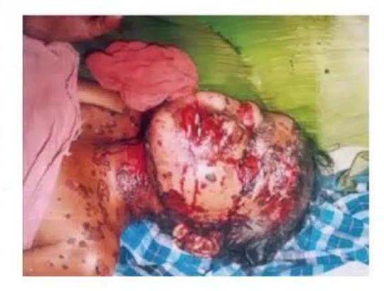
- Diagnosis of this condition.
- Describe the lesions shown in the figure.
- Causes: Two infections / two drugs.
- Management plan of this condition.
- Name two complications.
Answer
-
Diagnosis:
- Stevens-Johnson Syndrome (SJS) / Toxic Epidermal Necrolysis (TEN) overlap.
-
Description of lesions:
- Atypical targetoid maculopapular lesions with dusky purpuric centers, flaccid bullae, epidermal detachment, and extensive hemorrhagic crusting/erosions involving
mucous membranes (vermilion border of lips, oral mucosa, conjunctivae, genital mucosa).
- Atypical targetoid maculopapular lesions with dusky purpuric centers, flaccid bullae, epidermal detachment, and extensive hemorrhagic crusting/erosions involving
-
Causes:
- Infections (2): Mycoplasma pneumoniae, Herpes Simplex Virus (HSV), Cytomegalovirus.
- Drugs (2): Sulfonamides (Cotrimoxazole), Antiepileptics (Carbamazepine, Phenytoin, Lamotrigine, Phenobarbital), NSAIDs, Aminopenicillins.
-
Management plan:
- Immediate discontinuation of all potential offending/suspect drugs.
- Transfer to a burn intensive care unit or specialized pediatric ICU.
- Supportive care: Fluid and electrolyte maintenance, temperature regulation, nutritional support, meticulous non-adherent wound dressing.
- Ophthalmologic care: Lubricating eye drops, topical antibiotic/steroid drops, glass rodding / amniotic membrane transplantation to prevent symblepharon.
- Immunomodulatory therapy: IVIG (
), pulse Methylprednisolone, or Cyclosporine.
-
Two complications:
- Ocular: Cicatricial symblepharon, trichiasis, corneal ulceration, blindness.
- Systemic: Sepsis, Acute Tubular Necrosis (ATN) / renal failure, respiratory tract sloughing/failure, esophageal/urethral strictures.
MNJ23-028
A 4-year-old child came to you with complaints of fever with rash on face shown below:
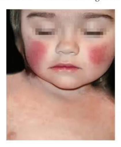
- What is the diagnosis?
- What is the causative organism?
- Name one serious hematological complication in this disease.
- Name one orthopedic complication.
- Name one cause for intrauterine fetal demise.
- What treatment is recommended for severe hematological complications?
Answer
-
Diagnosis:
- Erythema Infectiosum (Fifth disease / Slapped cheek rash).
-
Causative organism:
- Parvovirus B19.
-
Hematological complication:
- Transient Aplastic Crisis (TAC) / Pure Red Cell Aplasia.
-
Orthopedic complication:
- Post-infectious symmetric polyarthropathy / polyarthritis (involves small joints of hands, wrists, knees, ankles).
-
Cause of intrauterine fetal demise:
- Non-immune Hydrops Fetalis (due to severe viral tropism for fetal erythroid progenitor cells leading to severe fetal anemia, high-output congestive heart failure, and hydrops).
-
Treatment for severe hematological complications:
- Intravenous Immunoglobulin (IVIG) (
for 5 days or for 2 days) and Packed Red Blood Cell (PRBC) transfusion.
- Intravenous Immunoglobulin (IVIG) (
MNJ23-029
A 6-year-old female child Rani came to your clinic with her mother, concerned about red lesions on her anterior shin area. The mother reports that these lesions are tender:
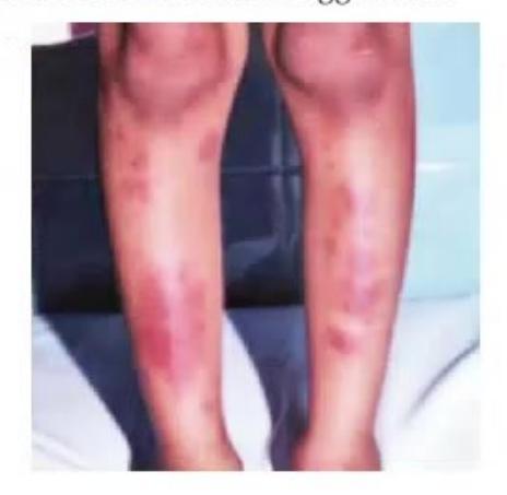
- What is the diagnosis?
- What 2 common infections and 2 drugs can trigger this?
- What 2 non-infectious systemic disorders can trigger this?
Answer
-
Diagnosis:
- Erythema Nodosum (Septal panniculitis characterized by tender, non-ulcerative, erythematous subcutaneous nodules over the anterior tibial surfaces).
-
Common Triggers:
- Infections (2): Group A
-hemolytic Streptococcus (pharyngitis), Mycobacterium tuberculosis (Primary TB), Mycoplasma pneumoniae, Yersinia, Histoplasmosis. - Drugs (2): Sulfonamides, Oral Contraceptives, Penicillins, Bromides/Iodides.
- Infections (2): Group A
-
Non-infectious systemic disorders (2):
- Sarcoidosis (Löfgren's syndrome)
- Inflammatory Bowel Disease (Crohn's disease, Ulcerative Colitis)
- Behçet's disease
- Malignancies (Lymphomas/Leukemia).
MNJ23-030
Study the microscopic image:
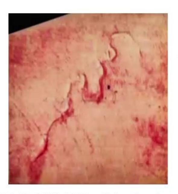
- Identify this parasite.
- What is the route of entry of this organism?
- Name 2 conditions that are high-risk for severe infection.
- What hematological clue will occur?
- Drug of choice.
- One severe complication.
Answer
-
Identification:
- Strongyloides stercoralis larva (Rhabditiform / Filariform larva with short buccal cavity and prominent genital primordium).
-
Route of entry:
- Transcutaneous penetration of intact skin by infective filariform larvae from contaminated soil (walking barefoot).
-
Two high-risk conditions:
- Immunosuppressive therapy (especially systemic corticosteroids)
- HTLV-1 infection
- Malnutrition / PEM
- Hematologic malignancies / Solid organ transplantation / HIV infection.
-
Hematological clue:
- Marked Peripheral Blood Eosinophilia.
-
Drug of choice:
- Oral Ivermectin:
for 1–2 days (repeated in 2 weeks; continued for in hyperinfection syndrome). Alternative: Albendazole.
- Oral Ivermectin:
-
Severe complication:
- Strongyloides Hyperinfection Syndrome / Disseminated Strongyloidiasis (with secondary Gram-negative bacterial sepsis, meningitis, and ARDS).
MNJ23-031
A 17-month-old irritable child had high-grade fever for 6 days and redness over palms and soles. She also has redness over eyes:
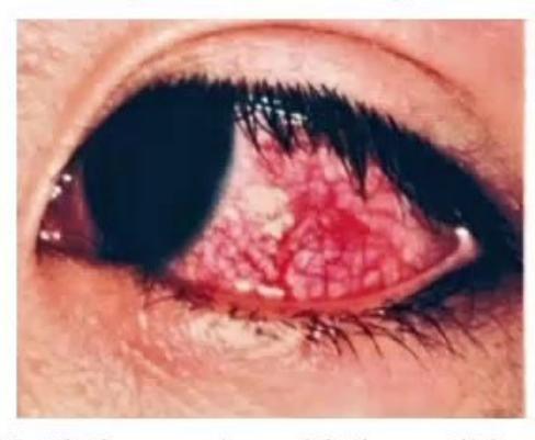
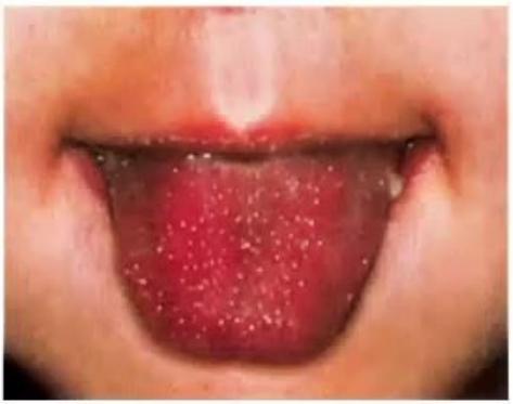
- What is the most probable diagnosis of this condition?
- What are the predictors of severity of this disease (coronary artery aneurysm formation)?
Answer
-
Most probable diagnosis:
- Kawasaki Disease (showing strawberry tongue, non-exudative bulbar conjunctival injection, and erythema of extremities).
-
Predictors of severity / Kobayashi & Harada Risk Criteria:
- Demographic: Male sex, age
or - Clinical: Prolonged fever (
), recurrence/recrudescence of fever after initial defervescence, presence of hepatomegaly or cardiomegaly - Laboratory parameters:
- Serum Albumin
(hypoalbuminemia) - Serum Sodium
(hyponatremia) - Platelet count
(early thrombocytopenia) or extreme thrombocytosis ( ) later - C-Reactive Protein (CRP)
or markedly elevated ESR - Peripheral blood absolute neutrophil count (ANC)
/ high band count - Serum ALT/SGPT
- Low hemoglobin for age.
- Serum Albumin
- Demographic: Male sex, age
MNJ23-032
Match the organism with the condition associated:
| Condition | Organism |
|---|---|
| 1. Melioidosis | a. HHV-8 |
| 2. Pontiac fever | b. Coxsackievirus B |
| 3. Oroya fever | c. Coronavirus |
| 4. Swimming pool granuloma | d. Bartonella bacilliformis |
| 5. Bornholm disease | e. Treponema pallidum |
| 6. Ecthyma gangrenosum | f. Aspergillus fumigatus |
| 7. Condyloma lata | g. Mycobacterium marinum |
| 8. Malt worker's lung | h. Pseudomonas aeruginosa |
| 9. SARS | i. Burkholderia pseudomallei |
| 10. Kaposi sarcoma | j. Legionella micdadei / pneumophila |
Answer
- Melioidosis
i. Burkholderia pseudomallei - Pontiac fever
j. Legionella micdadei / pneumophila - Oroya fever
d. Bartonella bacilliformis - Swimming pool granuloma
g. Mycobacterium marinum - Bornholm disease
b. Coxsackievirus B (Epidemic pleurodynia) - Ecthyma gangrenosum
h. Pseudomonas aeruginosa - Condyloma lata
e. Treponema pallidum (Secondary syphilis) - Malt worker's lung
f. Aspergillus fumigatus / clavatus - SARS
c. Coronavirus (SARS-CoV) - Kaposi sarcoma
a. HHV-8 (Human Herpesvirus 8 / KSHV)
MNJ23-033
HIV infection in newborn:
- Mother has HIV infection and if baby is PCR positive within 48 hours, what does it imply?
- Another baby born to an HIV-positive mother tests negative for PCR at 48 hours but PCR turns positive between 7–90 days. What does it imply?
- How early can the p24 antigen test be done?
- When do you label a newborn as HIV infected?
- After what age is HIV ELISA considered the best test for diagnosis, and what is its sensitivity and specificity?
Answer
-
PCR positive within 48 hours:
- Implies in utero (transplacental / antenatal) transmission; typically associated with higher viral load and more rapidly progressive disease.
-
PCR negative at 48 hours but positive at 7–90 days:
- Implies intrapartum (during delivery) or early postpartum (breastfeeding) transmission; associated with relatively slower disease progression.
-
p24 antigen testing:
- Can be detected by 4 to 6 weeks of age (not recommended before 1 month due to high false-negative rates and circulating maternal antibodies; HIV DNA PCR is the gold standard).
-
Labeling a newborn as HIV infected:
- Requires two positive virological tests (HIV DNA PCR, HIV RNA PCR, or viral culture) performed on separate blood samples.
-
HIV ELISA diagnostic age, sensitivity & specificity:
- Considered definitive after 18 months of age (when passively acquired maternal anti-HIV IgG antibodies have completely cleared).
- Sensitivity and specificity are both
.
MNJ23-034
- Dispose the following biomedical wastes into suitable color-coded bags/containers:
- i. Blood agar media
- ii. Used injection needle
- iii. Amikacin injection with expiry
- iv. Blood-soaked cotton swab
- v. IV set
- vi. Incinerated ash
- Identify the organism shown in the stool smear and state its medical treatment:
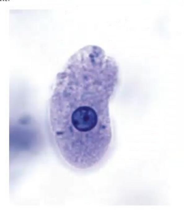

Answer
-
Biomedical Waste Segregation:
- i. Blood agar media: Yellow bag (microbiology and biotechnological waste)
- ii. Used injection needle: White translucent puncture-proof container (Sharps)
- iii. Amikacin injection with expiry: Yellow bag / Cardboard box with blue marking (expired/discarded pharmaceutical waste)
- iv. Blood-soaked cotton swab: Yellow bag (soiled anatomical waste)
- v. IV set: Red bag (recyclable contaminated plastic tubing)
- vi. Incinerated ash: Black container / landfill (disposal in secure landfill)
-
Organism Identification & Treatment:
- Organism: Entamoeba histolytica (Trophozoite demonstrating single nucleus with central karyosome and ingested erythrocytes).
- Treatment:
- Tissue amebicide: Metronidazole (
in 3 divided doses for 7–10 days) or Tinidazole ( once daily for 3 days). - Followed by a luminal amebicide: Diloxanide furoate (
for 10 days) or Paromomycin ( in 3 divided doses for 7 days) to eliminate luminal cyst carriage.
- Tissue amebicide: Metronidazole (
MNJ23-035
Study the picture of lesions in a neonate:
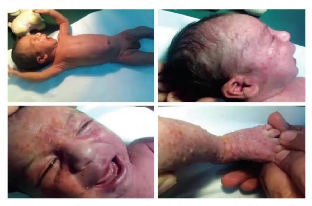
- Diagnose the condition in a 7-day-old child.
- What are the complications of this condition? (Write any 4 organ systems)
Answer
-
Diagnosis:
- Neonatal Varicella (Perinatal varicella infection acquired when mother develops varicella 5 days before to 2 days after delivery).
-
Complications across 4 organ systems:
- Hematological: Disseminated Intravascular Coagulation (DIC), purpura fulminans, severe thrombocytopenia.
- Central Nervous System (CNS): Varicella meningoencephalitis, acute cerebellar ataxia, transverse myelitis.
- Respiratory: Varicella pneumonitis / Acute Respiratory Distress Syndrome (ARDS).
- Hepatic: Acute hepatitis, fulminant hepatic failure / Reye's syndrome.
- Ophthalmic: Chorioretinitis, keratitis, optic neuritis.
- Dermatological: Secondary severe invasive bacterial infection (Staphylococcus, Group A Streptococcus) with toxic shock syndrome.
MNJ23-036
A 3-year-old boy Sachin presented to you with complaints of fever, drooling of saliva, stridor, and respiratory distress. Child looks toxic on examination:
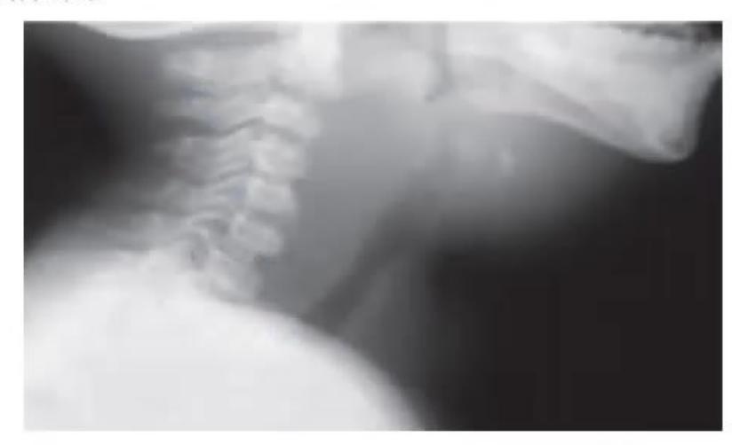
- What is the X-ray finding?
- What is the possible diagnosis?
- What is the treatment?
Answer
-
X-ray finding:
- Lateral soft tissue neck X-ray demonstrates marked widening and thickening of the retropharyngeal soft tissue prevertebral space (
at C2, at C6, or width greater than the adjacent vertebral body diameter) with anterior displacement of the airway and loss of normal cervical lordosis.
- Lateral soft tissue neck X-ray demonstrates marked widening and thickening of the retropharyngeal soft tissue prevertebral space (
-
Possible diagnosis:
- Retropharyngeal Abscess (RPA).
-
Treatment:
- Airway management: Maintain airway, keep patient in a position of comfort, avoid agitation, prepare for emergency intubation or tracheostomy if needed.
- Intravenous Antibiotics: High-dose IV broad-spectrum coverage:
- Ampicillin-Sulbactam (
6-hourly) OR Ceftriaxone + Clindamycin / Metronidazole Vancomycin (for MRSA).
- Ampicillin-Sulbactam (
- Surgical drainage: Urgent transoral or external surgical incision and drainage if large abscess, airway compromise, lack of clinical improvement after 24–48 hours of IV antibiotics, or evidence of fluctuance/gas.
MNJ23-037
An 18-month-old child presented to the OPD with complaints of high fever, cough, and intense irritability. On examination, you find a red throat. When you examine the ear with an otoscope, you observe this picture:
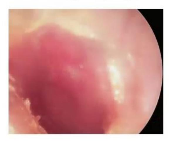
- What are the findings in the ear picture on otoscopy? (Any 2)
- What is the diagnosis on the basis of history and otoscopic findings?
- What is the pathogenesis behind this condition?
- What are the risk factors for this in children 6 years or younger?
Answer
-
Otoscopic findings:
- Marked erythema (hyperemia) and vascular injection of the tympanic membrane
- Bulging / outward fullness of the tympanic membrane
- Loss of normal landmarks (loss of cone of light, obscured handle of malleus)
- Decreased or absent mobility of the tympanic membrane on pneumatic otoscopy.
-
Diagnosis:
- Acute Otitis Media (AOM).
-
Pathogenesis:
- Viral Upper Respiratory Infection (URI)
Eustachian tube inflammation, mucosal edema, and dysfunction failure of middle ear clearance resulting in negative middle ear pressure accumulation of middle ear effusion secondary colonization and bacterial proliferation (Streptococcus pneumoniae, Haemophilus influenzae, Moraxella catarrhalis) acute purulent middle ear inflammation.
- Viral Upper Respiratory Infection (URI)
-
Risk factors in children
years: - Age-dependent anatomical factors (shorter, wider, and more horizontally oriented Eustachian tubes in infants/young children)
- Day care attendance and crowding
- Absence of exclusive breastfeeding / supine bottle propping
- Exposure to environmental tobacco smoke (second-hand smoke)
- Pacifier use
- Craniofacial anomalies (e.g., cleft palate, Down syndrome)
- Allergic rhinitis and recurrent viral URIs.
MNJ23-038
A female neonate after normal delivery developed respiratory distress after birth and a diagnosis of PPHN (Persistent Pulmonary Hypertension of the Newborn) was made after ECHO and treated with iNO and HFOV ventilator. She was extubated on Day 4 and commenced on oral feeds. She developed progressive jaundice on Day 10 of life. Other examination findings include broad forehead, pointed chin, deep-set eyes, a soft systolic murmur, and hepatosplenomegaly:
- What is the possible diagnosis?
- What are the differential diagnoses?
- What tests will you order in this child?
- What nutritional deficiencies is this child at risk for and how will you prevent them?
Answer
-
Possible diagnosis:
- Alagille Syndrome (Arteriohepatic Dysplasia / JAG1 mutation).
-
Differential diagnoses:
- Biliary Atresia
- Idiopathic Neonatal Hepatitis
- Congenital TORCH infections (CMV, Rubella)
- Progressive Familial Intrahepatic Cholestasis (PFIC)
- Alpha-1 Antitrypsin Deficiency
- Galactosemia / Inborn errors of bile acid metabolism.
-
Diagnostic tests:
- Liver Function Tests: Conjugated hyperbilirubinemia, elevated serum bile acids, marked elevation of serum GGT and alkaline phosphatase.
- Liver Biopsy: Demonstrates paucity of intrahepatic interlobular bile ducts (bile duct ductopenia).
- Echocardiography: Assess for Peripheral Pulmonary Artery Stenosis (PPS), Tetralogy of Fallot, or VSD.
- Ophthalmologic Examination (Slit-lamp): Evaluate for posterior embryotoxon (prominent Schwalbe's line).
- Spinal X-ray: Check for butterfly vertebrae (anterior sagittal clefting).
- Genetic testing: Sequence analysis for JAG1 (97% of cases) or NOTCH2 gene mutations.
-
Deficiencies & Prevention:
- Deficiencies: Severe deficiency of fat-soluble vitamins (Vitamins A, D, E, and K) and essential fatty acids due to chronic severe cholestasis and impaired micellar fat absorption.
- Prevention & Management:
- High-dose daily oral supplementation of water-soluble / water-miscible forms of Vitamins A, D, E, and K.
- High-calorie formula supplemented with Medium-Chain Triglycerides (MCT oil) (absorbed directly into portal circulation without requiring bile salts).
- Ursodeoxycholic Acid (UDCA):
to stimulate bile flow and treat pruritus.