MNJ06-001
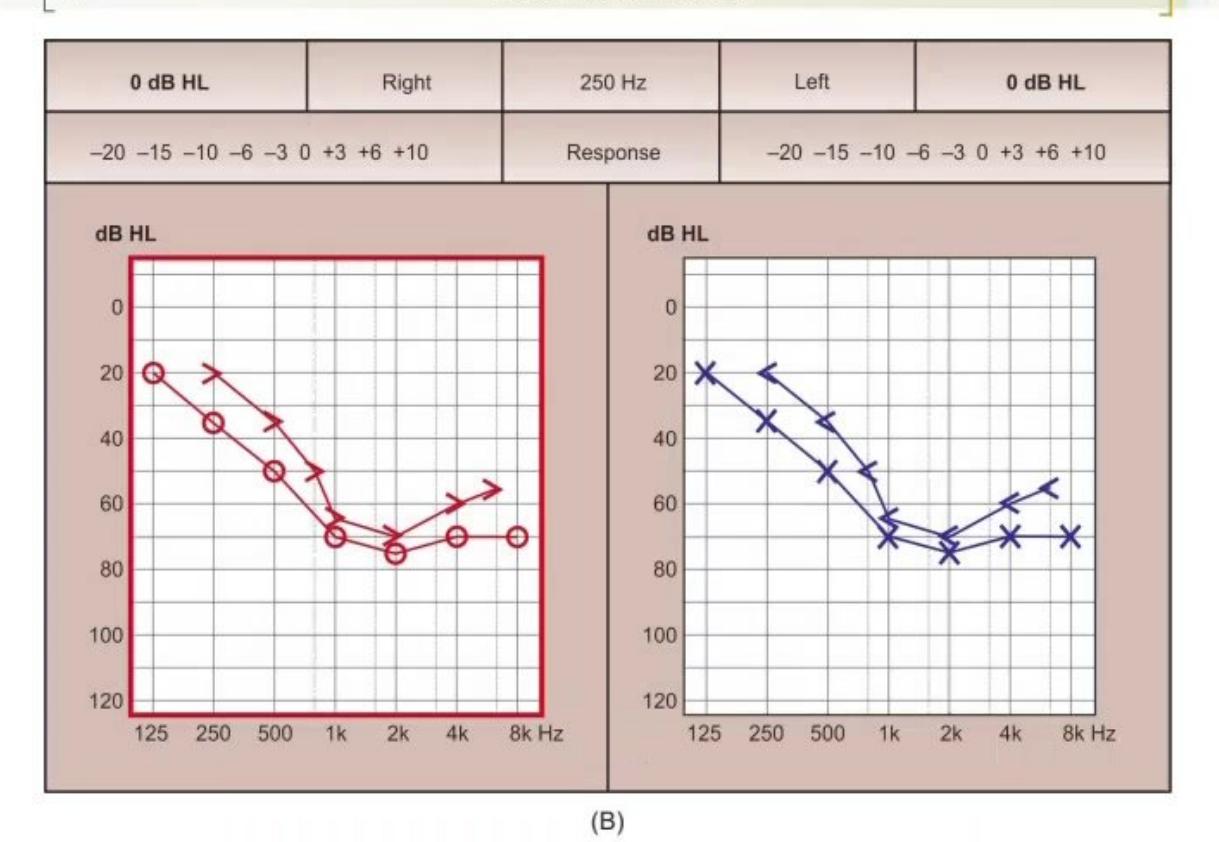
Identify the audiogram patterns shown in Image A and Image B:
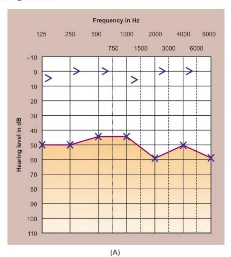
- Identify the hearing defect shown in Image A.
- Identify the hearing defect shown in Image B.
Answer
- Image A: Conductive Hearing Loss (Left Ear) — Air-bone gap present (bone conduction normal, air conduction impaired).
- Image B: Bilateral Sensorineural Hearing Loss (SNHL) — Both air and bone conduction thresholds depressed equally without air-bone gap.
MNJ06-002
Review the instrument shown below:
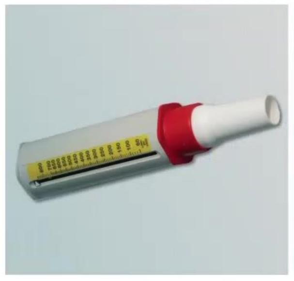
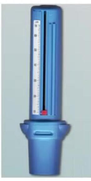
- Identify the instrument.
- Which respiratory illness is it primarily used for home monitoring?
- What are the three color-coded zones and their corresponding Peak Expiratory Flow (PEF) percentages?
Answer
- Peak Expiratory Flow Meter (PEFR meter / Wright peak flow meter).
- Bronchial Asthma (monitoring airflow obstruction, response to bronchodilators, and asthma action plan).
- Three Zones (Asthma Action Plan):
- Green Zone: >80% of personal best PEF (Good control; continue baseline controller medications).
- Yellow Zone: 50–80% of personal best PEF (Caution; worsening symptoms/exacerbation; step up reliever therapy).
- Red Zone: <50% of personal best PEF (Medical emergency; immediate short-acting bronchodilators and seek urgent hospital care).
MNJ06-003
Review the waveform tracings below:
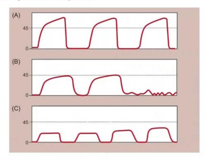
- What is this monitoring modality / graphical technique?
- What are the interpretations for wave patterns A, B, and C?
Answer
- Capnography / Capnogram (end-tidal carbon dioxide
monitoring). - Interpretations:
- A (Shark-fin waveform): Bronchospasm / Airway obstruction (e.g., Bronchial Asthma, COPD, prolonged expiratory upstroke).
- B (Loss of waveform / Flatline): Endotracheal tube (ET) displacement / accidental extubation, esophageal intubation, or complete airway obstruction / circuit disconnection.
- C (Progressive rise in
baseline during CPR): Return of Spontaneous Circulation (ROSC) / effective cardiac output during CPR.
MNJ06-004
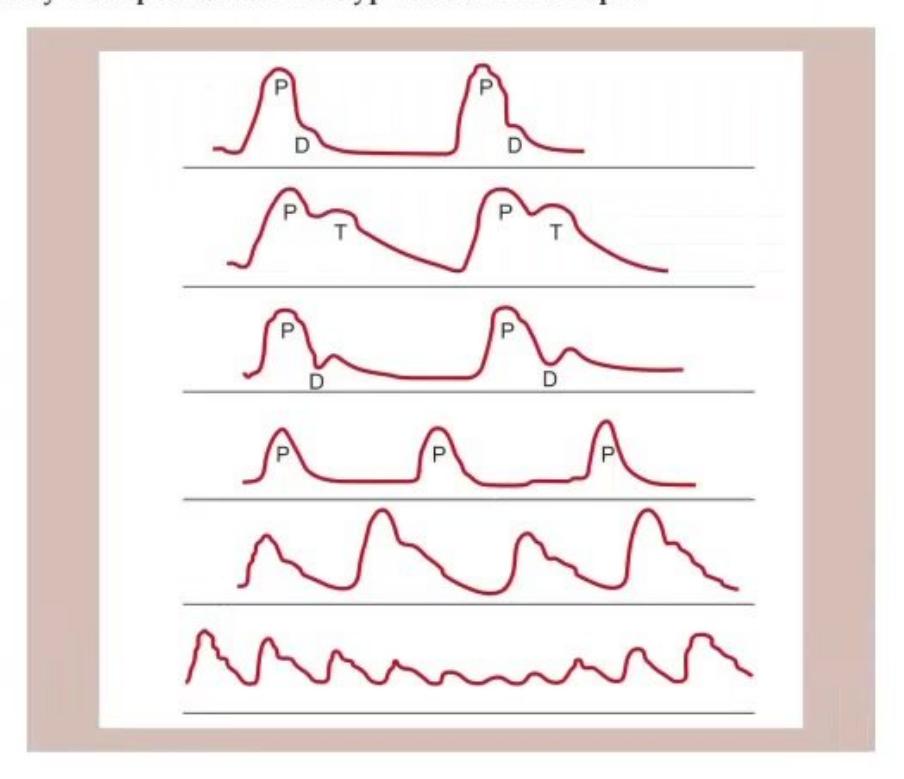
Identify the arterial pulse waveforms shown below and give one clinical condition for each:
- Waveform 1
- Waveform 2
- Waveform 3
- Waveform 4
- Waveform 5
- Waveform 6
Answer
- Collapsing Pulse (Water-hammer / Corrigan pulse): Aortic Regurgitation (AR), Patent Ductus Arteriosus (PDA), Large arteriovenous fistula, Thyrotoxicosis.
- Pulsus Bisferiens (Biphasic pulse with two systolic peaks): Combined Aortic Stenosis + Aortic Regurgitation (AS + AR), Hypertrophic Obstructive Cardiomyopathy (HOCM).
- Dicrotic Pulse (Two peaks, one in systole and one in diastole): Typhoid fever, Severe dilated cardiomyopathy, Septic shock.
- Small Volume Collapsing / Weak Pulse (Pulsus Parvus et Tardus): Severe Aortic Stenosis (AS), Mitral Regurgitation (MR).
- Pulsus Alternans (Alternating strong and weak beats): Severe Left Ventricular Systolic Failure (severe CHF).
- Pulsus Paradoxus (Exaggerated drop in systolic BP > 10 mm Hg during inspiration): Cardiac Tamponade, Severe acute asthma (status asthmaticus), Constrictive pericarditis, Superior Vena Cava (SVC) obstruction.
MNJ06-005
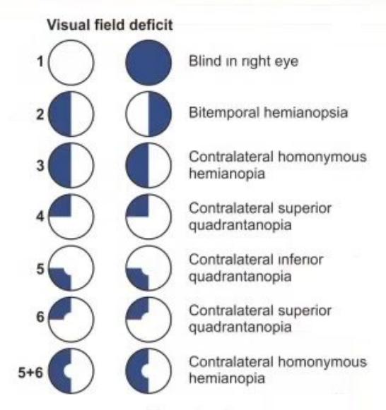
Give the type of visual field defect according to the anatomical lesion at sites 1 to 6 on the visual pathway:
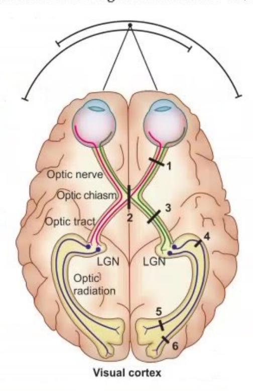
- Lesion at Site 1 (Optic nerve)
- Lesion at Site 2 (Optic chiasm - midline)
- Lesion at Site 3 (Optic chiasm - lateral margin / uncrossed fibers)
- Lesion at Site 4 (Optic tract)
- Lesion at Site 5 (Meyer loop / Temporal optic radiation)
- Lesion at Site 6 (Visual / Occipital cortex with macular sparing)
Answer
- Site 1 (Optic Nerve): Ipsilateral Monocular Anopia / Complete Blindness in that eye.
- Site 2 (Optic Chiasm - central): Bitemporal Hemianopia (e.g., Pituitary adenoma, Craniopharyngioma compressing decussating nasal fibers).
- Site 3 (Optic Chiasm - lateral): Binasal Hemianopia (calcified carotid arteries compressing uncrossed temporal fibers).
- Site 4 (Optic Tract): Contralateral Homonymous Hemianopia.
- Site 5 (Meyer Loop / Temporal Optic Radiation): Contralateral Superior Homonymous Quadrantanopia ("Pie-in-the-sky" defect).
- Site 6 (Occipital Visual Cortex / Calcarine Sulcus): Contralateral Homonymous Hemianopia with Macular Sparing (due to dual blood supply from MCA and PCA).
MNJ06-006
Review the electrophysiological test tracing below:
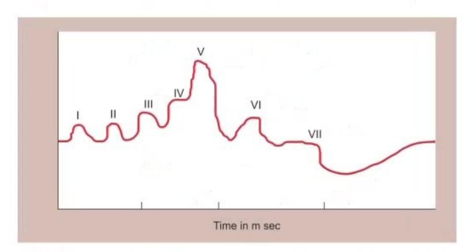
- What is this test?
- Identify the anatomical generators for Waves I through VII (using the mnemonic E. COLI).
Answer
- Brainstem Evoked Response Audiometry (BERA / ABR - Auditory Brainstem Response).
- Anatomical Generators (Mnemonic: E. COLI):
- Wave I & II: Cochlear nerve / distal and proximal cranial nerve VIII (Eighth nerve)
- Wave III: Cochlear nucleus (pons)
- Wave IV: Superior Olivary complex (pons)
- Wave V: Lateral lemniscus (most prominent wave, used for threshold estimation)
- Wave VI & VII: Inferior colliculus (midbrain)
MNJ06-007
Review the pulmonary function flow-volume loop below:
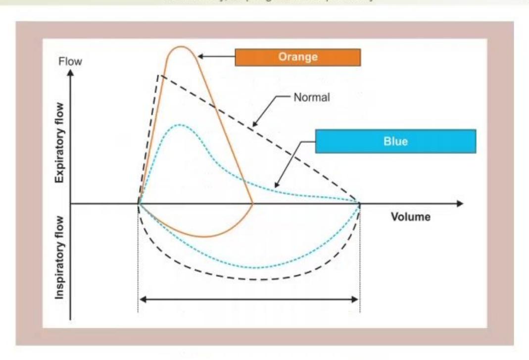
- Identify the graphical plot and state its clinical utility.
- Differentiate the Red loop and Green loop patterns with an example of each.
Answer
- Flow-Volume Loop (Spirometry): Used to differentiate obstructive vs. restrictive ventilatory defects, evaluate upper airway obstruction (fixed vs. variable intra-/extra-thoracic), and monitor response to bronchodilators.
- Patterns:
- Green Loop (Obstructive pattern): Scooped-out expiratory curve ("coving") with reduced peak expiratory flow and concave descending limb (e.g., Bronchial Asthma, Cystic Fibrosis, Bronchiectasis).
- Red Loop (Restrictive pattern): Miniature / narrow "witch's hat" appearance with preserved shape but symmetrically reduced lung volumes (FVC and TLC reduced with normal/high FEV1/FVC ratio) (e.g., Interstitial lung disease, Kyphoscoliosis, Neuromuscular weakness).
MNJ06-008
Audiogram from a 7-year-old boy Ronit. His mother was worried about his hearing in the left ear. He had passed his routine audiometry screening at 5 years and there was no family history of deafness. On physical and otoscopic examination, no abnormalities were found:
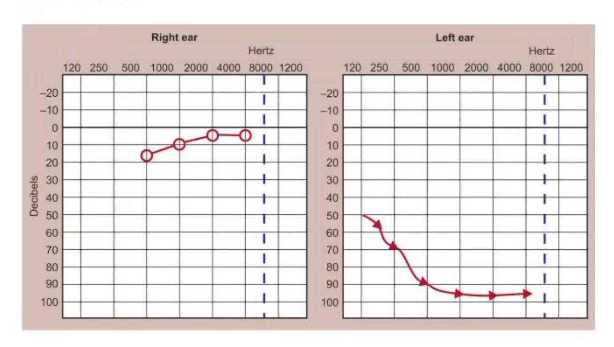
- Diagnose the audiogram findings.
- List 4 possible acquired causes of this hearing loss in childhood.
Answer
- Diagnosis: Profound Unilateral Sensorineural Hearing Loss (SNHL) affecting the left ear (both air and bone conduction thresholds severely elevated > 90 dB on the left, with normal hearing in the right ear).
- Causes:
- Viral labyrinthitis (e.g., Mumps [most common acquired cause of unilateral profound SNHL], Measles, Rubella)
- Post-meningitic labyrinthitis / bacterial meningitis
- Temporal bone trauma / skull base fracture
- Ototoxic drug exposure (Aminoglycosides, Cisplatin)
- Acoustic neuroma / Vestibular schwannoma
- Autoimmune inner ear disease / Sudden sensorineural hearing loss
MNJ06-009
This is the pure-tone audiogram of a 4-year-old girl who was referred because of severe speech delay:
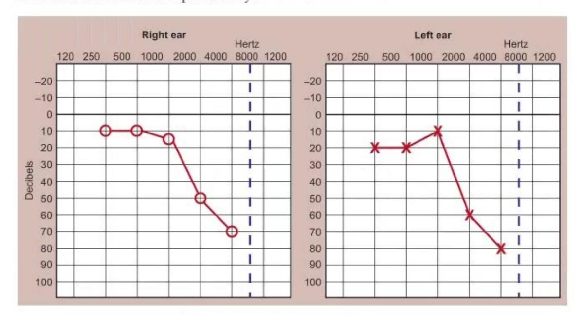

- What does this audiogram show?
- What is the most likely cause of her speech delay?
- What is the next step in auditory habilitation for this child?
Answer
- Diagnosis: Bilateral Moderate-to-Severe Sensorineural Hearing Loss (SNHL) across all speech frequencies (500–4000 Hz) with superimposed threshold loss.
- Cause of Speech Delay: Congenital or early-onset bilateral sensorineural hearing impairment impairing auditory input during critical periods of speech and language acquisition.
- Auditory Habilitation:
- Prompt fitting of bilateral digital hearing aids.
- Comprehensive speech and language therapy.
- Evaluation for Cochlear Implantation if hearing aids provide inadequate benefit.
- Genetic and etiology workup (Connexin 26/GJB2 mutation testing, CMV screening, CT/MRI temporal bone for inner ear anomalies).