THG19-001
A 6-month-old child was brought with history of feeding difficulty, chest retractions, and poor weight gain. His weight was 4 kg. SpO2 was 98%. X-ray showed cardiomegaly. ECG is given here.
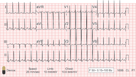
a. Write down the ECG characteristics.
b. What is the likely diagnosis?
c. What is the management you would advise for this child?
Answer
- a. ECG characteristics:
- Sinus tachycardia with HR ~130 bpm
- Katz-Wachtel phenomenon: Large equiphasic (biphasic) QRS complexes (R + S >60 mm) in the midprecordial leads (V2, V3, V4), indicative of biventricular hypertrophy (BVH).
- Typically seen in large post-tricuspid left-to-right shunts (VSD, large PDA, AP window).
- b. Large post-tricuspid left-to-right shunt (e.g., large Ventricular Septal Defect / Patent Ductus Arteriosus / Aortopulmonary Window) presenting with congestive heart failure.
- c. Management:
- Confirm anatomy and shunt size with 2D Echocardiogram.
- Medical management of heart failure (diuretics, ACE inhibitors).
- Early surgical closure / corrective repair (ideal age: 3–6 months of age before irreversible pulmonary vascular disease develops).
THG19-002
This is the ECG of a 7-year-old asymptomatic boy.
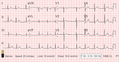
a. What is the ECG diagnosis?
b. Explain the physiologic basis.
c. What is your advice regarding management?
Answer
- a. Sinus Arrhythmia (respiratory sinus arrhythmia).
- Regular P wave morphology and constant PR interval with phasic variation in P-P and R-R intervals (heart rate varies with respiration).
- b. Normal physiological variation in children and adolescents mediated by vagal tone:
- During inspiration: Venous return increases, vagal tone decreases -> Heart rate increases.
- During expiration: Vagal tone increases -> Heart rate decreases.
- c. Benign physiological finding; reassure parents, no medical therapy or restriction required.
THG19-003
A 12-year-old child was diagnosed to have congenital heart disease and planned for cardiac surgery. This is the ECG of the child.
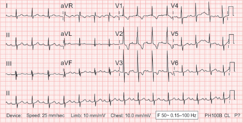
a. What is the ECG finding?
b. What are the congenital heart defects which can be associated with this?
c. What is the treatment?
Answer
- a. First-degree Atrioventricular (AV) Block (PR interval prolonged to 240 ms, constant PR, 1:1 AV conduction) with incomplete Right Bundle Branch Block (RBBB / rsR' in V1).
- b. Associated conditions:
- Congenital: Secundum ASD, Ostium primum ASD / AV canal defect, Ebstein anomaly, Congenitally corrected transposition of great arteries (L-TGA).
- Acquired: Acute Rheumatic Fever (Jones minor criterion), Lyme disease, myocarditis, digitalis toxicity.
- c. No specific treatment required for first-degree AV block per se; manage underlying congenital heart defect surgically/interventionally as indicated.
THG19-004
Given below is the ECG of an asymptomatic neonate with bradycardia. Mother gives history of intrauterine death during previous pregnancy.
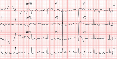
a. What is the diagnosis?
b. What is the most likely etiology?
c. What is the definitive treatment?
Answer
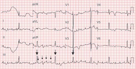
- a. Congenital Complete Heart Block (Third-degree AV block): Marked sinus/atrial rate (~170 bpm) with slow regular escape ventricular rate (42 bpm) and complete AV dissociation (P waves and QRS complexes have no relationship).
- b. Maternal autoimmune connective tissue disease (Systemic Lupus Erythematosus / Sjögren syndrome with transplacental passage of maternal anti-Ro/SSA and anti-La/SSB autoantibodies causing immune-mediated fibrosis of the fetal AV node).
- c. Definitive treatment: Permanent Pacemaker Implantation (PPI).
- Indications in congenital CHB:
- Symptomatic bradycardia / heart failure / syncope
- Ventricular rate <50–55 bpm in neonates/infants (or <70 bpm with associated structural CHD)
- Wide QRS escape rhythm, prolonged QTc, ventricular ectopy, or ventricular dysfunction
- Indications in congenital CHB:
THG19-005
A 3-month-old infant brought to the emergency room (ER) with poor feeding, lethargy, and cold peripheries. On examination, he was drowsy. He had tachycardia, feeble peripheral pulses, and poor perfusion.
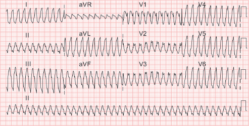
a. Interpret the ECG.
b. What are the possible etiologies?
c. How do you manage this child?
d. What are the investigations you would like to do?
Answer
- a. Interpretation: Regular Wide-Complex Tachycardia (WCT) with HR >200 bpm and AV dissociation -> Ventricular Tachycardia (VT).
- b. Etiologies: Acute viral myocarditis, dilated/hypertrophic cardiomyopathy, congenital heart disease, cardiac tumors (e.g., rhabdomyoma), channelopathies (Long QT syndrome, catecholaminergic polymorphic VT), severe dyselectrolytemia (hyperkalemia, hypomagnesemia), myocardial ischemia.
- c. Management (Unstable VT with pulses / altered mental status & shock):
- Maintain airway, breathing, high-flow oxygen, IV access.
- Immediate synchronized electrical cardioversion: 0.5–1.0 J/kg (increase to 2 J/kg if refractory).
- Antiarrhythmic infusion post-conversion: IV Amiodarone (5 mg/kg over 20–60 min) or IV Lidocaine (1 mg/kg bolus followed by 20–50 µg/kg/min infusion).
- Correct electrolytes, acidosis, and hypoxemia.
- d. Investigations: 12-lead ECG post-conversion, serum electrolytes (K+, Ca++, Mg++), troponin-I/T, CK-MB, blood gas, echocardiography, and cardiac MRI/genetic testing once stable.
THG19-006
A 10-month-old asymptomatic child referred in view of murmur. ECG is given here.
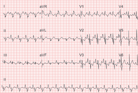
a. Interpret the ECG. What is the likely diagnosis?
b. What are the likely clinical signs in this condition?
c. What are the indications for surgery in this condition?
Answer
- a. ECG Interpretation:
- Sinus rhythm, HR ~125 bpm
- "Himalayan" P waves: Giant, tall peaked P waves (amplitude >5 mm in lead II, taller than the corresponding QRS complex) indicating massive right atrial enlargement.
- Wide, fragmented/splintered QRS complexes with RBBB pattern.
- Diagnosis: Ebstein Anomaly of the tricuspid valve.
- b. Clinical signs:
- Cyanosis (due to right-to-left shunt via ASD/PFO)
- Multiple heart sounds ("sail sound", wide split S1, wide split S2, S3/S4 giving a quadruple rhythm)
- Pansystolic murmur of tricuspid regurgitation at lower left sternal border
- Hepatomegaly, elevated JVP with prominent 'v' wave
- Chest X-ray: "Box-shaped" cardiac silhouette with ballooning right atrium
- c. Indications for surgery:
- NYHA Class III-IV heart failure symptoms / progressive exercise intolerance
- Severe cyanosis (resting SpO2 <85–90%)
- Refractory tachyarrhythmias (WPW / SVT / atrial flutter)
- Progressive cardiomegaly (cardiothoracic ratio >0.65) or RV dysfunction / dilation
THG19-007
A 5-year-old asymptomatic child was evaluated for an incidentally detected murmur and wide split-second sound. Her ECG is given below.
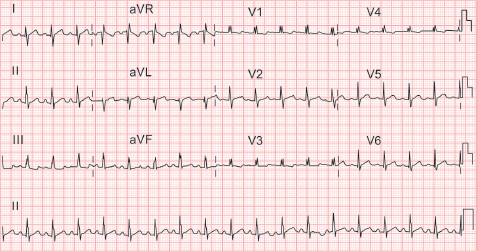
a. Interpret the ECG. What is the likely diagnosis?
b. What are the options available for treatment? What is the ideal age to intervene?
Answer
- a. ECG Interpretation:
- Sinus rhythm, HR ~120 bpm, right axis deviation
- rsR' pattern in lead V1 (incomplete RBBB due to RV volume overload)
- Crochetage sign: Notching near the apex of the R wave in inferior leads (II, III, aVF)
- Diagnosis: Ostium Secundum Atrial Septal Defect (ASD).
- b. Treatment & timing:
- Percutaneous transcatheter device closure (e.g., Amplatzer septal occluder) if adequate rims present (>5 mm except aortic rim).
- Surgical patch/direct closure if rims inadequate or associated defects present.
- Ideal age of intervention: 3–5 years of age (before starting formal schooling).
THG19-008
A 6-month-old infant was brought with history of irritable cry, tachypnea and bluish discoloration of lips, fingers, and toes. His oxygen saturation was 65%. On auscultation, S2 was single. There was no murmur. There was no history of fever. There was history of similar episodes in the previous week. His X-ray is given here.
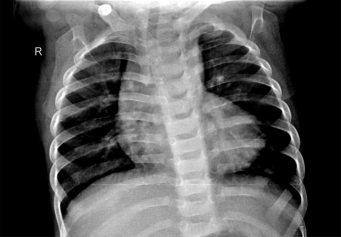
a. Describe the X-ray finding. What is the likely diagnosis?
b. What is the acute management in this child?
c. What is the definitive management after stabilizing the child?
Answer
- a. X-ray findings: "Boot-shaped" heart (coeur en sabot) with upturned apex (RV hypertrophy), concave pulmonary arterial segment/bay, and decreased pulmonary vascularity (pulmonary oligemia).
- Diagnosis: Tetralogy of Fallot (TOF) presenting with hypercyanotic / anoxic ("Tet") spell (infundibular spasm reducing pulmonary blood flow, causing murmur disappearance and severe hypoxia).
- b. Acute management of Tet spell:
-
- Knee-chest / squatting position (increases SVR, decreases right-to-left shunt)
-
- High-flow 100% humidified oxygen via face mask
-
- IV Morphine (0.1–0.2 mg/kg SC/IV) to suppress respiratory center and relieve infundibular spasm
-
- IV Fluid bolus (Normal Saline 10–20 mL/kg) to augment preload
-
- IV Sodium Bicarbonate (1–2 mEq/kg) to correct metabolic acidosis
-
- Refractory spell: IV Phenylephrine (0.02 mg/kg) to elevate SVR, IV Esmolol/Propranolol, or Ketamine (sedation + SVR preservation).
-
- c. Definitive management:
- Maintenance oral Propranolol (1–4 mg/kg/day) and oral Iron therapy (target Hb >14 g/dL to prevent microcytosis-induced stroke).
- Corrective cardiac surgery: Complete intracardiac repair (VSD closure + RVOT reconstruction) or palliative modified Blalock-Taussig-Thomas (mBTT) shunt / RVOT stenting if branch pulmonary arteries are hypoplastic.
THG19-009
A 6-year-old girl was referred for symptoms of exertional dyspnea. Her oxygen saturation was 87%. On auscultation, she had a wide split second heart sound and ejection systolic murmur. Her X-ray is given here.
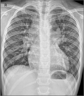
a. Interpret the X-ray.
b. What is the diagnosis?
c. What is the reason for this X-ray finding?
d. What is the management?
Answer
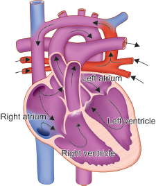
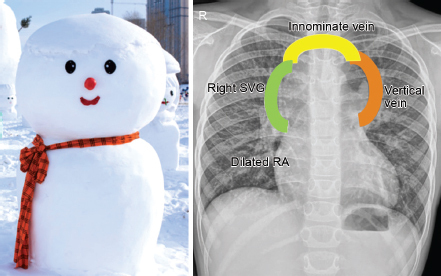
- a. X-ray interpretation: Classic "Snowman" sign or "Figure-of-8" appearance with marked cardiomegaly and increased pulmonary vascular markings (pulmonary plethora).
- b. Diagnosis: Supracardiac Total Anomalous Pulmonary Venous Connection (TAPVC).
- c. Anatomical basis of Snowman configuration:
- Head / upper bulb: Formed by the dilated left vertical vein, dilated left innominate (brachiocephalic) vein, and dilated right superior vena cava (SVC).
- Body / lower bulb: Formed by dilated right atrium and right ventricle.
- d. Management: Corrective open-heart surgery: Anastomosis of common pulmonary venous confluence to the posterior wall of the left atrium, ligation of the vertical vein, and patch closure of the ASD.
THG19-010
An adolescent was referred for cardiac evaluation as the heart sounds were better heard on the right side during a routine auscultation. Her X-ray is given here.
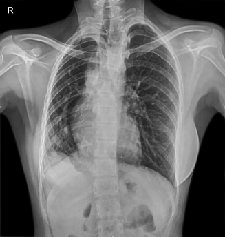
a. Mention the X-ray finding.
b. What is the likely diagnosis?
c. What are the associated lesions in this condition? What is the clinical presentation?
Answer
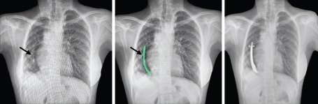
- a. X-ray findings:
- Mediastinal shift to the right with dextroversion of the heart.
- Reduced right lung volume (hypoplastic right lung).
- Scimitar sign: Curved, vertically oriented vascular density resembling a Turkish curved sword (scimitar) along the right heart border extending below the diaphragm into the IVC.
- b. Diagnosis: Scimitar Syndrome (Congenital pulmonary venolobar syndrome / Halasz syndrome).
- c. Associated features & presentation:
- Partial anomalous pulmonary venous drainage (PAPVD) of right pulmonary veins into the IVC.
- Hypoplasia of right pulmonary artery and right lung, systemic collateral arterial supply from abdominal aorta to right lung base.
- Associated ASD, VSD, or tetralogy of Fallot.
- Presentation ranges from severe pulmonary hypertension and heart failure in infancy to asymptomatic murmur or exertional dyspnea in older children.
THG19-011
A 2-month-old neonate was admitted with history of fever, respiratory distress, poor feeding, and lethargy. He was one of twins and had a long neonatal intensive care unit (NICU) stay for 6 weeks. His chest X-ray was normal. A screening echocardiogram was done. The echo image is given here.
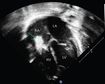
a. What do you see on the echocardiographic image? What is the likely diagnosis?
b. What are the investigations you will do?
c. How will you manage this child?
Answer
- a. Echocardiogram finding: Large mobile, echogenic mass (vegetation) attached to the tricuspid valve leaflets.
- Diagnosis: Infective Endocarditis (Right-sided / Tricuspid valve endocarditis, common after central venous catheterization in NICU).
- b. Investigations:
- Three separate sets of blood cultures obtained from different venipuncture sites over 24 hours (for bacteria and fungi).
- Inflammatory markers: CBC (leukocytosis), CRP, ESR.
- Serial 2D Echocardiography to evaluate vegetation size, leaflet destruction, and regurgitation.
- Urinalysis (hematuria/proteinuria), renal function test, liver function tests.
- c. Management:
- Prompt initiation of parenteral bactericidal antibiotics based on likely pathogens (e.g., IV Cefotaxime/Ceftriaxone or Vancomycin plus Gentamicin; antifungal if Candida suspected).
- Tailor antibiotics according to culture and sensitivity results for 4–6 weeks.
- Surgical intervention if refractory heart failure, persistent fungal infection, large mobile vegetation (>10–15 mm) with recurrent pulmonary emboli, or uncontrolled bacteremia.
THG19-012
A 14-year-old boy with structurally normal heart had three episodes of supraventricular tachycardia (SVT) in the last 1 year. During every episode, the rhythm reverted to sinus with injection adenosine. This is his baseline ECG during sinus rhythm.
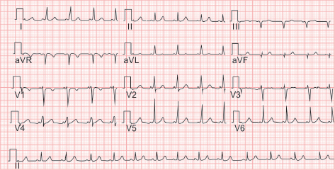
a. Interpret the ECG finding. What is the diagnosis?
b. What treatment would you advise in the given situation?
Answer
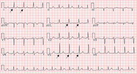
- a. ECG Interpretation:
- Sinus rhythm, HR ~80 bpm
- Short PR interval (<120 ms)
- Delta wave: Slurred upstroke of the initial QRS complex
- Widened QRS duration (>100–120 ms) and secondary ST-T changes
- Diagnosis: Wolff-Parkinson-White (WPW) Syndrome (ventricular pre-excitation via accessory pathway / bundle of Kent).
- b. Treatment:
- Definitive therapy in an adolescent with recurrent symptomatic SVT: Electrophysiology (EP) study and Catheter Radiofrequency Ablation (RFA) of the accessory pathway.
- Interim maintenance therapy: Oral beta-blockers or class IC agents (Flecainide) (AV nodal blockers like verapamil/digoxin contraindicated in WPW with pre-excited AF).
THG19-013
A 9-year-old boy presented with recurrent syncope. He was evaluated and subsequently underwent a cardiac procedure. This is his ECG after the procedure.
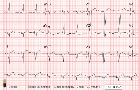
a. What is the ECG finding?
b. What is the procedure he has undergone?
Answer
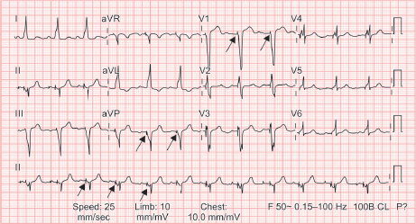
- a. ECG finding: Narrow sharp vertical deflection spikes (pacemaker spikes) preceding each wide QRS complex with left bundle branch block (LBBB) morphology -> Ventricular-paced rhythm (atrial-sensed, ventricular-paced / VDD or DDD mode).
- b. Permanent Pacemaker Implantation (PPI) (endocardial or epicardial dual/single chamber pacemaker).
THG19-014
A 12-year-old girl presented with exertional dyspnea. She was diagnosed to have congenital heart defect in her infancy, but did not undergo treatment and was lost to follow-up. On auscultation, she has a loud P2 and early diastolic murmur at the left upper sternal border. Oxygen saturation in the right upper limb and lower limb was 97% and 78% respectively. Her chest X-ray is given here.
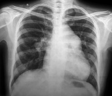
a. Discuss her clinical signs and X-ray features.
b. What is the diagnosis?
Answer
- a. Clinical & X-ray discussion:
- Differential cyanosis and clubbing: Normal SpO2 (97%) in upper limbs with marked desaturation (78%) in lower limbs occurs because deoxygenated blood from the pulmonary artery is shunted right-to-left through the PDA entering the descending aorta distal to the left subclavian artery.
- Loud P2 and Graham Steell murmur (early diastolic murmur of pulmonary regurgitation) signify severe suprasystemic Pulmonary Arterial Hypertension (PAH).
- Chest X-ray: Dilated main and central pulmonary arteries with abrupt tapering/attenuation of peripheral pulmonary vasculature ("peripheral pruning").
- b. Diagnosis: Patent Ductus Arteriosus (PDA) with Eisenmenger Syndrome (reversed right-to-left shunt with irreversible pulmonary vascular disease).
THG19-015
A 1-year-old male child is brought to the pediatric ER with history of lethargy and irritability. On examination, airway was stable; tachycardia, respiratory rate (RR)—50 breaths/min, normal blood pressure (BP), and good peripheral pulses. Electrocardiogram was done.
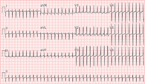
a. Calculate heart rate (HR)?
b. What is the diagnosis?
c. In a resource limited setting with no cardiac drugs what will you do?
d. What is the drug that you will use to restore normal rhythm? What is the mechanism of its action and its half-life? What is the recommended dose of the drug?
Answer
- a. Heart Rate: ~250 beats/min (RR interval is approximately 6 small boxes: 1500/6 = 250 bpm).
- b. Diagnosis: Supraventricular Tachycardia (SVT) (Narrow QRS complex tachycardia with absent/hidden P waves and no beat-to-beat variability).
- c. Non-pharmacological measures (Vagal maneuvers):
- Application of an ice-water bag to the upper face (forehead and nose) for 10–15 seconds (diving reflex), or rectal stimulation/modified Valsalva. Avoid eyeball pressure.
- d. Pharmacotherapy:
- Drug: Adenosine rapid IV push.
- Mechanism of action: Stimulates cardiac A1 adenosine receptors -> activates potassium efflux channels and inhibits adenylate cyclase, transiently slowing/blocking conduction through the AV node.
- Half-life: <10 seconds (rapidly taken up by erythrocytes and vascular endothelium).
- Dose: Initial dose 0.1 mg/kg (max 6 mg) rapid IV push followed immediately by 5–10 mL saline flush; can double to 0.2 mg/kg (max 12 mg) if no response.
THG19-016
A 3-month-old girl is brought to the pediatric ER with complaints of breathlessness and feeding difficulty. On examination, baby was irritable with HR—170 beats/min, RR—55 breaths/min, SpO2—99%, normal BP, poor perfusion, and hepatomegaly. Chest X-ray was taken. Electrocardiogram was taken few hours after stabilization.
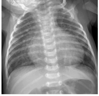
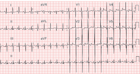
a. What are the findings in chest X-ray?
b. What are the probable congenital cardiac lesions that can present in this manner?
c. What are the findings in ECG?
Answer
- a. Chest X-ray findings: Marked cardiomegaly (cardiothoracic ratio >60%) and prominent pulmonary plethora (increased pulmonary vascular markings extending to periphery).
- b. Probable lesions: Large left-to-right shunts:
- Large Ventricular Septal Defect (VSD)
- Large Patent Ductus Arteriosus (PDA)
- Complete Atrioventricular Septal Defect (AVSD / AV canal)
- Large Aortopulmonary Window (AP window)
- c. ECG findings: Sinus tachycardia (HR ~140 bpm), left axis deviation, and biventricular hypertrophy (tall R in V5-V6 and deep S in V1 / Katz-Wachtel phenomenon in midprecordial leads).
THG19-017
A 4-month-old infant weighing 3.5 kg was brought to pediatric OPD with history of feeding difficulty, forehead sweating, and poor weight gain. Two months back the baby was diagnosed to have episodes of abdominal colic and advised oral medication. On examination, infant had HR—140 beats/min, RR—60 breaths/min, normal BP, and normal perfusion. Auscultation revealed basal crepitations and S3 at apex. Chest X-ray showed cardiomegaly. Screening ECHO revealed decreased left ventricular function.
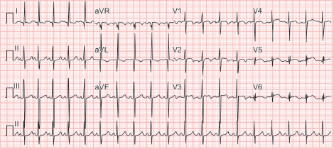
a. What are the salient findings in ECG?
b. What could be the probable diagnosis?
c. What is the definitive treatment?
Answer
- a. ECG findings:
- Sinus rhythm, HR ~140 bpm, Left axis deviation
- Anterolateral myocardial infarction pattern: Deep and wide abnormal Q waves (>2 mm, >30 ms) with ST-segment elevation or T-wave inversion in leads I, aVL, V5, and V6.
- b. Diagnosis: ALCAPA (Anomalous Origin of Left Coronary Artery from the Pulmonary Artery) / Bland-White-Garland syndrome.
- c. Definitive treatment: Surgical correction: Direct reimplantation of the anomalous left coronary artery into the ascending aorta (or Takeuchi intrapulmonary baffle procedure if direct transfer not feasible).
THG19-018
An 8-year-old boy came with history of palpitations of 1 hour duration. He had past history of recurrent palpitations over the last 3 months period. He had one episode of syncope lasting for less than a minute 2 months back. On examination, child was lethargic with HR—140 beats/min, RR—20 breaths/min, weak peripheral pulses, and hypotension.
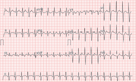
a. What is the ECG finding?
b. What is the preferred treatment?
c. What are the cardiac channelopathies which can present with arrhythmias?
Answer
- a. ECG finding: Narrow QRS complex tachycardia with characteristic sawtooth flutter waves (F waves) in inferior leads (II, III, aVF) with 2:1 / 3:1 AV conduction -> Atrial Flutter.
- b. Preferred treatment (hemodynamically unstable with hypotension/shock): Synchronized DC Cardioversion (0.5–1 J/kg, up to 2 J/kg).
- c. Cardiac channelopathies:
- Congenital Long QT Syndrome (LQTS - KCNQ1, KCNH2, SCN5A)
- Short QT Syndrome (SQTS)
- Brugada Syndrome (SCN5A)
- Catecholaminergic Polymorphic Ventricular Tachycardia (CPVT - RyR2, CASQ2)
THG19-019
A male infant aged 3 months came with history of breathlessness and feeding difficulty from 2 weeks of age. There was history of recurrent respiratory tract infections. Birth weight was 3 kg, present weight was 3.7 kg. On examination, baby had HR—150 beats/min, RR—45 breaths/min, normal BP, and SpO2—98%. Chest X-ray showed cardiomegaly with pulmonary plethora. Echocardiogram was done.
Consider the following echocardiographic findings:
- i. Both atrioventricular (AV) valves insert at the same level at the cardiac crux
- ii. Cleft in the left-sided AV valve component
- iii. Large ostium primum atrial septal defect (ASD)
- iv. Large inlet ventricular septal defect (VSD)
a. Which of the above findings on echocardiogram are characteristic of complete atrioventricular septal defect (AVSD)?
- A. i and ii only
- B. i, ii, and iv only
- C. i, ii, and iii only
- D. i, ii, iii, and iv
b. Which of the above findings would be suggestive of partial AV canal defect?
- A. i and ii only
- B. i, ii, and iv only
- C. i, ii, and iii only
- D. i, ii, iii, and iv
c. What is the ideal age of surgical repair for complete AVSD?
d. Familial occurrence of complete AVSD is rare—True/False?
Answer
- a. D (i, ii, iii, and iv: Complete AVSD has common AV valve inserting at same level, cleft left AV valve, primum ASD, and inlet VSD).
- b. C (i, ii, and iii: Partial AVSD has primum ASD and cleft mitral valve with intact ventricular septum).
- c. 3 to 6 months of age (before irreversible pulmonary vascular obstructive disease develops, especially in Down syndrome).
- d. True (most cases occur sporadically or associated with Trisomy 21 / Down syndrome).
THG19-020
Child aged 5 years was admitted in pediatric ward with history of fever >10 days. On examination, child had tachycardia, tachypnea, and high-grade fever. Total count was raised with neutrophilic predominance. Acute phase reactants were elevated. Chest X-ray showed cardiomegaly. Echocardiogram was done and the fixed frame image is given here.
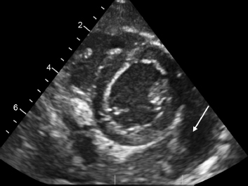
a. What is the diagnosis from echocardiogram?
b. What are the most common causes of the above mentioned condition? List out the other infectious causes.
c. What are the noninfectious causes for the above mentioned condition?
Answer
- a. Diagnosis: Large pericardial effusion with thickened pericardium and internal echoes/fibrinous strands suggestive of Pyopericardium (Purulent Pericarditis).
- b. Infectious causes:
- Bacterial: Staphylococcus aureus, Streptococcus pneumoniae, Haemophilus influenzae, Streptococcus pyogenes, Pseudomonas, Mycobacterium tuberculosis.
- Viral: Enteroviruses (Coxsackie B), Adenovirus, CMV, EBV, Parvovirus B19, HIV.
- Fungal / Parasitic: Candida, Aspergillus, Entamoeba histolytica (amoebic pericarditis).
- c. Non-infectious causes:
- Systemic Juvenile Idiopathic Arthritis (sJIA), SLE, Rheumatic fever
- Uremic pericarditis (chronic kidney disease)
- Malignancy / neoplastic infiltration (leukemia, lymphoma)
- Severe hypothyroidism (myxedema)
- Post-pericardiotomy syndrome
THG19-021
Baby was delivered by emergency lower segment cesarean section (LSCS) for obstetric indication [gestational age (GA): 39 weeks] at a primary health center (PHC) located in a remote village. Birth weight was 3.2 kg. Baby was in postnatal ward with mother. On day 4 of life, baby was found to be lethargic with HR—130 beats/min, RR—40 breaths/min, normal perfusion status, and BP—65/40 mm Hg. Capillary blood glucose was 90 mg/dL. SpO2 (right upper limb) was 70%. System examination was done. Cardiovascular system (CVS): S1—normal and S2—difficult to interpret with no murmur; Respiratory system (RS): normal breath sounds; Abdomen: no organomegaly; Central nervous system (CNS): anterior fontanelle (AF)—normal, pupils equal and reacting to light; and No focal neurological deficit. Within few minutes baby developed apnea and required intubation. Chest X-ray was done.
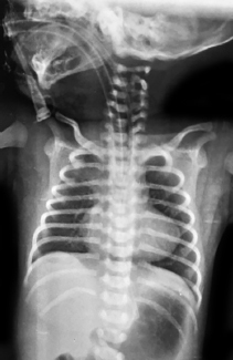
a. What are the findings in chest X-ray?
b. What could be the probable cardiac condition in this baby?
c. What should the medical officer at PHC do to ensure safe referral of baby to the nearest tertiary neonatal care unit located 100 km away?
d. What should be done by the neonatologist at the tertiary neonatal care unit?
e. What will be the general line of management for such cardiac conditions?
Answer
- a. Chest X-ray findings: Boot-shaped heart (coeur en sabot) with normal heart size and marked pulmonary oligemia (decreased vascular markings).
- b. Duct-dependent Pulmonary Blood Flow lesion (e.g., Pulmonary Atresia with intact ventricular septum, Critical Pulmonary Stenosis, Severe TOF with ductus closure).
- c. Emergency referral stabilization:
- Immediate initiation of continuous IV Prostaglandin E1 (Alprostadil) infusion (starting dose 0.05–0.1 µg/kg/min) to reopen and maintain ductal patency.
- Endotracheal intubation and mechanical ventilation (PGE1 frequently causes apnea).
- Secure thermal support and vascular access.
- d. Emergency bedside echocardiography to confirm cardiac anatomy, assess ductal patency and pulmonary blood flow; contact pediatric cardiac surgical team.
- e. Definitive management:
- Maintain PGE1 at lowest effective dose to achieve SpO2 80–85%.
- Transcatheter intervention (PDA stenting or balloon pulmonary valvotomy) or surgical systemic-to-pulmonary shunt (modified Blalock-Taussig shunt).
THG19-022
A 3-week-old baby was brought to a PHC with history of refusal of feeds for 2 days and breathlessness for 1 day. Baby was delivered by LSCS in the PHC and was given to mother for breastfeeding soon after birth. Baby and mother were discharged after 5 days. On examination, baby general condition (GC) was poor, mild subcostal retractions, HR—180 beats/min, RR—65 breaths/min, and weak dorsalis pedis and femoral pulses. Soles were dusky in color. Noninvasive blood pressure (NIBP) recorded by staff nurse was 80/60 mm Hg (right upper limb). Capillary blood glucose (CBG) was 75 mg/dL. Medical officer at PHC diagnosed the baby to have shock and started IV saline boluses 5 mL/kg. In spite of two saline boluses, baby did not improve. Dorsalis pedis pulse was still weak and liver span had increased.
a. Can a cardiac condition be suspected in this baby? If so, what could be the probable cardiac diagnosis?
b. What drug should be available in PHC to stabilize the baby before referral to a tertiary neonatal care facility?
c. What are the structural cardiac defects that can present in this manner?
Answer
- a. Yes, Duct-Dependent Systemic Circulation. As the ductus arteriosus closes in the 2nd to 3rd week of life, systemic perfusion falls abruptly leading to cardiogenic shock, absent femoral pulses, and worsening hepatomegaly upon fluid administration.
- b. Prostaglandin E1 (Alprostadil) infusion (to restore ductal patency and systemic perfusion).
- c. Structural lesions with duct-dependent systemic flow:
- Critical Coarctation of the Aorta
- Interrupted Aortic Arch
- Hypoplastic Left Heart Syndrome (HLHS)
- Critical Aortic Stenosis
THG19-023
Male baby aged 5 weeks weighing 3.5 kg was brought to the pediatric casualty with history of breathlessness for past 1 week, difficulty in breastfeeding for past 3 days and failure to gain weight. No history of fever/cough/coryza. Baby was delivered by LSCS and birth weight was 2.9 kg. On examination, baby had HR—170 beats/min, RR— 65 breaths/min, SpO2—99%, normal perfusion, and normal BP. Baby had mild subcostal retractions and an audible systolic murmur at left lower sternal border. Chest X-ray revealed mild cardiomegaly with plethora. Suspecting a cardiac disease, the pediatrician requested for ECHO.
a. What did the pediatrician have in mind when he wanted a cardiologist opinion?
b. What are the common structural cardiac lesions that can present at this age with the above findings?
c. What is the initial line of management for the cardiac lesion diagnosed other than general management? What are the drugs that can be used?
d. Which left-to-right shunt is unlikely to present at this age?
Answer
- a. Pediatrician suspects a large left-to-right shunt lesion presenting with congestive heart failure as pulmonary vascular resistance drops at 4–6 weeks of age.
- b. Common lesions: Large Ventricular Septal Defect (VSD), large Patent Ductus Arteriosus (PDA), Complete AV Canal defect, Aortopulmonary Window.
- c. Medical management of heart failure:
- Loop diuretic: Furosemide (1–2 mg/kg/day)
- Aldosterone antagonist: Spironolactone (1–2 mg/kg/day)
- Afterload reducer: ACE inhibitor (Enalapril / Captopril)
- Inotropic support: Digoxin (selected cases)
- d. Atrial Septal Defect (ASD) (pre-tricuspid shunts rarely cause heart failure in infancy due to high RV compliance).
THG19-024
A newborn baby was delivered in a PHC by labour naturale at GA of 36 weeks. Birth weight was 2.4 kg. Baby was noted to have fast breathing with mild retractions at about 6 hours of life. Chest X-ray was done. Baby was given surfactant suspecting hyaline membrane disease. However, baby’s condition did not improve and baby was intubated. SpO2 did not pick up and remained at 80% despite FiO2 >90%.
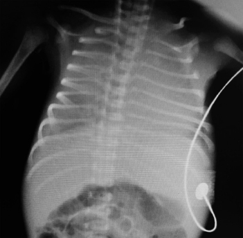
a. What is the chest X-ray finding?
b. What cardiac lesion should be suspected if X-ray findings remain the same after surfactant therapy?
c. What should the medical officer do next?
d. What will be the definitive line of management for the suspected cardiac lesion?
Answer
- a. Chest X-ray findings: Diffuse, ground-glass reticulogranular haze with severe pulmonary venous congestion and normal/small cardiac silhouette (resembling RDS/HMD).
- b. Infradiaphragmatic / Obstructed Total Anomalous Pulmonary Venous Connection (TAPVC).
- c. Next steps: Arrange immediate emergency echocardiogram (or transfer to a pediatric cardiac surgical center) while providing mechanical ventilation and inotropic support; avoid excessive fluid administration.
- d. Definitive management: Emergency surgical repair (reconnection of pulmonary venous confluence to left atrium and closure of ASD).
THG19-025
A 2-year-old male child was brought to pediatric OPD with complaints of two episodes of sudden loss of consciousness in the past 1 week. Each episode lasted for approximately 1 minute. Child regained consciousness in few minutes. On examination, vitals were normal and CVS examination revealed normal S1 and S2 with no murmur. Electrocardiogram was done.
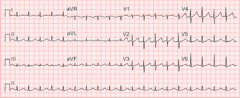
a. What are the ECG findings? What is the diagnosis?
b. What is Bazett formula?
c. What are the syndromes associated with this condition?
Answer
- a. ECG findings: Sinus rhythm, HR ~100 bpm, normal axis, marked prolongation of corrected QT interval (QTc >500 ms; measured ~534 ms) with T-wave abnormalities -> Long QT Syndrome (LQTS).
- b. Bazett's Formula:
(Normal QTc: <440 ms in males, <460 ms in females; >480–500 ms is distinctly abnormal). - c. Associated Syndromes:
- Romano-Ward Syndrome: Autosomal dominant LQTS with normal hearing.
- Jervell and Lange-Nielsen Syndrome: Autosomal recessive LQTS with congenital sensorineural deafness.
- Timothy Syndrome (LQT8): LQTS with syndactyly, facial dysmorphism, autism, and immune deficiency.
THG19-026
A 6-year-old boy complained of left- and right-sided chest pain which was pinpricking in nature with no radiation. He plays football regularly. There was no history of palpitations/syncope. General and system examination was normal. Vital signs were normal. ECG was taken.
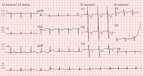
a. Interpret the ECG? What could be the probable diagnosis?
b. What are the supportive findings?
Answer
- a. Dextrocardia (with Situs Inversus / isolated Dextrocardia) (or lead reversal).
- b. ECG criteria supportive of Dextrocardia:
- Inverted P wave, negative QRS complex, and inverted T wave in lead I
- Positive P wave and upright tall QRS in lead aVR
- Right axis deviation
- Progressive reduction in R wave amplitude from V1 through V6 (reverse R-wave progression across precordium)