MNJ22-001
A 9-year-old girl Ritu presents with high irregular fever for 3 weeks, each episode associated with erythematous rashes over the trunk and proximal extremities, pain and swelling of right knee and easy fatigability. Has leucocytosis, raised ESR, normal chest skiagram and negative Mantoux test:
- Give most probable diagnosis.
- List other expected systemic clinical findings.
- Give value of serology in diagnosis.
- Outline steps in management.
- List prognostic factors.
Answer
-
Most probable diagnosis:
- Systemic-onset Juvenile Idiopathic Arthritis (SoJIA) / Still's disease (Systemic JRA)
-
Expected systemic clinical findings:
- Hepatosplenomegaly
- Generalized lymphadenopathy
- Serositis (pericarditis, pleuritis, peritonitis / serous cavity effusions)
- Evanescent, salmon-pink macular rash (accompanies fever spikes)
- Myalgias, arthralgias, and polyserositis
-
Value of serology in diagnosis:
- Antinuclear antibodies (ANA): Usually negative
- Rheumatoid Factor (RF): Usually negative
- Markedly elevated inflammatory markers (ESR, CRP, serum ferritin, leukocytosis, thrombocytosis)
-
Steps in management:
- First-line: NSAIDs (e.g., indomethacin, naproxen)
- Systemic Corticosteroids: Oral prednisolone or IV pulse methylprednisolone for severe systemic illness/serositis
- Disease-Modifying Antirheumatic Drugs (DMARDs): Methotrexate, Leflunomide
- Biologic agents:
- IL-1 antagonists (Anakinra, Canakinumab)
- IL-6 receptor antagonists (Tocilizumab)
- TNF-alpha inhibitors (Etanercept, Infliximab, Adalimumab) for persistent arthritis
-
Prognostic factors:
- Number and severity of joint involvement (polyarticular extension)
- Persistent systemic inflammation / persistent thrombocytosis or leukocytosis
- Development of Macrophage Activation Syndrome (MAS)
- Early radiographic joint damage / persistent active disease at 6–12 months
MNJ22-002
A child has chronic polyarthritis of 4 joints and is ANA positive:
- What arthritis is it likely to be?
- What complication should we anticipate?
- In which type of JRA is HLA B27 positive?
- A child with JRA presents with fever, leukopenia, hepatosplenomegaly, and lymphadenopathy. What is the diagnosis?
- What drug is indicated in treatment of Q4?
Answer
-
Likely arthritis:
- Oligoarticular (Pauciarticular) JIA Type 1 (involves
joints, early onset, female predominance, ANA positive)
- Oligoarticular (Pauciarticular) JIA Type 1 (involves
-
Complication to anticipate:
- Chronic anterior uveitis (iridocyclitis) — usually asymptomatic/insidious; requires regular slit-lamp screening to prevent visual impairment/blindness
-
Type of JRA with HLA-B27 positivity:
- Enthesitis-Related Arthritis (ERA) / Oligoarticular JIA Type 2 (typically older boys, axial/lower limb involvement)
-
Diagnosis for Q4:
- Macrophage Activation Syndrome (MAS) / Secondary HLH
-
Drug indicated in MAS:
- Cyclosporine (along with high-dose pulse IV Methylprednisolone; Anakinra/biologics in refractory cases)
MNJ22-003
A 35-day-old male child came with complaints of rash over the face for the last 4 weeks. On examination, HR was 58/min but child is not in CHF. Face picture is given below:
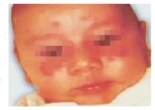
- What is the diagnosis?
- What is the clinical condition in mother you will see?
- What is the prognosis of skin and cardiac disease?
- When can you diagnose this earliest in the antenatal period? And what test will you do?
- What treatment will you offer to mother if condition is diagnosed antenatally?
- What is the risk in the next pregnancy?
Answer
-
Diagnosis:
- Neonatal Lupus Erythematosus (NLE) with Congenital Complete Heart Block (CCHB)
-
Clinical condition in mother:
- Systemic Lupus Erythematosus (SLE), Sjögren's syndrome, or undifferentiated autoimmune connective tissue disease (may be completely asymptomatic carrier of autoantibodies)
-
Prognosis of skin and cardiac disease:
- Skin lesions: Benign and transient; resolve spontaneously within 6 months as transplacentally acquired maternal IgG antibodies clear from neonatal circulation.
- Cardiac disease: Congenital heart block is irreversible and permanent (often requires permanent pacemaker placement).
-
Earliest antenatal diagnosis and test:
- At 16–18 weeks of gestation
- Test: Fetal echocardiography (evaluates fetal heart rate, PR interval, and rhythm) and Maternal serum anti-Ro/SSA & anti-La/SSB antibody titers
-
Antenatal maternal treatment:
- Fluorinated corticosteroids (Dexamethasone or Betamethasone) that cross the placenta; IVIG; plasmapheresis
-
Risk of recurrence in next pregnancy:
- Approximately 15–20% (increases to ~50% after two affected infants)
MNJ22-004
A child presents with skin lesions shown in the picture below:

- What is the probable diagnosis and what are the diagnostic criteria?
- What is the indication for steroids?
- Unit head advised at discharge to monitor urine protein. What is the importance of this?
- What antibiotic/drug is used for chronic/refractory skin lesions in this condition?
- Write 2 diseases with small vessel vasculitis.
Answer
-
Probable diagnosis & Diagnostic criteria:
- Diagnosis: Henoch-Schönlein Purpura (HSP) / IgA Vasculitis
- Criteria (EULAR/PRINTO/PRES):
- Mandatory criterion: Palpable purpura (non-thrombocytopenic, predominantly lower extremities/buttocks)
- Plus at least ONE of the following:
- Diffuse abdominal pain
- Predominant IgA deposition on histopathology / immunofluorescence
- Arthritis (acute) or arthralgia
- Renal involvement (hematuria and/or proteinuria)
-
Indication for systemic steroids:
- Severe, debilitating abdominal pain / GI bleeding / bowel ischemia / intussusception
- Severe scrotal swelling/orchitis
- Severe renal involvement (crescentic HSP nephritis)
- Pulmonary hemorrhage or CNS vasculitis
-
Importance of monitoring urine protein:
- To monitor for HSP nephritis, which can develop or progress up to 6 months after disease onset. Early detection and treatment of persistent proteinuria/nephritis prevents progressive renal impairment.
-
Drug for chronic/refractory lesions:
- Dapsone (or colchicine)
-
Two small-vessel vasculitides:
- Granulomatosis with polyangiitis (GPA / Wegener's)
- Eosinophilic granulomatosis with polyangiitis (EGPA / Churg-Strauss)
- Microscopic polyangiitis (MPA)
MNJ22-005
A 4-year-old male child presents with complaints of fever for 5 days with all features shown in picture. Antibiotic was given but no response. Coronary angiogram was done:

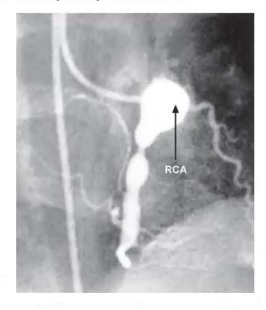
- What is the probable diagnosis?
- What are the diagnostic criteria?
- What are the ECHO criteria for this disease?
- What are the DDs? (At least 4)
- What is the treatment?
- What is the follow-up plan for a child with coronary artery involvement?
Answer
-
Probable diagnosis:
- Kawasaki Disease (Mucocutaneous Lymph Node Syndrome)
-
Diagnostic criteria (Classic Kawasaki Disease):
- High-grade fever for
days plus at least 4 of the 5 principal clinical criteria: - Bilateral bulbar conjunctival injection without exudate
- Erythema and cracking of lips, strawberry tongue, erythema of oral and pharyngeal mucosa
- Polymorphous rash (maculopapular, erythema multiforme-like, or scarlatiniform)
- Changes in extremities: Acute erythema and edema of hands/feet, subacute periungual desquamation
- Cervical lymphadenopathy (usually unilateral,
)
- High-grade fever for
-
ECHO criteria:
- Z-score of LAD (Left Anterior Descending) or RCA (Right Coronary Artery)
- Perivascular brightness/cuffing
- Lack of normal tapering of coronary arteries
- Decreased left ventricular systolic function
- Mitral regurgitation / pericardial effusion
- Z-score of LAD (Left Anterior Descending) or RCA (Right Coronary Artery)
-
Differential diagnoses (DDs):
- Scarlet fever
- Staphylococcal Scalded Skin Syndrome (SSSS) / Toxic Shock Syndrome (TSS)
- Toxic Epidermal Necrolysis (TEN) / Stevens-Johnson Syndrome (SJS)
- Measles
- Rocky Mountain spotted fever
- Multisystem Inflammatory Syndrome in Children (MIS-C)
- Systemic JIA
-
Treatment:
- IVIG:
single IV infusion given over 10–12 hours - High-dose Aspirin:
in 4 divided doses until child is afebrile for 48–72 hours - Low-dose Aspirin:
once daily for 6–8 weeks (continued indefinitely if coronary aneurysms develop) - Corticosteroids / Infliximab / Anakinra in IVIG-resistant Kawasaki disease
- IVIG:
-
Follow-up plan (AHA Risk Stratification):
| Risk Level | Coronary Artery Involvement | Pharmacological Therapy | Physical Activity | Follow-up | Invasive Testing |
|---|---|---|---|---|---|
| I | No coronary artery involvement | None beyond 1st 6–8 weeks | No restrictions beyond 1st 6–8 weeks | Cardiovascular risk assessment, counseling at 5-year intervals | None |
| II | Transient coronary artery ectasia disappears within 1st 6–8 weeks | None beyond 1st 6–8 weeks | No restrictions beyond 1st 6–8 weeks | Cardiovascular risk assessment, counseling at 3–5 year intervals | None |
| III | 1 small-to-medium coronary artery aneurysm / major coronary artery | Low-dose aspirin ( |
Age |
Annual cardiology follow-up with ECHO + ECG; biennial stress test / myocardial perfusion scan | Angiography if non-invasive test suggests ischemia |
| IV | 1 large or giant aneurysm ( |
Long-term antiplatelet (Aspirin) + Warfarin (target INR 2.0–2.5) or LMWH (target anti-Xa 0.5–1.0 U/mL) | Avoid contact or high-impact sports (bleeding risk); other activity guided by stress test / myocardial perfusion scan | Biannual follow-up with ECHO + ECG; annual stress test / perfusion scan | 1st angiography at 6–12 months or sooner if clinically indicated; repeated if ischemia suspected |
| V | Coronary artery obstruction | Long-term low-dose aspirin; Warfarin or LMWH if giant aneurysm persists; consider beta-blockers | Avoid contact or high-impact sports; activity guided by stress test / perfusion scan | Biannual follow-up with ECHO + ECG; annual stress test / perfusion scan | Angiography recommended to evaluate therapeutic options |
MNJ22-006
A 7-year-old male child presents with complaints of knee joint swelling for 2 weeks duration with limitation of movement. There is history of gastroenteritis 2 weeks back. CBC: Normal, X-ray knee: normal:
- What is the probable diagnosis?
- What HLA is associated with this? And what % of patients are positive for this?
- What are the causative agents (4 bacteria and 2 viruses)?
- What is the prognosis?
Answer
-
Probable diagnosis:
- Post-infectious Reactive Arthritis (Reiter's Syndrome)
-
HLA association:
- HLA-B27; positive in ~75% of affected patients
-
Causative agents:
- Bacteria: Campylobacter jejuni, Salmonella enterica, Shigella flexneri, Yersinia enterocolitica, Chlamydia trachomatis
- Viruses: Cytomegalovirus (CMV), Varicella Zoster Virus (VZV), Herpes Simplex Virus (HSV), Rubella, Parvovirus B19
-
Prognosis:
- Good; usually resolves completely within 3–6 months without long-term joint damage or permanent sequelae.
MNJ22-007
A 13-year-old female presents with fever for 10 days, swelling of feet, and pain in abdomen. One episode of seizure present 2 days back. On examination, free fluid is present in abdominal cavity with pallor and rash over face given below:
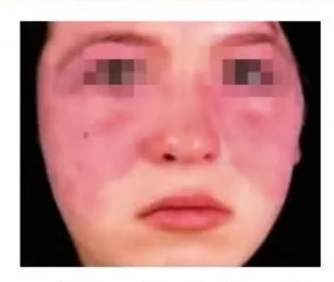
- What is the diagnosis?
- What investigations will you order to confirm the diagnosis?
- What are 4 drugs that lead to this disease?
- What are the treatment options?
- What HLA is associated with this?
Answer
-
Diagnosis:
- Systemic Lupus Erythematosus (SLE)
-
Investigations to confirm diagnosis:
- Anti-dsDNA antibody: High specificity for SLE; correlates with disease activity and renal involvement
- Anti-Smith (anti-Sm) antibody: Most specific antibody for SLE
- ANA (Antinuclear antibody): Highly sensitive screening test
- Serum complement levels (
): Decreased in active disease / flare - Complete blood count & Urinalysis: Leukopenia, thrombocytopenia, proteinuria, active urinary casts
-
4 drugs that lead to drug-induced lupus (DIL):
- S - Sulfonamides / Sulfasalazine
- H - Hydralazine
- I - Isoniazid (INH)
- P - Procainamide / Penicillamine
-
Treatment options:
- Mild/Skin/Joint disease: Hydroxychloroquine (HCQ), NSAIDs, low-dose corticosteroids
- Severe/Organ-threatening (Lupus nephritis, CNS lupus):
- Pulse IV Methylprednisolone followed by high-dose oral corticosteroids
- Cyclophosphamide or Mycophenolate Mofetil (MMF)
- Azathioprine, Methotrexate
- Biologics: Rituximab (anti-CD20), Belimumab (anti-BAFF)
-
HLA associations:
- HLA-DR2, HLA-DR3, HLA-B8
MNJ22-008
True/False:
- Congenital deficiency of C1q, C2, C4 is associated with SLE.
- Positive lupus anticoagulant / anticardiolipin antibodies are also associated with SLE.
- ANA is a good screening test for SLE because it is associated with >95% cases of SLE.
- Serum level of
decrease in active disease. - MC causes of death in SLE are infection, glomerulonephritis, and neuropsychiatric disease.
- Hip joint is never involved in oligoarticular arthritis.
Answer
- True (Early complement component deficiencies impair clearance of immune complexes and apoptotic cells, predisposing to SLE).
- True (Antiphospholipid antibodies are present in 30–40% of pediatric SLE patients).
- True (ANA has high sensitivity of >95%, making it the premier screening test, though specificity is lower).
- True (Classic pathway complement activation and consumption occurs during active flares).
- True (Infections secondary to immunosuppression, active renal disease, and severe CNS lupus are leading causes of mortality).
- True (Hip involvement at presentation is essentially absent in oligoarticular JIA and points to spondyloarthropathy, septic arthritis, or avascular necrosis).
MNJ22-009
Match the following drugs according to their action:
| Drug | Mechanism / Action |
|---|---|
| 1. Methotrexate | A. Anti-CD20 |
| 2. Adalimumab | B. Interleukin-1 receptor antagonist |
| 3. Abatacept | C. DMARD |
| 4. Rituximab | D. Anti-tumor necrosis factor-alpha |
| 5. Anakinra | E. Anti-cytotoxic T-lymphocyte associated antigen 4 Ig |
Answer
- Methotrexate
C. DMARD (Dihydrofolate reductase inhibitor / antimetabolite) - Adalimumab
D. Anti-tumor necrosis factor alpha (Anti-TNF-α mAb) - Abatacept
E. Anti-cytotoxic T-lymphocyte associated antigen 4 Ig (CTLA-4 Ig / T-cell costimulation modulator) - Rituximab
A. Anti CD20 (B-cell depleting monoclonal antibody) - Anakinra
B. Interleukin 1 receptor antagonist (Recombinant IL-1Ra)
MNJ22-010
A 4-year-old child presents with complaints of fatigue and weakness during walking, child also developed red rash around eyes and hands. He has history of gastroenteritis 4 weeks back:
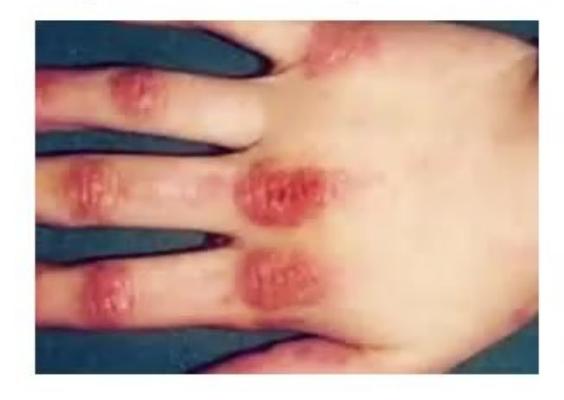
- What is the probable diagnosis?
- What is the sign shown in this picture? And what is present over face?
- What serum enzyme levels will you advise for diagnosis?
- What antibody is present in serum?
- What drug is used for these skin lesions with dose and side effects?
Answer
-
Probable diagnosis:
- Juvenile Dermatomyositis (JDM)
-
Signs shown:
- Hand sign shown in picture: Gottron papules (violaceous, scaly papules over metacarpophalangeal and interphalangeal joints)
- Facial signs: Heliotrope rash (violaceous reddish-purple discoloration of upper eyelids with periorbital edema) and malar erythema
-
Serum enzyme levels to order:
- Creatine Phosphokinase (CPK)
- Serum Glutamic Oxaloacetic Transaminase (SGOT / AST)
- Lactate Dehydrogenase (LDH)
- Aldolase
-
Antibodies present in serum:
- Myositis-specific antibodies (Anti-TIF1-gamma / p155/140, Anti-NXP-2 / MJ, Anti-MDA5)
- Myositis-associated antibodies (Anti-RNP, Anti-PM/Scl, Anti-Ro)
- ANA (positive in 70–80%)
-
Drug for skin lesions, dose, and side effects:
- Drug: Hydroxychloroquine
- Dose:
PO once daily (max ) - Side effects: Retinopathy (maculopathy / bull's eye retinopathy, color vision impairment), gastrointestinal upset, skin pigmentation. Requires periodic ophthalmological monitoring.
MNJ22-011
A 9-year-old female child presents with complaints of fever, malaise, weight loss, headache, and claudication for the last 2 months. On examination, child has high BP and low pulse. Angiography done is shown below:
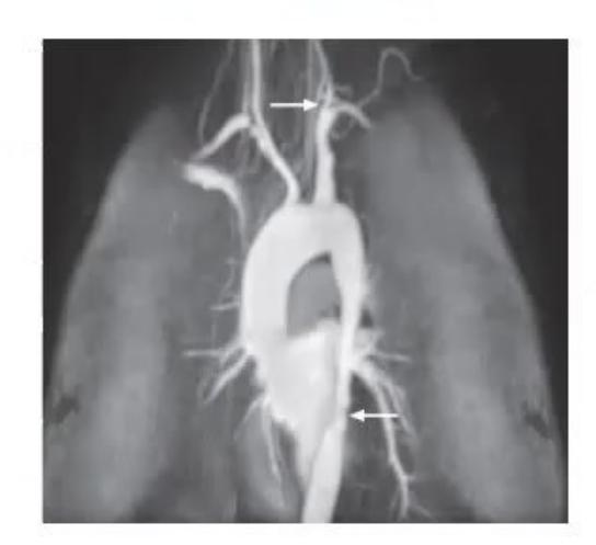
- What is the probable diagnosis?
- What is the most common artery involved in this?
- What are the criteria for diagnosis?
- What is the prognosis?
Answer
-
Probable diagnosis:
- Takayasu Arteritis (Pulseless Disease / Non-specific Aortoarteritis)
-
Most common artery involved:
- Aorta and its primary branches (subclavian arteries, carotid arteries, renal arteries)
-
Diagnostic criteria (EULAR/PRINTO/PRES Criteria):
- Mandatory: Angiographic abnormalities (conventional, CT, or MR angiography) of the aorta or its main branches (stenosis, occlusion, or aneurysm)
- Plus at least ONE of the following:
- Pulse deficit or claudication of an extremity
- Systolic blood pressure discrepancy
between limbs - Bruits over aorta or its major branches
- Hypertension (compared with age- and height-appropriate norms)
- Elevated acute phase reactants (ESR
or elevated CRP)
-
Prognosis:
- Variable; ~20% have a monophasic, self-limiting course, but the majority (~80%) have a chronic relapsing or progressive disease course requiring long-term immunosuppressive therapy. Morbidity is determined by vascular complications (hypertension, retinopathy, stroke, heart failure, renal impairment).