MNJ04-001
EEG of a six-year-old child:
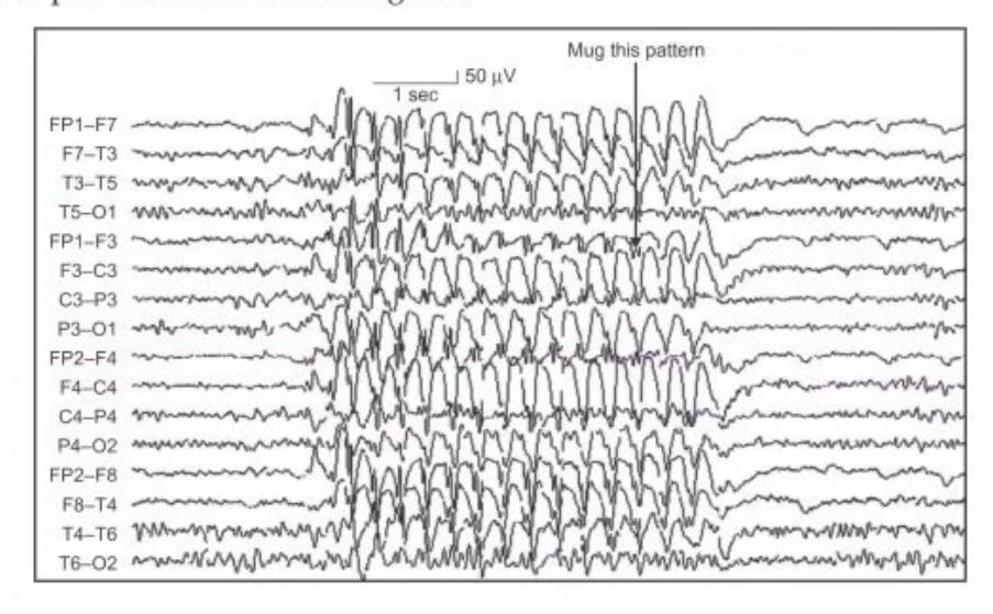
- What does this EEG show?
- What is the drug of choice (write two drugs)?
- What is the prognosis?
- Name one OPD procedure to confirm / provoke the diagnosis.
Answer
- Generalized 3 Hz spike-and-wave discharges characteristic of Absence Seizures (Childhood / Juvenile Absence Epilepsy).
- Valproate, Ethosuximide (or Lamotrigine).
- Good (most children outgrow childhood absence epilepsy by adolescence).
- Hyperventilation (or intermittent photic stimulation).
MNJ04-002
A 10-year-old child came with history of deterioration in school performance and abnormal body movements for the last 3 months. The child now has loss of interest in surroundings. Past history suggests febrile illness with rash around 1 year of age:
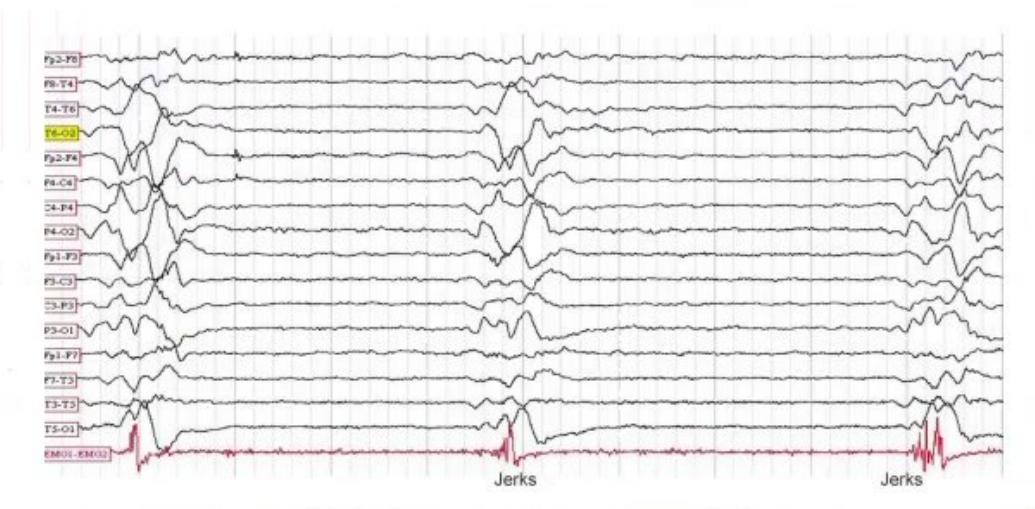
- Diagnosis of clinical condition and EEG pattern.
- Staging of disease.
- Diagnostic modalities of disease.
- Treatment options and recent change in immunization prevention strategy.
Answer
- Diagnosis: Subacute Sclerosing Panencephalitis (SSPE) following measles infection.
- EEG pattern: Periodic high-amplitude slow-wave complexes / burst-suppression pattern (Radermecker complexes) recurring every 4–10 seconds.
- Staging (Jabbour Staging):
- Stage I: Insidious mental deterioration, behavioral/personality changes, decline in school performance.
- Stage II: Myoclonic jerks, choreoathetosis, seizures, motor and language deficits.
- Stage III: Severe dementia, rigidity, decorticate/decerebrate posturing, loss of vision/speech, vegetative state.
- Stage IV: Mutism, coma, loss of autonomic control, death.
- Diagnostic modalities:
- Characteristic EEG (periodic complexes)
- CSF and serum measles antibody titers (elevated anti-measles IgG with reduced serum-to-CSF ratio, oligoclonal bands)
- MRI brain (periventricular white matter hyperintensities)
- Treatment & Prevention:
- Treatment: Inosiplex (isoprinosine), intraventricular/intrathecal interferon-alpha, ribavirin, amantadine.
- Prevention: Measles vaccination (MR/MMR vaccine at 9 and 15 months).
MNJ04-003
A 9-month-old male child admitted with complaints of abnormal movements of head and hand, more than 100 times per day, occurring predominantly when just waking from sleep. Baby is now unable to sit without support (developmental regression over past 2 months). EEG was done:
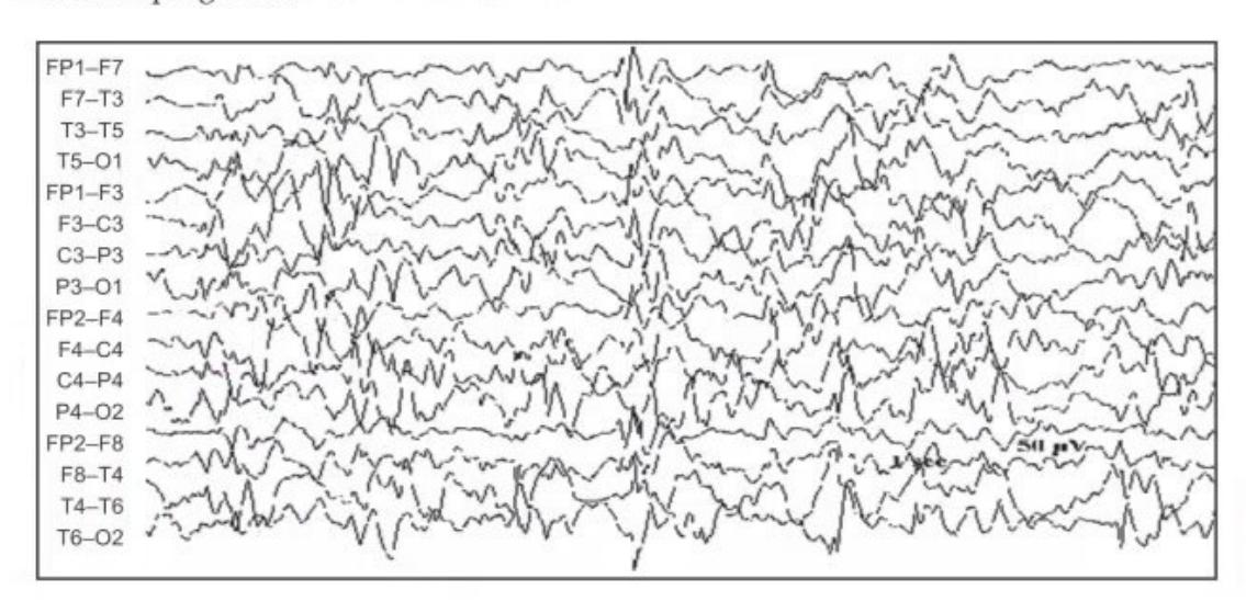
- What is the diagnosis and characteristic EEG pattern?
- What are the types of disease and causes?
- What are the treatment options?
- What is the prognosis?
Answer
- Diagnosis: West syndrome (Infantile spasms with hypsarrhythmia and developmental regression).
- EEG pattern: Hypsarrhythmia (chaotic, high-voltage, disorganized background with multifocal sharp waves, spikes, and polyspikes).
- Types & Causes:
- Symptomatic (Identifiable etiology): Tuberous sclerosis, hypoxic-ischemic encephalopathy (HIE), perinatal stroke, CNS infections, cortical dysplasia, inborn errors of metabolism.
- Cryptogenic / Genetic: Unknown etiology with normal development prior to onset.
- Treatment:
- Adrenocorticotropic hormone (ACTH) (75 U/m²/day or 150 U/m²/day)
- Vigabatrin (first-line especially for tuberous sclerosis)
- Oral corticosteroids (prednisolone)
- Ketogenic diet, valproate, topiramate, or surgery if focal lesion.
- Prognosis: Poor (high risk of cognitive impairment, developmental delay, and evolution into Lennox-Gastaut syndrome).
MNJ04-004
A 3-year-old child admitted with complaints of high-grade fever with seizures, rash, and loss of consciousness for the last 2 days. EEG was done:
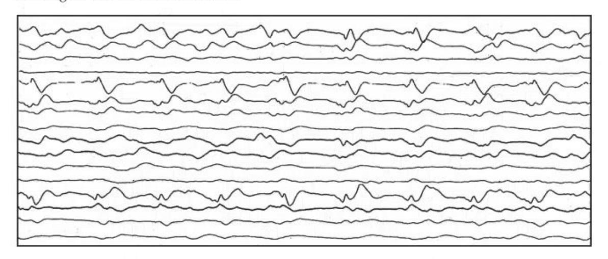
- Diagnosis of EEG pattern and clinical condition.
- Characteristic site of lesion in MRI brain in this disease.
- Drug used in treatment with dose.
Answer
- EEG pattern: Periodic Lateralized Epileptiform Discharges (PLEDs).
- Diagnosis: Herpes Simplex Encephalitis (HSE).
- MRI brain site: Frontotemporal lobes (asymmetric frontotemporal hyperintensity on T2/FLAIR).
- Treatment: Intravenous Acyclovir 10–15 mg/kg/dose (or 500 mg/m²/dose) IV every 8 hours (TDS) for 14–21 days.
MNJ04-005
A 7-year-old boy presents with nocturnal seizures, jerky movements of the lips, eyes wide open, inability to speak, and hypersalivation. EEG was done:
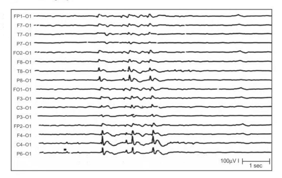
- Diagnosis of the condition.
- Two important clinical features affecting the face/mouth.
- Treatment and prognosis.
Answer
- Diagnosis: Benign Childhood Epilepsy with Centrotemporal Spikes (BECTS) / Benign Rolandic Epilepsy.
- Clinical features:
- Unilateral facial clonic twitching / sensorimotor seizures
- Oropharyngeal symptoms: hypersalivation (drooling), guttural sounds, speech arrest / inability to speak.
- Treatment & Prognosis:
- Treatment: Often observation only without antiepileptic therapy; if frequent/daytime seizures, Carbamazepine, Levetiracetam, or Oxcarbazepine.
- Prognosis: Excellent; spontaneously remits in almost all children by 14–16 years of age with normal neurocognition.
MNJ04-006
An 8-year-old boy with history of delayed milestones (walking at 2 years, speech at 3 years), multiple types of seizures several times a day for 2 months (tonic seizures, atonic drop attacks, atypical absences, myoclonic seizures), and regression of milestones:
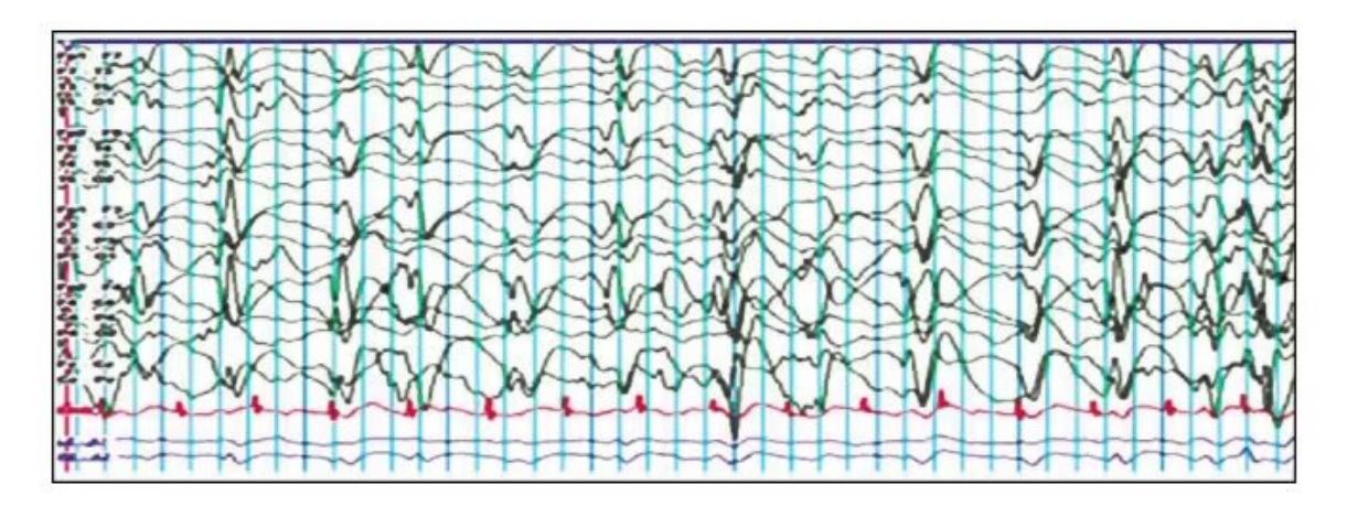
- What is the EEG pattern and diagnosis of the disease?
- Management options and name of surgery.
- What test is done to evaluate hemispheric language and memory dominance before epilepsy surgery?
Answer
- Diagnosis: Lennox-Gastaut Syndrome (LGS).
- EEG pattern: Slow generalized spike-and-wave discharges (<2.5 Hz) with disorganized, slow background and paroxysmal fast activity during sleep.
- Management:
- Medical: Broad-spectrum AEDs (Valproate, Clobazam, Lamotrigine, Topiramate, Rufinamide, Cannabidiol), Ketogenic diet, IVIG.
- Surgical: Corpus callosotomy (effective for atonic drop attacks), Vagus Nerve Stimulation (VNS).
- Pre-surgical evaluation: Wada test (intracarotid sodium amobarbital procedure).
MNJ04-007
A 4-year-old girl presented with fever and recurrent focal seizures for 2 days, presenting in status epilepticus. Seizures stopped after IV lorazepam and phenytoin. However, the patient remains comatose >24 hours after all motor seizures have stopped:
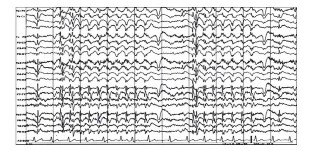

- Diagnose the EEG pattern and clinical condition.
- What is the best way to diagnose and monitor this problem?
Answer
- Diagnosis: Non-Convulsive Status Epilepticus (NCSE).
- EEG features: Continuous rhythmic spikes, sharp waves, or spike-wave discharges without overt convulsive motor movements, showing electrical improvement upon IV antiepileptic administration.
- Diagnostic modality: Continuous 24-hour video-EEG monitoring (cEEG).
MNJ04-008
A 2-month-old infant presents with hypotonia. CXR and ECG are shown:
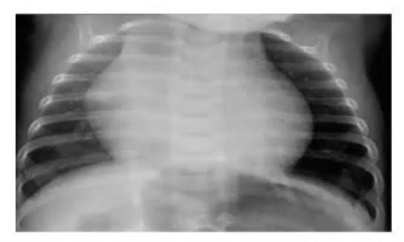
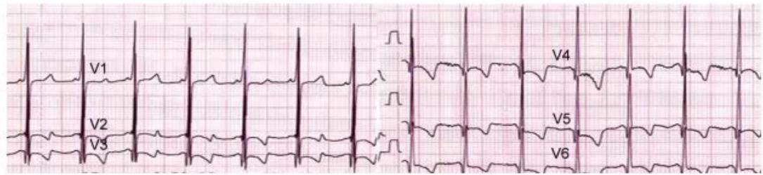
- What findings in CXR and ECG suggest the diagnosis?
- What are the differential diagnoses?
- What test confirms the diagnosis?
- What is the specific treatment?
Answer
- Diagnosis: Pompe disease (Glycogen Storage Disease Type II / Acid maltase deficiency).
- CXR: Massive cardiomegaly (globular heart) in a floppy infant.
- ECG: Extremely tall QRS voltages (giant biventricular hypertrophy) requiring half-standard calibration, short PR interval, and deep T-wave inversions.
- Differential diagnosis: Spinal muscular atrophy (SMA type 1), congenital muscular dystrophy, hypotonic cerebral palsy, Down syndrome, viral myocarditis.
- Confirmatory test: Acid alpha-glucosidase (GAA) enzyme activity assay in dried blood spots/leukocytes/skin fibroblasts; GAA gene mutation analysis.
- Treatment: Enzyme Replacement Therapy (ERT) with recombinant human acid alpha-glucosidase (Alglucosidase alfa / Myozyme / Lumizyme) 20 mg/kg IV every 2 weeks.
MNJ04-009
Review the ECG tracing below:
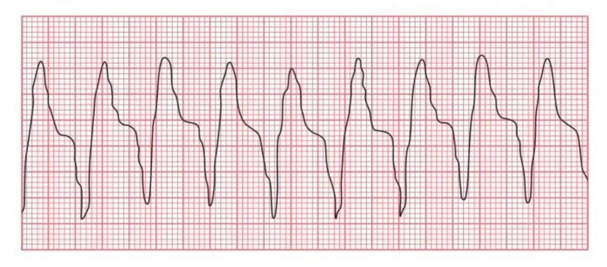
- What is the heart rate?
- Is the QRS complex wide or narrow? What is the rhythm diagnosis?
- List 4 possible causes.
- What is the treatment?
Answer
- Rate: >150–200 beats/min (tachycardia).
- QRS & Diagnosis: Wide QRS complex (>0.08–0.09 s); Ventricular Tachycardia (VT).
- Causes: Myocarditis, congenital heart disease (post-operative repair, e.g. TOF repair), ALCAPA, Long QT syndrome, electrolyte disturbance (hypokalemia/hypomagnesemia), drug toxicity (digoxin, antiarrhythmics), cardiac tumors (rhabdomyoma).
- Treatment:
- Hemodynamically unstable (pulseless/hypotensive): Immediate synchronized cardioversion (0.5–1 J/kg, up to 2 J/kg) or defibrillation if pulseless.
- Hemodynamically stable: IV Amiodarone (5 mg/kg over 20–60 min), IV Lidocaine (1 mg/kg bolus), or IV Procainamide.
MNJ04-010
Review the ECG below:
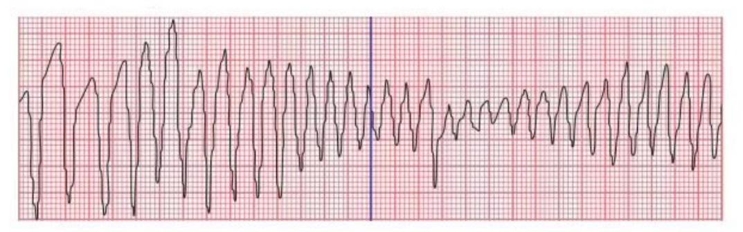
- Diagnose the condition shown in the ECG.
- List 3 causes leading to this condition, including 2 electrolyte disturbances.
- What is the immediate treatment of choice?
Answer
- Diagnosis: Torsades de Pointes (polymorphic ventricular tachycardia characterized by twisting of QRS points around the isoelectric line, rate 150–250 bpm).
- Causes:
- Prolonged QT interval (congenital Long QT syndromes: Jervell-Lange-Nielsen, Romano-Ward)
- Electrolyte disturbances: Hypokalemia, Hypomagnesemia, Hypocalcemia
- Drugs: Macrolides, fluoroquinolones, ondansetron, antiarrhythmics (Class Ia, III), psychotropics.
- Treatment:
- IV Magnesium sulphate (25–50 mg/kg IV over 10–15 min).
- If unstable: Immediate defibrillation/cardioversion.
- Overdrive pacing / Isoproterenol infusion.
MNJ04-011
Diagnose these 2 ECG findings and give one possible cause for each:
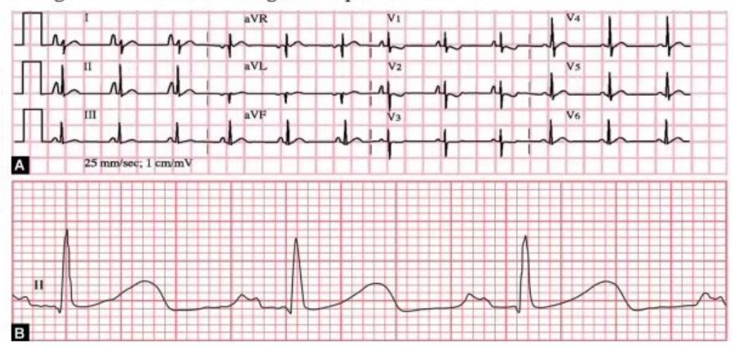
- ECG finding in Image 1 and cause.
- ECG finding in Image 2 and cause.
Answer
- P-pulmonale: Tall, peaked P waves (>2.5 mm in lead II / >3 SD) indicating Right Atrial Enlargement (e.g., Pulmonary hypertension, severe pulmonary stenosis, tricuspid atresia, chronic lung disease).
- P-mitrale: Broad, bifid/notched P waves (>0.10 s duration / >3 SD) indicating Left Atrial Enlargement (e.g., Mitral stenosis, mitral regurgitation, severe left ventricular failure).
MNJ04-012
Diagnose these 2 ECG conduction abnormalities:
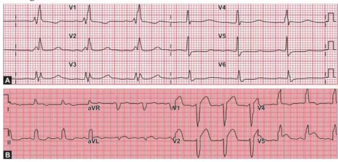
- Diagnosis and key morphology in Image 1.
- Diagnosis and key morphology in Image 2.
Answer
- Right Bundle Branch Block (RBBB):
- Wide QRS complex (>0.12 s in older children / >0.09 s in infants).
- "M" shaped rsR' / rSR' pattern in right precordial leads (V1, V2, V3R).
- Wide slurred S wave in lateral leads (I, aVL, V5, V6).
- Left Bundle Branch Block (LBBB):
- Wide QRS complex (>0.12 s).
- "M" shaped broad notched R wave in lateral leads (V5, V6, lead I, aVL).
- "W" shaped QS / rS pattern in lead V1.
MNJ04-013
A 20-day-old infant presents with fussiness and poor feeding. You are not sure about P waves as quality is poor. BP: 80/50 mm Hg, CFT: <2 seconds:

- What is the diagnosis and ECG features of this condition?
- Calculate heart rate on the ECG and what should be the next diagnostic step?
- Name one drug which is contraindicated in infants <1 year with this rhythm, name a simple non-pharmacological vagal maneuver, and state how many attempts can be made.
- If vagal maneuvers fail in a stable patient, what drug would you give? Give name, dose for a 4 kg baby and adult dose, and 2 administration precautions.
- Prior to giving medication, the patient becomes hypotensive, mottled, and cold with HR 300 bpm. What is the immediate next therapeutic step?
Answer
- Diagnosis: Supraventricular Tachycardia (SVT) (narrow complex tachycardia, regular rhythm, absent/retrograde P waves).
- Heart rate: ~300 bpm (1500 / 5 small boxes). Next step: 12-lead ECG recording at double speed (50 mm/s) to differentiate P waves.
- Contraindicated drug: Verapamil (causes profound hypotension, cardiovascular collapse, and asystole in infants <1 year).
- Vagal maneuver: Ice bag / ice-cold water bag applied to forehead and mid-face for 10–15 seconds (without occluding airway). Max 2 attempts.
- Pharmacological therapy:
- Adenosine: Initial dose 0.1 mg/kg (0.4 mg for 4 kg baby; max first dose 6 mg for adult/70 kg), followed by 0.2 mg/kg if needed.
- Precautions: Rapid IV push via a large proximal vein (antecubital/central) followed immediately by rapid normal saline flush (5–10 mL); continuous ECG monitoring.
- Immediate step for unstable patient: Synchronized cardioversion (0.5–1 J/kg, increasing to 2 J/kg if refractory) with appropriate airway management and oxygenation.
MNJ04-014
Identify arrhythmias A, B, C, and D:
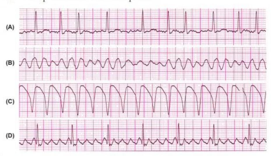
- Diagnose patterns A, B, C, and D.
- What is the treatment of choice for pattern D in an unstable patient?
- List 3 drugs useful in pharmacological management of pattern D.
- What major complication can occur secondary to chronic rhythm D?
Answer
- Identifications:
- A: Atrial Fibrillation (irregularly irregular baseline, absent P waves)
- B: Ventricular Fibrillation (chaotic, disorganized waveforms)
- C: Ventricular Tachycardia (wide regular monomorphic complexes)
- D: Atrial Flutter (regular "sawtooth" flutter waves / F waves)
- Treatment for unstable Atrial Flutter (D): Synchronized electrical cardioversion (0.5–1 J/kg).
- Drugs for Atrial Flutter: Beta-blockers (e.g., Esmolol, Propranolol), Digoxin, Calcium channel blockers (in older children), Amiodarone, Flecainide.
- Complication: Intracardiac thrombus formation leading to thromboembolism and ischemic stroke.
MNJ04-015
Review the ECG below:

- What is the diagnosis of this ECG pattern? Give reason.
- List 3 indications for investigating further in patients with premature ventricular complexes.
- What is the emergency intravenous medication if suppression is needed?
Answer
- Diagnosis: Premature Ventricular Contractions (PVCs) in a ventricular bigeminy pattern (alternating normal sinus beat and premature wide bizarre QRS complex).
- Indications for further investigation:
- Multifocal / multiform PVCs
- Runs of consecutive PVCs (couplets, triplets, non-sustained VT)
- PVCs that increase with exercise / exertion
- Presence of underlying structural heart disease or LV dysfunction
- "R-on-T" phenomenon
- Emergency medication: Intravenous Lidocaine (1 mg/kg bolus followed by continuous infusion) or Amiodarone.
MNJ04-016
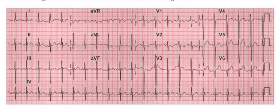
A 2-month-old infant presents with left ventricular dysfunction, mitral regurgitation, and cardiomegaly:
- Identify the key abnormalities in this ECG.
- What is the diagnosis and differential diagnosis?
Answer
- ECG Abnormalities:
- Deep, wide Q waves in lateral leads (I, aVL, V5, V6)
- ST segment elevation / depression and T wave inversion in anterolateral leads
- Signs of anterolateral myocardial ischemia / infarction and left ventricular hypertrophy
- Diagnosis & Differential:
- Diagnosis: Anomalous Left Coronary Artery from the Pulmonary Artery (ALCAPA / Bland-White-Garland syndrome).
- Differential Diagnosis: Dilated cardiomyopathy (DCM), acute viral myocarditis, severe infantile colic / intussusception (due to episodic irritability/crying during angina).
MNJ04-017
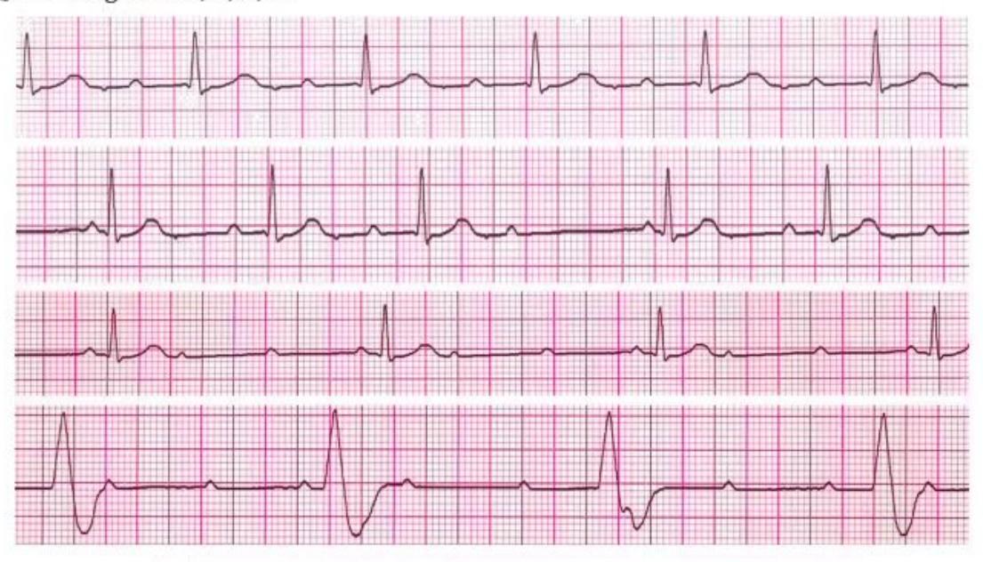
Identify the four degrees of atrioventricular (AV) block shown in A, B, C, and D:
- Diagnose pattern A.
- Diagnose pattern B.
- Diagnose pattern C.
- Diagnose pattern D.
Answer
- A: First-degree AV block (Fixed, prolonged PR interval > 0.20 s; every P wave conducted to a QRS).
- B: Second-degree AV block, Mobitz Type I (Wenckebach) (Progressive PR prolongation until a P wave is non-conducted / dropped).
- C: Second-degree AV block, Mobitz Type II (Constant PR interval with intermittent non-conducted P waves).
- D: Third-degree (Complete) AV block (Complete AV dissociation; P waves and QRS complexes beat independently).
MNJ04-018
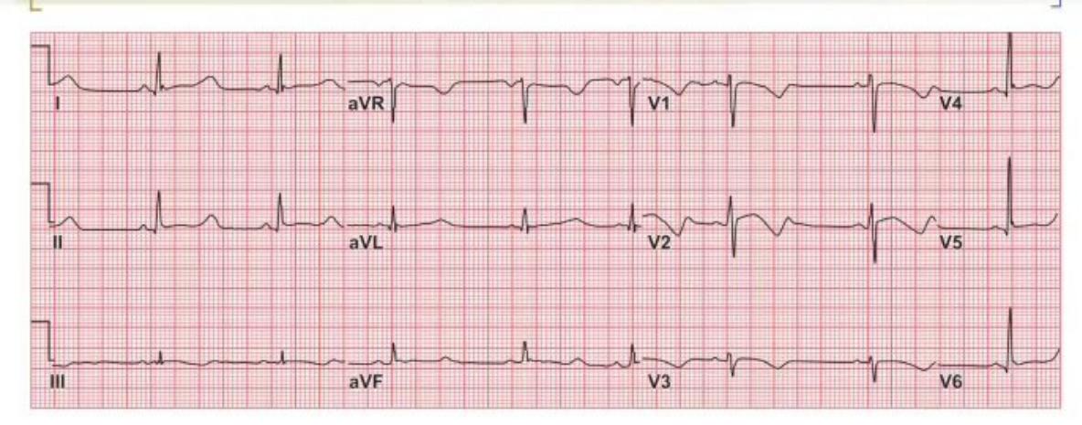
Review the ECG below:
- Identify the abnormality in the ECG.
- What electrolyte abnormality can cause this?
- Why is it important to identify this rhythm disturbance?
Answer
- Abnormality: Prolonged QT interval (QTc > 0.46 s, specifically ST segment lengthening).
- Electrolyte cause: Hypocalcemia (causes lengthening of the ST segment leading to QT prolongation without T wave widening); also hypomagnesemia, hypokalemia.
- Clinical importance: Prolonged QT interval predisposes to life-threatening polymorphic ventricular tachycardia (Torsades de Pointes), ventricular fibrillation, syncope, and sudden cardiac death.
MNJ04-019
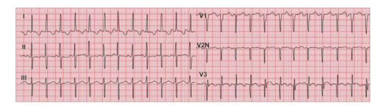
A 2-month-old baby admitted with recurrent cough, cold, irritability, dyspnea, and sweating:
- What is the diagnosis?
- Name 4 EKG findings that help in diagnosis.
- What is the definitive diagnostic test?
- Name the treatment options.
Answer
- Diagnosis: ALCAPA (Anomalous Left Coronary Artery from the Pulmonary Artery / Bland-White-Garland syndrome).
- EKG findings:
- Deep and wide Q waves in leads I, aVL, V5, V6
- ST-segment elevation or depression in leads I, aVL, V5, V6
- Inverted T waves in anterolateral leads (I, aVL, V5-V6)
- Diminished R wave progression or left ventricular hypertrophy / strain pattern
- Diagnostic test: Transthoracic Echocardiography with Doppler (demonstrating retrograde flow into PA) / Coronary CT Angiography / Cardiac Catheterization & Aortography.
- Treatment:
- Medical: Anti-failure therapy (diuretics, ACE inhibitors, inotropic support) and afterload reduction.
- Surgical (Definitive): Direct coronary reimplantation into the aorta (preferred) or Takeuchi procedure (intrapulmonary baffle) / ligation of ALCAPA.
MNJ04-020
Identify the acid-base disorders in columns A, B, C, and D and fill in the missing delta calculations:
| ABG Parameter | A | B | C | D |
|---|---|---|---|---|
| pH | 7.40 | 7.29 | 7.38 | 7.10 |
| pCO₂ (mm Hg) | 40 | 30 | 35 | 20 |
| HCO₃⁻ (mEq/L) | 24 | 14 | 20 | 6 |
| Anion Gap (AG) | 12 | 20 | 26 | 22 |
| ΔAG (AG - 12) | ||||
| ΔHCO₃⁻ (24 - HCO₃⁻) | ||||
| Delta Ratio (ΔAG / ΔHCO₃⁻) |
- Calculate ΔAG, ΔHCO₃⁻, and Delta Ratio for A, B, C, and D.
- State the final acid-base diagnosis for each column (A, B, C, and D).
Answer
- Calculations:
| Calculation | A | B | C | D |
|---|---|---|---|---|
| ΔAG (AG - 12) | 0 | +8 (20-12) | +14 (26-12) | +10 (22-12) |
| ΔHCO₃⁻ (24 - HCO₃⁻) | 0 | +10 (24-14) | +4 (24-20) | +18 (24-6) |
| Delta Ratio (ΔAG / ΔHCO₃⁻) | - | 0.8 (8/10) | 3.5 (14/4) | 0.55 (10/18) |
- Diagnoses:
- A: Normal acid-base status.
- B: High Anion Gap Metabolic Acidosis (HAGMA) with expected respiratory compensation (Winter's formula: 1.5×14 + 8 = 29 ± 2). Delta ratio ~0.8–1.0 represents pure high AG metabolic acidosis.
- C: Mixed High Anion Gap Metabolic Acidosis + Metabolic Alkalosis (Delta ratio 3.5 > 2.0 indicates concurrent metabolic alkalosis elevating HCO₃⁻).
- D: Mixed High Anion Gap Metabolic Acidosis + Normal Anion Gap Metabolic Acidosis (Delta ratio 0.55 < 0.8 indicates additional non-AG metabolic acidosis).
MNJ04-021
A child with short stature presents to the OPD. ABG and laboratory reports are given below:
| Parameter | Value |
|---|---|
| FiO₂ | 100% |
| PaO₂ | 477 mm Hg |
| PaCO₂ | 47 mm Hg |
| pH | 7.23 |
| HCO₃⁻ | 19 mEq/L |
| Hb | 11 g/dL |
| Serum Albumin | 2.0 g/dL |
| Serum Na⁺ | 131 mEq/L |
| Serum K⁺ | 3.4 mEq/L |
| Serum Cl⁻ | 104 mEq/L |
| Urine Na⁺ | 146 mEq/L |
| Urine K⁺ | 27.6 mEq/L |
| Urine Cl⁻ | 146 mEq/L |
| Urine pH | 6.1 |
- What is the ABG diagnosis?
- Calculate the corrected expected normal Anion Gap accounting for the serum albumin level.
- Calculate ΔAG, ΔHCO₃⁻, and Delta Ratio.
- Calculate the Urinary Anion Gap (UAG).
- What is the most likely clinical diagnosis?
Answer
- ABG diagnosis: Mixed Respiratory Acidosis (elevated PaCO₂ 47) and Metabolic Acidosis (low pH 7.23, low HCO₃⁻ 19).
- Corrected Anion Gap:
- Measured AG = Na - (Cl + HCO₃) = 131 - (104 + 19) = 8 mEq/L
- Expected Normal AG = 12 - 2.5 × (4.0 - Albumin) = 12 - 2.5 × (4.0 - 2.0) = 7 mEq/L.
- Measured AG (8) equals the expected corrected AG, so this is a Normal Anion Gap (Hyperchloremic) Metabolic Acidosis (NAGMA).
- Delta calculations:
- ΔAG = 8 - 8 = 0
- ΔHCO₃⁻ = 24 - 19 = 5
- Delta ratio = 0 (Confirms pure NAGMA).
- Urinary Anion Gap (UAG):
(Positive UAG indicates impaired urinary ammonium excretion).
- Clinical Diagnosis: Distal Renal Tubular Acidosis (Type 1 RTA) (positive urinary anion gap + inability to acidify urine with urine pH > 6.0 in the presence of systemic metabolic acidosis + hypokalemia + short stature / growth failure).
MNJ04-022
The ABG of a patient breathing room air (FiO₂: 0.21) is given below:
| Parameter | Value |
|---|---|
| FiO₂ | 0.21 (Room air) |
| PaO₂ | 48 mm Hg |
| PaCO₂ | 40 mm Hg |
| pH | 7.40 |
| HCO₃⁻ | 24 mEq/L |
- What is the abnormality on this ABG?
- How severe is it?
- What is the immediate management?
Answer
- Abnormality: Severe Hypoxemia / Type 1 Respiratory Failure (normal PaCO₂ 40 with PaO₂ 48 mm Hg on room air; normal PaO₂ should be 80–100 mm Hg).
- Severity: Moderately severe to severe hypoxemia (PaO₂ < 60 mm Hg corresponds to SpO₂ < 90%).
- Management:
- Immediately administer supplemental oxygen (increase FiO₂ via face mask / high-flow nasal cannula) to achieve PaO₂ > 80 mm Hg / SpO₂ 94–98%.
- Investigate underlying pulmonary etiology (e.g., CXR for pneumonia, atelectasis, pneumothorax, pulmonary edema).
MNJ04-023
ABG of a patient on mechanical ventilation is given below:
| Parameter | Value |
|---|---|
| FiO₂ | 0.80 (80%) |
| PaO₂ | 220 mm Hg |
| PaCO₂ | 34.6 mm Hg |
| pH | 7.48 |
| HCO₃⁻ | 26 mEq/L |
- What is the abnormality on this ABG?
- How are the lungs functioning?
- Can the patient be weaned directly to room air?
- What should you do next?
Answer
- Abnormality: Hyperoxemia (PaO₂ 220 mm Hg on 80% oxygen) and mild respiratory alkalosis (pH 7.48, PaCO₂ 34.6).
- Lung function: The expected PaO₂ on 80% oxygen in healthy lungs is ~5 × FiO₂% = 5 × 80 = 400 mm Hg. The PaO₂/FiO₂ ratio is 220 / 0.80 = 275 (indicating mild-to-moderate lung injury/V-Q mismatch).
- Weaning to room air: No, the patient cannot be extubated/weaned directly to room air. If placed on room air (FiO₂ 0.21), the predicted PaO₂ would be
(hypoxemic). - Action:
- Stepwise reduce FiO₂ from 0.80 to 0.35–0.40 to target a safe PaO₂ of ~80–100 mm Hg and prevent oxygen toxicity.
- Slightly reduce minute ventilation to normalize pH and PaCO₂.
MNJ04-024
ABG of a patient on mechanical ventilator with FiO₂: 40%, Tidal Volume (TV): 500 mL, Respiratory Rate (RR): 12 breaths/min is given below:
| Parameter | Value |
|---|---|
| FiO₂ | 0.40 |
| PaO₂ | 128 mm Hg |
| PaCO₂ | 60 mm Hg |
| pH | 7.21 |
| HCO₃⁻ | 24 mEq/L |
- What is the acid-base and respiratory abnormality?
- What ventilator adjustment should you make to achieve a target PaCO₂ of 40 mm Hg?
Answer
- Abnormality: Uncompensated Acute Respiratory Acidosis with hypercapnia (pH 7.21, PaCO₂ 60 mm Hg) due to alveolar hypoventilation, with adequate oxygenation (PaO₂ 128 mm Hg on 40% FiO₂).
- Ventilator adjustment:
- Increase minute ventilation by increasing the Respiratory Rate (RR).
- Formula:
. - Increase ventilator Respiratory Rate from 12 to 18 breaths/min (and slightly titrate down FiO₂ to 30–35%).
MNJ04-025
A baby is admitted to the NICU with persistent pulmonary hypertension of the newborn (PPHN). He is on a mechanical ventilator with FiO₂ 100%, PIP 35 cm H₂O, PEEP 6 cm H₂O, SpO₂ 85%, and has the following ABG values:
- pH: 7.22, PaCO₂: 50 mm Hg, PaO₂: 50 mm Hg, Na⁺: 136 mEq/L, K⁺: 4.0 mEq/L, Cl⁻: 103 mEq/L
- Calculate the Alveolar-arterial oxygen gradient (
). Assume Respiratory Quotient (RQ) = 0.8, barometric pressure , water vapor pressure . - What are the indications for Extracorporeal Membrane Oxygenation (ECMO) in severe neonatal respiratory failure / PPHN?
Answer
-
Alveolar-arterial oxygen gradient (
) Calculation: - Alveolar
:
Gradient:
- Alveolar
-
Indications for ECMO:
- Oxygenation Index (OI) > 40 on 100% oxygen for > 2–4 hours, or OI > 25 for > 12–24 hours.
for > 8–12 hours (>80% predicted mortality without ECMO). - Intractable severe hypoxemia / acidosis unresponsive to maximal medical therapy (inhaled nitric oxide, HFOV, surfactant, inotropic support).
- Acute reversible respiratory/cardiac failure in gestational age ≥ 34 weeks and birth weight ≥ 2.0 kg without major intracranial hemorrhage (Grade III/IV) or lethal chromosomal anomalies.
MNJ04-026
A 5-month-old boy with developmental delay presents with the following ABG and electrolyte report:
- pH: 6.64, PaCO₂: 25.8 mm Hg, PaO₂: 396.4 mm Hg (on high FiO₂), Na⁺: 140 mEq/L, K⁺: 4.3 mEq/L, Cl⁻: 95 mEq/L, HCO₃⁻: 5 mEq/L
- List the abnormalities in this ABG report.
- Calculate the Serum Anion Gap.
- List 4 conditions associated with high anion gap metabolic acidosis in infants.
- List 2 conditions associated with a decreased / low anion gap.
Answer
- ABG abnormalities:
- Severe uncompensated / partially compensated Metabolic Acidosis with marked acidemia (pH 6.64, HCO₃⁻ 5 mEq/L, compensatory hyperventilation with PaCO₂ 25.8 mm Hg).
- Hyperoxia (PaO₂ 396.4 mm Hg).
- Anion Gap Calculation:
(Markedly elevated; normal is ). - Causes of High Anion Gap Acidosis in infants:
- Inborn errors of metabolism (organic acidemias, maple syrup urine disease, methylmalonic acidemia)
- Lactic acidosis (sepsis, mitochondrial disorders, severe tissue hypoxia)
- Diabetic ketoacidosis (DKA)
- Renal failure / Uremia
- Toxic ingestions (salicylates, methanol, ethylene glycol)
- Causes of Decreased / Low Anion Gap (< 4–8 mEq/L):
- Hypoalbuminemia (most common)
- Severe hypercalcemia, hypermagnesemia
- Lithium toxicity
- Multiple myeloma (monoclonal IgG paraproteinemia)
- Severe bromide / iodide ingestion
MNJ04-027
Interpret the following three ABG scenarios:
- Patient 1: pH: 7.60, PaCO₂: 25 mm Hg, PaO₂: 160 mm Hg, HCO₃⁻: 24 mEq/L (FiO₂: 50% / 0.50).
- Calculate
( ) and write the formula used.
- Calculate
- Patient 2: pH: 7.38, PaCO₂: 65 mm Hg, PaO₂: 48 mm Hg, HCO₃⁻: 34 mEq/L (FiO₂: 60% / 0.60).
- a. What is the ABG diagnosis?
- b. What is the expected normal PaO₂ if a child is breathing room air with healthy lungs?
- Patient 3: pH: 7.45, PaCO₂: 40 mm Hg, PaO₂: 120 mm Hg, HCO₃⁻: 28.5 mEq/L, SpO₂: 99%, Hb: 8.2 g/dL.
- Calculate the total Arterial Oxygen Content (
) and write the formula.
- Calculate the total Arterial Oxygen Content (
Answer
- Patient 1 (
Calculation): - Formula:
.
- Formula:
- Patient 2:
- a. Diagnosis: Fully compensated Chronic Respiratory Acidosis (pH normal 7.38, elevated PaCO₂ 65 mm Hg with compensatory renal retention of HCO₃⁻ 34 mEq/L) with severe hypoxemia / V-Q mismatch (PaO₂ 48 mm Hg on 60% oxygen).
- b. Expected PaO₂ on room air: 80–100 mm Hg.
- Patient 3 (Arterial Oxygen Content
Calculation): - Formula:
of blood.
- Formula: