THG34-001
A 2-year-old boy fell from height and presented to emergency room (ER) with complaints of vomiting thrice with altered sensorium. CT brain done is displayed here.
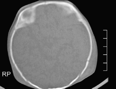
a. Identify the CT findings.
b. What is the primary survey in trauma resuscitation? List the parameters you assess in primary survey.
c. What is PECARN rule used for?
d. What is the PECARN recommendation for child <2 years of age?
Answer
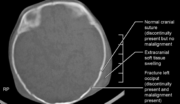
- a. CT brain plain (Bone window): Left occipital bone fracture line with slight diastasis and overlying extracranial scalp soft tissue swelling.
- b. Primary survey in trauma resuscitation: Order of priority in initial assessment and resuscitation:
- A: Airway maintenance with cervical spine stabilization/protection
- B: Breathing and ventilation
- C: Circulation with bleeding/hemorrhage control
- D: Disability — neurologic status evaluation (GCS, pupils)
- E: Exposure (complete visualization) and Environmental control (hypothermia prevention)
- c. PECARN rule: Pediatric Emergency Care Applied Research Network rule clinically predicts/identifies children at very low risk of clinically important traumatic brain injury (ciTBI) to avoid unnecessary CT head scans.
- d. PECARN Clinical Prediction Rule:
For age < 2 years:
| CT Recommendation | Clinical Conditions |
|---|---|
| CT Recommended | GCS < 15, Altered mental status, or Palpable skull fracture |
| Observation vs CT (CT if multiple findings, worsening signs, age < 3 months, or parental preference) | Non-frontal scalp hematoma, Loss of consciousness (LOC) > 5 seconds, Severe mechanism of injury, or Not acting normally per parent |
For age ≥ 2 years:
| CT Recommendation | Clinical Conditions |
|---|---|
| CT Recommended | GCS < 15, Altered mental status, or Signs of basilar skull fracture (hemotympanum, CSF otorrhea/rhinorrhea, raccoon eyes, Battle sign) |
| Observation vs CT (CT if multiple findings, worsening signs, or parental preference) | History of LOC, History of vomiting, Severe mechanism of injury, or Severe headache |
THG34-002
A 2-year-old girl met with a road traffic accident (RTA), had loss of consciousness for 5 minutes following which she regained consciousness but became drowsy after 2 hours. She sustained a lacerated wound in left temporal region. This is the CT brain image of this child.
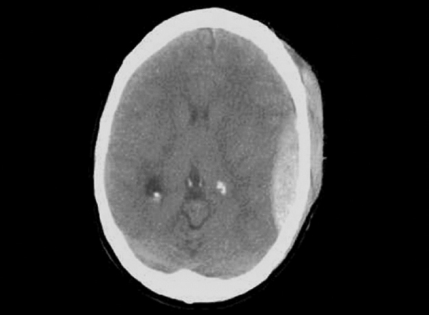
a. Mention the CT findings and diagnosis.
b. Which vessel injury occurs in this condition?
c. What is the complication you expect here?
d. What clinical sign described in the above scenario gives a clue for this diagnosis?
e. What will be your management steps?
Answer
- a. CT brain plain (Brain window): Soft tissue swelling in left temporal region, biconvex / lenticular hyperdense extra-axial lesion in left temporoparietal region — suggestive of extradural hemorrhage (EDH).
- Mnemonic (BCCT for hyperdense lesion on CT brain): Blood, Calcification, Contrast-enhanced ring lesion, Tuber.
- Mnemonic for EDH: Biconvex / lens-shaped (side view of idli).
- b. Tearing of branches of the middle meningeal artery (less commonly middle meningeal vein or dural venous sinus) between the dura mater and inner table of the skull.
- c. Uncal/transtentorial herniation, raised intracranial pressure, seizures, and respiratory arrest.
- d. Lucid interval: Transient initial loss of consciousness after trauma followed by temporary recovery of normal consciousness, and subsequent rapid neurological deterioration as the arterial hematoma expands.
- e. Management:
- Airway, breathing, and circulation stabilization.
- Neuroprotective measures: 30° head-end elevation, maintain normothermia, normoglycemia, normal electrolytes, and avoid hypovolemia/hypotension.
- Urgent neurosurgical consultation for craniotomy and surgical evacuation of hematoma if volume > 30 mL, thickness > 15 mm, midline shift > 5 mm, or GCS < 9 / neurological deterioration.
THG34-003
A 10-year-old boy fell down from a height and following which he complained headache and had nonprojectile vomiting. He was brought to ER 10 hours after injury with active seizure for 20 minutes.
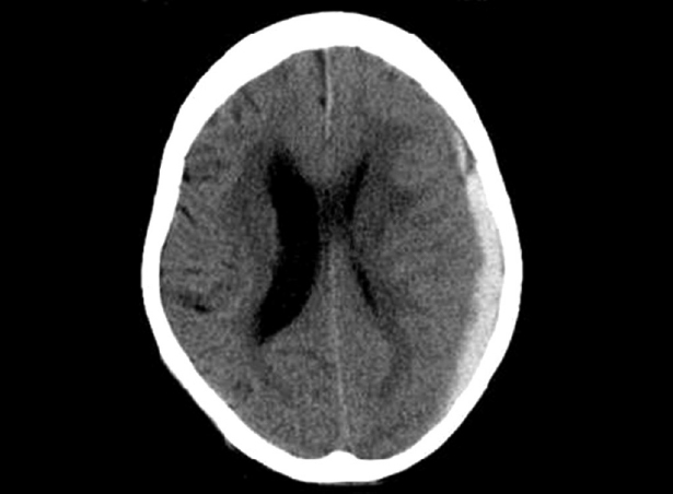
a. What is the CT finding here and what is the diagnosis?
b. Which vessel injury will cause this type of CT image?
c. How do you differentiate this injury between acute and chronic?
d. How will you manage this case?
Answer
- a. CT brain plain: Extra-axial hyperdense crescent-shaped (concavo-convex) collection along the left cerebral hemisphere with compression of left lateral ventricle and significant midline shift — Acute Subdural Hemorrhage (SDH).
- b. Tearing of bridging cortical veins traversing the subdural space between arachnoid and dural venous sinuses.
- c.
- Acute SDH: Hyperdense (bright white, 0–3 days).
- Subacute SDH: Isodense (3 days – 3 weeks).
- Chronic SDH: Hypodense (dark, > 3 weeks).
- d. ABC stabilization, emergency seizure termination (IV benzodiazepine followed by Levetiracetam/Fosphenytoin loading), hyperosmolar therapy (3% NaCl / Mannitol) for raised ICP, and urgent neurosurgical craniotomy/burr hole evacuation.
THG34-004
This 2-month-old boy previously well infant presented with sudden onset of seizures and lethargy with irritability and refusal of feeds. He was delivered at home and birth vaccination was given at 2 weeks of birth. On arrival to hospital, he had active seizures, bulging anterior fontanelle, and pallor. The child was intubated, seizure controlled with medication, and CT brain was taken.
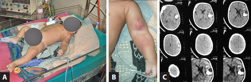
a. Any clue from the images A and B? What is the clinical finding and probable diagnosis?
b. What is the CT finding?
c. What are the possible causes you consider for this condition?
d. Can you correlate the clinical image with radiology and give a diagnosis?
e. What measures could have prevented this?
f. What is the biomarker used in this condition?
Answer
- a. Cutaneous ecchymosis and bruising on the left arm and forearm with acute neurological deterioration, indicating a systemic bleeding diathesis with intracranial hemorrhage.
- b. CT brain plain: Hyperdense intraparenchymal hemorrhage in the left temporal lobe with surrounding hypodense perilesional edema.
- c. Late Vitamin K Deficiency Bleeding (VKDB), inherited bleeding disorders (Hemophilia A/B, Factor VII deficiency), severe immune thrombocytopenia, child abuse / non-accidental trauma, and vascular malformations (AVM).
- d. Late-onset Vitamin K Deficiency Bleeding (VKDB) in an exclusively breastfed, home-delivered infant who missed neonatal prophylactic vitamin K.
- e. Routine administration of intramuscular Vitamin K1 (1 mg IM) at birth to all neonates.
- f. PIVKA-II (Proteins Induced by Vitamin K Absence or Antagonist-II) / undercarboxylated prothrombin.
THG34-005
A 16-year-old boy met with a road traffic accident (RTA). He was riding a bike and it collided with a lorry. This CT brain was taken after 12 hours (image A). He remained unconscious for 3 days, hence MRI brain was done on day 4 (images B and C).
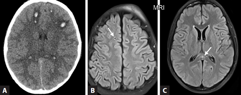
a. What are the radiological findings?
b. What is the diagnosis?
c. What is the mechanism of injury and extent of the lesion?
Answer
- a.
- CT brain plain (Image A): Bilateral scalp hematomas with punctate microhemorrhages at the grey-white matter junction without focal mass lesion or midline shift.
- MRI brain (Images B & C): Multiple punctate hyperintense foci on T2/FLAIR in the subcortical white matter of the right frontal lobe and the splenium of the corpus callosum.
- b. Diffuse Axonal Injury (DAI).
- c. Shearing and rotational acceleration-deceleration forces that stretch and tear axonal cytoskeleton and microvasculature across grey-white matter boundaries, corpus callosum, and upper brainstem.
THG34-006
A 10-month-old infant presented with developmental delay and microcephaly presented with recurrent seizures. CT brain image is displayed here.
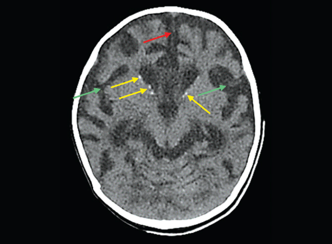
a. What are the CT findings?
b. What is the diagnosis?
c. What finding will you expect in ophthalmological evaluation?
Answer
- a. CT brain plain: Prominent periventricular punctate and linear calcifications outlining the anterior horns of the lateral ventricles, along with cerebral volume loss (widened interhemispheric and Sylvian fissures).
- b. Congenital Cytomegalovirus (CMV) infection.
- c. Chorioretinitis (and optic atrophy).
THG34-007
An 8-year-old, developmentally normal girl with no previous history of seizures, presented with complaints of left focal seizures in school for 20 minutes following which she regained consciousness. CT brain plain (A) and contrast (B) images are displayed here.
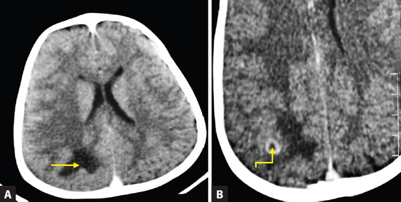
a. Identify the CT findings.
b. Give differential diagnoses based on the image findings and points to differentiate among the differentials.
c. What is your treatment plan here?
Answer
- a.
- Plain CT (Image A): Well-defined hypodense lesion in the right parietal cortex.
- CECT (Image B): Thick-walled, conglomerate ring-enhancing lesion with extensive surrounding hypodense vasogenic edema.
- b. Differential diagnosis: Intracranial Tuberculoma vs. Neurocysticercosis (NCC).
| Feature | Neurocysticercosis (NCC) | Tuberculoma |
|---|---|---|
| Size | Usually < 20 mm | Often > 20 mm |
| Wall | Thin, smooth, regular ring | Thick, irregular, nodular ring |
| Scolex | Often visible as eccentric bright nodule | Absent |
| Edema | Mild to moderate perifocal edema | Severe, extensive vasogenic edema |
| Location | Corticomedullary junction | Supratentorial or infratentorial |
- c. Treatment plan for CNS Tuberculoma:
- Standard Antitubercular Therapy (ATT): 2 months intensive HRZE followed by 7–10 months continuation HRE.
- Adjuvant systemic corticosteroids (prednisolone 1–2 mg/kg/day or dexamethasone 0.6 mg/kg/day for 4 weeks, then tapered over 4 weeks) to reduce cerebral edema and prevent paradoxical expansion.
- Antiseizure medication (e.g., Levetiracetam, Carbamazepine).
THG34-008
These are the neuroimaging of three different children with same diagnosis.
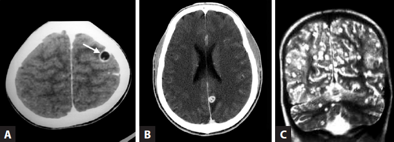
a. What is the most likely diagnosis and can you mention the difference among them?
b. What are the findings in each image?
c. What is the causative organism?
d. A person consumes only vegetarian diet cannot get this disease—True or False?
Answer
- a. Diagnosis: Neurocysticercosis (NCC) illustrating different evolutionary stages of parenchymal larval infection.
- b.
- Image A (CECT): Thin-walled cyst with central non-enhancing fluid and hyperdense eccentric scolex without surrounding edema — Vesicular stage (viable, quiescent).
- Image B (MRI T2/FLAIR): Thick enhancing cyst wall with turbid fluid content and extensive perilesional vasogenic edema — Colloidal vesicular stage (degenerating parasite, highly symptomatic).
- Image C (MRI / CT): Small, punctate, calcified nodule without perilesional edema — Nodular calcified stage (healed, inactive scar).
- c. Taenia solium (larval stage of pork tapeworm / Cysticercus cellulosae).
- d. False. Humans acquire cysticercosis by ingesting Taenia solium eggs via contaminated water, vegetables, or fecal-oral contamination from a human tapeworm carrier (not by eating pork itself, which causes intestinal taeniasis).
THG34-009
A 2-year-old girl presented with refractory left focal seizures not responding to antiepileptic drugs. She has a red birth mark in the right side of face involving forehead and eyelids.
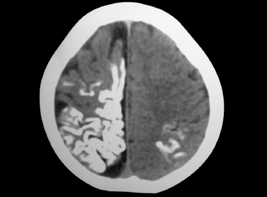
a. What is the CT finding?
b. What is the reason behind the CT finding?
c. What is the diagnosis?
d. What is the ophthalmological complication expected here?
e. Does this condition get inherited to the next generation?
Answer
- a. Prominent gyriform / serpentine cortical calcifications ("tram-track" sign) along the right cerebral cortex with ipsilateral cerebral hemiatrophy.
- b. Leptomeningeal capillary-venous angiomatosis causing chronic venous stasis, cortical hypoperfusion, ischemia, and subsequent cortical calcification and laminar necrosis.
- c. Sturge-Weber syndrome (Encephalotrigeminal angiomatosis).
- d. Glaucoma, buphthalmos, choroidal hemangioma, and episcleral hemangioma.
- e. No. It is caused by a non-hereditary, postzygotic somatic activating mutation in the GNAQ gene.
THG34-010
A 5-year-old boy presented with intellectual disability and seizure. He has multiple small red papular lesions over the cheeks and nose. CT brain was done during evaluation.
a. What is the CT finding?
b. What is the diagnosis?
c. What are the cutaneous manifestation in this condition?
d. What are the other systemic manifestations in this condition?
Answer
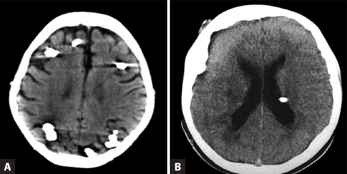
- a.
- Image A: Multiple hyperdense calcified cortical and subcortical tubers.
- Image B: Dense calcified subependymal nodules (SENs) projecting into the lateral ventricles ("candle-guttering" appearance).
- b. Tuberous Sclerosis Complex (Bourneville disease).
- c. Cutaneous manifestations: Hypomelanotic macules (ash-leaf spots), facial angiofibromas (adenoma sebaceum), shagreen patches (connective tissue nevi), ungual/periungual fibromas (Koenen tumors), and confetti skin lesions.
- d. Systemic manifestations:
- CNS: Subependymal giant cell astrocytoma (SEGA), epilepsy, intellectual disability, autism spectrum disorder.
- Cardiac: Cardiac rhabdomyomas.
- Renal: Renal angiomyolipomas, renal cysts, renal cell carcinoma.
- Pulmonary: Lymphangioleiomyomatosis (LAM), multifocal micronodular pneumocyte hyperplasia.
- Ocular: Retinal astrocytic hamartomas.
THG34-011
A 1.5-year-old boy child presented with 1 week of recurrent vomiting and irritability and a history of progressively increasing head size noted since early infancy.
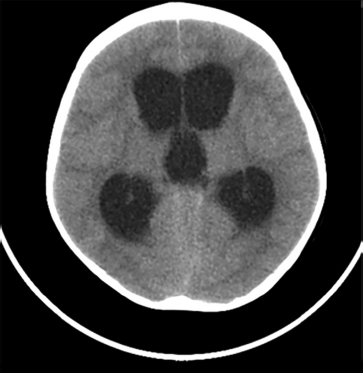
a. What are the CT findings?
b. What is the diagnosis?
c. Name one common cause.
d. Write the normal cerebrospinal fluid (CSF) pathway.
Answer
- a. Marked symmetric dilatation of both lateral ventricles and the third ventricle with a normal-sized, non-dilated fourth ventricle.
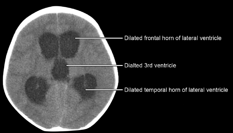
- b. Obstructive (non-communicating) hydrocephalus.
- c. Congenital aqueductal stenosis (Sylvian aqueduct narrowing).
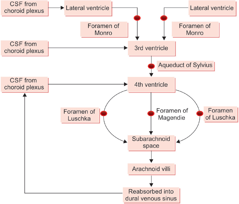
- d. Normal CSF Pathway:
- Choroid plexuses of lateral ventricles
Foramen of Monro Third ventricle Aqueduct of Sylvius Fourth ventricle Foramina of Luschka & Magendie Subarachnoid space & basal cisterns Absorption through arachnoid villi/granulations into superior sagittal sinus.
- Choroid plexuses of lateral ventricles
THG34-012
One-year-old child with increasing head size, irritability and vomiting. Hence CT brain was done.
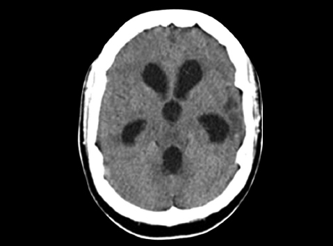
a. Identify the CT findings and diagnosis.
b. What is the pathophysiology here?
c. What is the management for the complication developed here?
Answer
- a. Generalized dilatation involving all ventricles (lateral, third, and fourth ventricles) with effacement of cortical sulci — Communicating hydrocephalus.
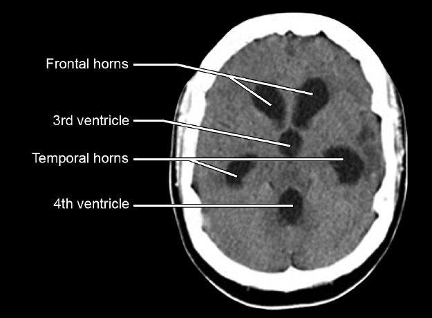
- b. Impaired CSF resorption at the arachnoid villi/granulations into dural venous sinuses (e.g., following pyogenic/tuberculous meningitis, intraventricular hemorrhage).
- c. Surgical diversion of CSF: Ventriculoperitoneal (VP) shunt placement or Endoscopic Third Ventriculostomy (ETV) with choroid plexus cauterization.
THG34-013
This 4-year-old girl, known case of congenital cyanotic heart disease, was admitted for 10 days of fever, left focal seizures, and left focal deficit.
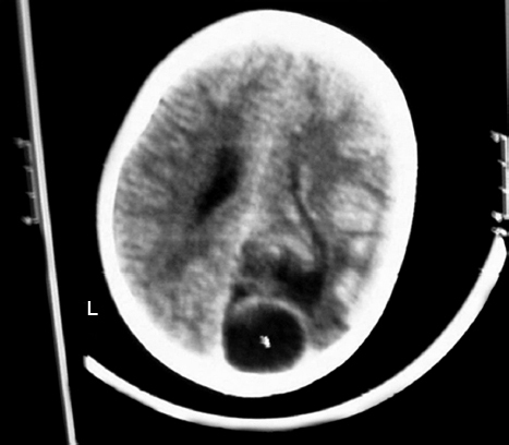
a. Identify the CT findings.
b. What is the probable diagnosis?
c. What are the risk factors/causes for this complication to occur?
d. What are the most common organisms to cause this complication?
e. What is the indication for surgery in these type of cases?
Answer
- a. Well-defined, rounded hypodense lesion in the right occipital lobe with thin surrounding ring and perilesional vasogenic edema.
- b. Brain Abscess in the right occipital lobe.
- c. Cyanotic congenital heart disease with right-to-left shunt (bypass of pulmonary capillary phagocytic filtration), chronic suppurative otitis media (CSOM), mastoiditis, paranasal sinusitis, dental infections, and meningitis.
- d. Streptococcus viridans, Streptococcus pneumoniae, Staphylococcus aureus, Enterococcus, Citrobacter koseri (neonates), and anaerobes (Bacteroides, Peptostreptococcus).
- e. Indications for surgical aspiration / excision:
- Abscess size
in diameter. - Significant mass effect, midline shift, or raised ICP.
- Location in posterior fossa / proximity to ventricular system (risk of intraventricular rupture).
- Presence of gas within the abscess cavity.
- Fungal or multiloculated abscess.
- Failure to improve after 2–3 weeks of targeted IV antimicrobial therapy.
- Abscess size
THG34-014
A 14-year-old boy presented with complaint of headache and progressive visual defect. On examination, he was short for his age and had bitemporal hemianopia. Plain CT showed hyperdense calcified lesion found anterior to pons.
a. Can you give a diagnosis based on the clinical features and CT findings mentioned?
b. What are the endocrinological issues you expect in this child?
Answer
- a. Craniopharyngioma (suprasellar/sellar calcified mass compressing the optic chiasm and pituitary stalk).
- b. Endocrinological manifestations:
- Growth hormone (GH) deficiency
Short stature / growth failure - Gonadotropin (LH/FSH) deficiency
Delayed / absent puberty - TSH deficiency
Central hypothyroidism - ACTH deficiency
Secondary adrenal insufficiency (lethargy, hypoglycemia, hypotension) - ADH / Vasopressin deficiency
Central diabetes insipidus (polyuria, polydipsia)
- Growth hormone (GH) deficiency
THG34-015
An 8-year-old girl child was brought with seizure. There is a history of skin lesion and reddish discoloration of urine 2 days prior to seizure. Her blood pressure (BP) was 160/100 mm Hg.
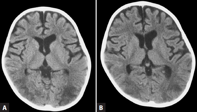
a. Identify the neuroimaging with its findings.
b. What is the diagnosis?
c. What is the predisposing condition for this complication to arise?
Answer
- a. CT brain plain: Symmetrical, patchy, subcortical hypodense lesions predominantly affecting the parieto-occipital white matter bilaterally.
- b. Posterior Reversible Encephalopathy Syndrome (PRES).
- c. Severe acute hypertension secondary to Acute Post-Streptococcal Glomerulonephritis (APSGN).
Basics of MRI Brain Interpretation
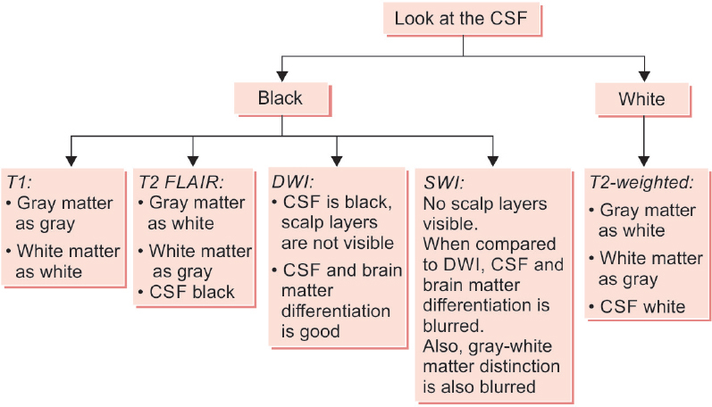
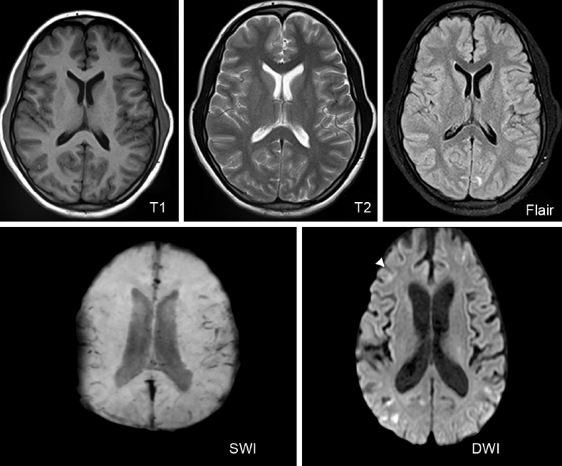
- Common Sequences:
- T1W (Plain): Best for anatomical detail (CSF is dark/hypointense, white matter is hyperintense).
- T1W + Contrast: Identifies breakdown of blood-brain barrier (infections, tumors).
- T2W: Best for visualizing pathology, edema, and demyelination (CSF is bright/hyperintense).
- FLAIR: T2 with CSF suppression (CSF is dark; periventricular lesions and edema stand out brightly).
- DWI: Restricts water movement; acute ischemic stroke/infarct appears bright.
- SWI / GRE: Identifies paramagnetic substances; microhemorrhages and calcifications appear black (signal drop-out).
Magnetic Resonance Spectroscopy (MRS):
Spectroscopy measures relative metabolite concentrations across chemical shifts (ppm):
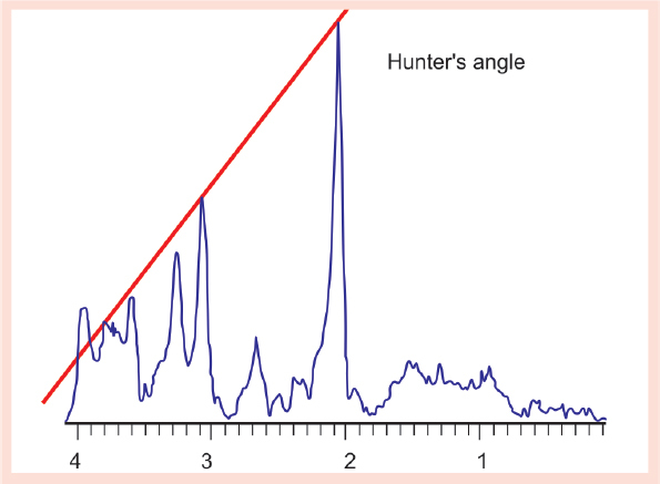
| Metabolite | Chemical Shift (ppm) | Clinical Significance |
|---|---|---|
| Lipid | 1.3 | Elevated in necrosis, tuberculoma, high-grade tumors |
| Lactate | 1.33 (inverted doublet) | Elevated in mitochondrial disorders, hypoxia/ischemia |
| N-acetylaspartate (NAA) | 2.02 | Neuronal integrity marker (decreased in brain injury; markedly elevated in Canavan disease) |
| Creatine | 3.03 & 3.93 | Energy metabolism reference peak |
| Choline | 3.22 | Cell membrane turnover (elevated in malignancies and active demyelination) |