VVK07-001
A 5-year-old child has a massive GI bleed. He weighs 20 kg.
His clotting time is 13 minutes. His PT test is 42 secs (control 15 secs) and aPTT test is 64 secs (control is 29 secs).
His Hb is 11 g/dL and platelet count is 2.5 lakhs/cumm.
a. What blood product would you transfuse?
b. How much of the above transfusion is required?
c. List 4 other indications for transfusion with this blood product.
Answer
- a. Fresh Frozen Plasma (FFP).
- b. 300 mL (15 mL/kg).
- c. Indications for FFP transfusion:
- i. Severe clotting factor deficiency and active bleeding.
- ii. Severe clotting factor deficiency prior to an invasive procedure.
- iii. Emergency reversal of warfarin effects.
- iv. Dilutional coagulopathy and active bleeding (massive transfusion).
- v. Thrombotic thrombocytopenic purpura (plasma exchange).
VVK07-002
An 11-year-old girl presents to ED with breathing difficulty since 1 day. She has been admitted with a similar condition 2 years back. This was associated with a bout of hemoptysis back then. She was found to have severe anemia and received 2 units of blood. She has been on iron therapy since due to persistently low Hb.
On examination, she is very pale with not much chest findings.
Investigations revealed a Hb of 5 g/dL, TLC and platelets are normal. Her MCV is 65 fl, RDW is 21% and RBC count is 2 million/cumm with low MCH and MCHC.
Chest X-ray shows bilateral infiltrates.
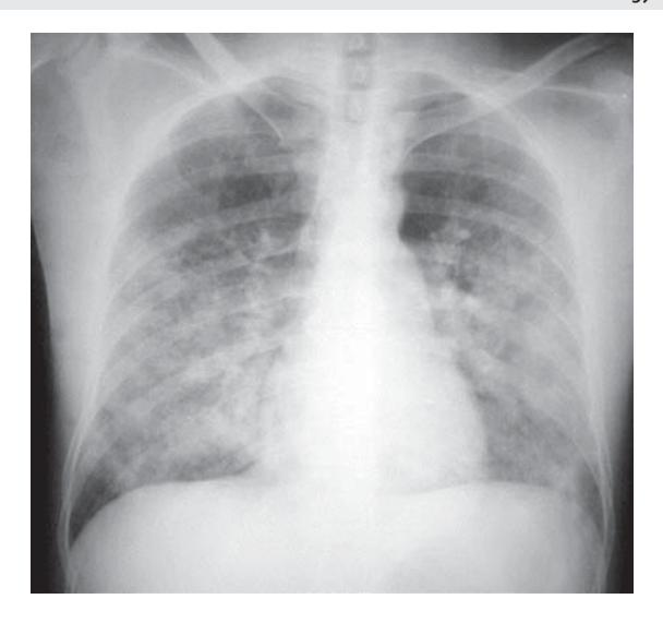
a. What is your diagnosis?
b. What are the various ways to confirm the diagnosis?
c. What is the drug commonly used to treat this?
Answer
- a. Idiopathic pulmonary hemosiderosis (IPH).
- b. Demonstration of hemosiderin-laden macrophages (siderophages) on:
- Bronchoalveolar lavage (BAL)
- Gastric lavage
- Lung biopsy (gold standard)
- c. Systemic corticosteroids (prednisolone/methylprednisolone) and immunosuppressive therapy (azathioprine/hydroxychloroquine).
VVK07-003
A 13-year-old girl presents with fever since last 3 months. This is associated with night sweats. She has lost 10 kg weight in last 2 months. On examination, she has supraclavicular node 3 × 3 cm, bilateral submandibular and jugulodigastric node 2 × 2 cm. No organomegaly is present. Biopsy of the lymph node shows:
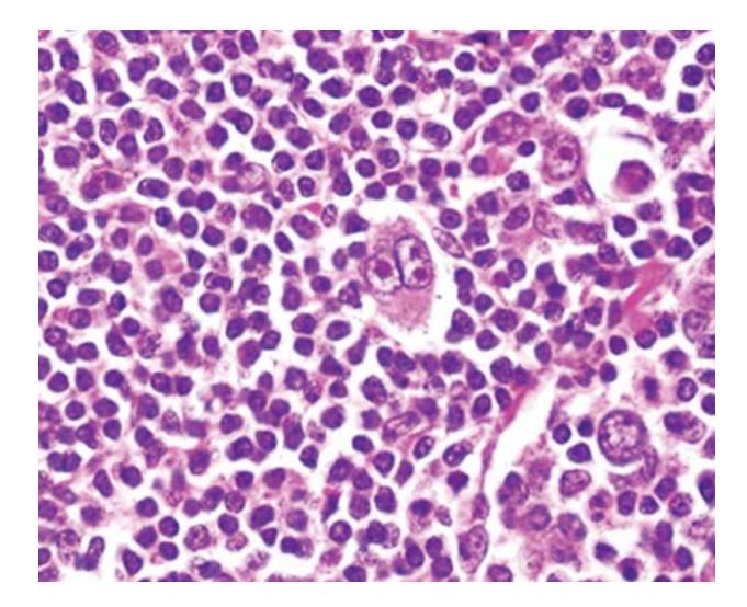
a. What is the diagnosis?
b. What is the classification?
c. What is the staging called?
d. Name 4 drugs commonly used to treat this condition.
Answer
- a. Hodgkin lymphoma (Reed-Sternberg cells seen on biopsy).
- b. WHO Classification:
- Classical Hodgkin Lymphoma:
- Nodular sclerosis
- Mixed cellularity
- Lymphocyte-depleted
- Lymphocyte-rich classical HL
- Nodular Lymphocyte Predominant Hodgkin Lymphoma (NLPHL)
- Classical Hodgkin Lymphoma:
- c. Ann Arbor Staging (Cotswolds modification).
- d. ABVD regimen: Adriamycin (Doxorubicin), Bleomycin, Vinblastine, Dacarbazine (or OEPA/COPP).
VVK07-004
A 2-year-old female child has fever and lymphadenopathy for 3 weeks:
- O/E: Has splenohepatomegaly.
- CBC: Shows Hb low, TLC normal, platelets low. No immature cells in the smear.
- Bone marrow: Shows extensive hemophagocytosis.
- Father says that the previous child died at 1 year of age with a similar illness. She too had a lack of blood formation and bleeding.
a. What is the likely diagnosis?
b. What are the other biochemical abnormalities that will lead to the diagnosis?
c. In a non-familial / secondary state, what infections can precipitate this?
Answer
- a. Familial Hemophagocytic Lymphohistiocytosis (HLH).
- b. Hyperferritinemia (> 500 ng/mL, often > 10,000 ng/mL), hypertriglyceridemia (fasting triglycerides ≥ 265 mg/dL), hypofibrinogenemia (≤ 1.5 g/L), elevated soluble CD25 (sIL-2 receptor), low/absent NK cell activity, elevated transaminases.
- c. Viral: EBV (most common), CMV, HIV, HSV, HHV-6, HHV-8, Rubella, Varicella, Parvovirus B19.
- Bacterial/Protozoal: Tuberculosis, Brucella, Mycoplasma, Leishmania donovani.
VVK07-005
A 1.5-year-old boy presents with swelling in the right ankle after a trivial fall. He has history of easy bruising and prolonged bleeding from injection sites. His maternal uncle died of a bleeding disorder.
a. Name a screening test to clinch the diagnosis.
b. What does RICE stand for in the management of this disease?
c. Name the drug used to control bleeding in case he suffers another joint bleed.
Answer
- a. Activated Partial Thromboplastin Time (aPTT) — prolonged with normal PT and platelet count.
- b. RICE:
- R — Rest
- I — Ice
- C — Compression
- E — Elevation
- c. Clotting factor concentrate:
- If Hemophilia A: Recombinant / Plasma-derived Factor VIII concentrate.
- If Hemophilia B: Recombinant / Plasma-derived Factor IX concentrate.
VVK07-006
A 6-month-old child is being evaluated for purpura. His platelet count is low (40,000/cumm). He has been on various skin ointments for a non-resolving dermatitis. He has been given antibiotics twice for management of otitis media and LRTI.
a. What is the diagnosis?
b. Name two screening tests.
c. What is the treatment?
d. What are the long term risks?
Answer
- a. Wiskott-Aldrich syndrome (triad of thrombocytopenia with small platelets, eczema, and recurrent infections).
- b.
- Platelet size / Mean Platelet Volume (Low MPV < 7 fl, small micro-thrombocytes).
- Serum immunoglobulin profile: Low IgM, elevated IgA and IgE, normal/variable IgG.
- WASP gene mutation analysis.
- c. Allogeneic Hematopoietic Stem Cell Transplant (HSCT) is the definitive cure.
- d. Untreated disease is fatal secondary to severe infections or intractable hemorrhage. Long-term risks include development of autoimmune diseases and malignancies (leukemias and B-cell lymphomas / EBV-related lymphomas).
VVK07-007
A 4-year-old boy presents with fever for 10 days, lethargy, poor oral intake, abdominal distension and decreased urine output. He is sick looking and has diffuse tenderness in his abdomen. USG abdomen reveals multiple lymph nodal masses with hepatosplenomegaly. His CBC is normal with LDH of 12,000 IU/L, blood urea of 120 mg/dL and serum creatinine of 2.2 mg/dL. His serum uric acid is 12.5 mg/dL.
a. What is the likely diagnosis?
b. What is the treatment to be initiated at the primary care center?
c. What is the novel agent used to treat this complication?
d. What is the characteristic histological appearance of this condition?
e. What is the overall prognosis of this condition?
Answer
- a. Burkitt lymphoma / B-cell Non-Hodgkin Lymphoma complicated by acute spontaneous Tumor Lysis Syndrome (TLS).
- b. Vigorous hyperhydration (double maintenance IV fluids without potassium), urinary alkalinization (if used cautiously), allopurinol, and close monitoring of urine output and renal function.
- c. Recombinant urate oxidase (Rasburicase).
- d. "Starry sky" appearance (sheets of uniform basophilic lymphoid cells interspersed with tingible body macrophages containing apoptotic debris).
- e. 70–90% event-free / overall survival with modern intensive multi-agent chemotherapy regimens.
VVK07-008
A 17-year-old boy presents with progressive lethargy and loss of appetite:
- O/E: Pallor, ecchymotic spots, no lymphadenopathy or organomegaly, no dysmorphology.
- Investigations:
- CBC: Hb 7 g/dL, TLC 2500/cumm, Platelet count 25,000/cumm, DLC: N35 L60 E3 M2, MCV 88 fl, RBC 2.2 million/cumm, RDW 14%
- Peripheral smear: Pancytopenia, no immature cells
- Reticulocyte count: 0.2%
- Bone marrow picture shows:
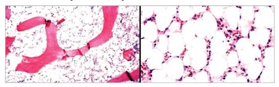
a. What is the diagnosis?
b. What will be important investigations before you embark on treatment?
c. What are the treatment options?
Answer
- a. Severe Aplastic Anemia (hypocellular marrow with replacement of hematopoietic tissue by fat cells).
- b.
- Bone marrow biopsy (cellularity < 25%).
- Chromosomal breakage analysis with DEB / Diepoxybutane / Mitomycin C (to rule out Fanconi anemia).
- PNH clone testing (Flow cytometry for CD55 / CD59 / FLAER).
- Viral serologies: Hepatitis B, Hepatitis C, HIV, Hepatitis A, EBV, CMV, Parvovirus B19.
- HLA typing of patient and siblings.
- c.
- Allogeneic Hematopoietic Stem Cell Transplant (HSCT) from matched sibling donor.
- Immunosuppressive therapy (IST): Antithymocyte globulin (ATG) + Cyclosporine + Eltrombopag.
VVK07-009
Study the photograph provided below:
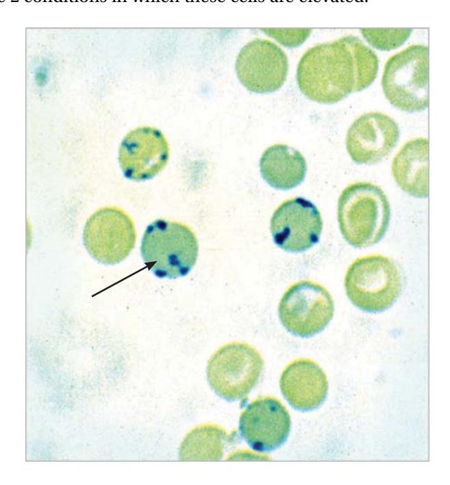
a. Name the cell marked by the arrow.
b. Name the dye used to stain the cell.
c. What is the normal value of this cell in a child?
d. Name conditions in which these cells are elevated.
Answer
- a. Reticulocyte (showing ribosomal RNA precipitates / reticular network).
- b. Supravital stain (New methylene blue or Brilliant cresyl blue).
- c. 0.5–2.0% in children (2–6% in neonates).
- d. Conditions with elevated reticulocytes (reticulocytosis):
- Hemolytic anemias (Autoimmune hemolytic anemia, Hereditary spherocytosis, G6PD deficiency, Sickle cell anemia).
- Acute blood loss / hemorrhage.
- Response to nutritional replacement therapy (iron, vitamin B12, or folic acid).
VVK07-010
A 12-year-old male presents with severe pancytopenia. His bone marrow biopsy confirms severe aplastic anemia. He is referred for a BMT to higher center. On examination, he is found to have the following:
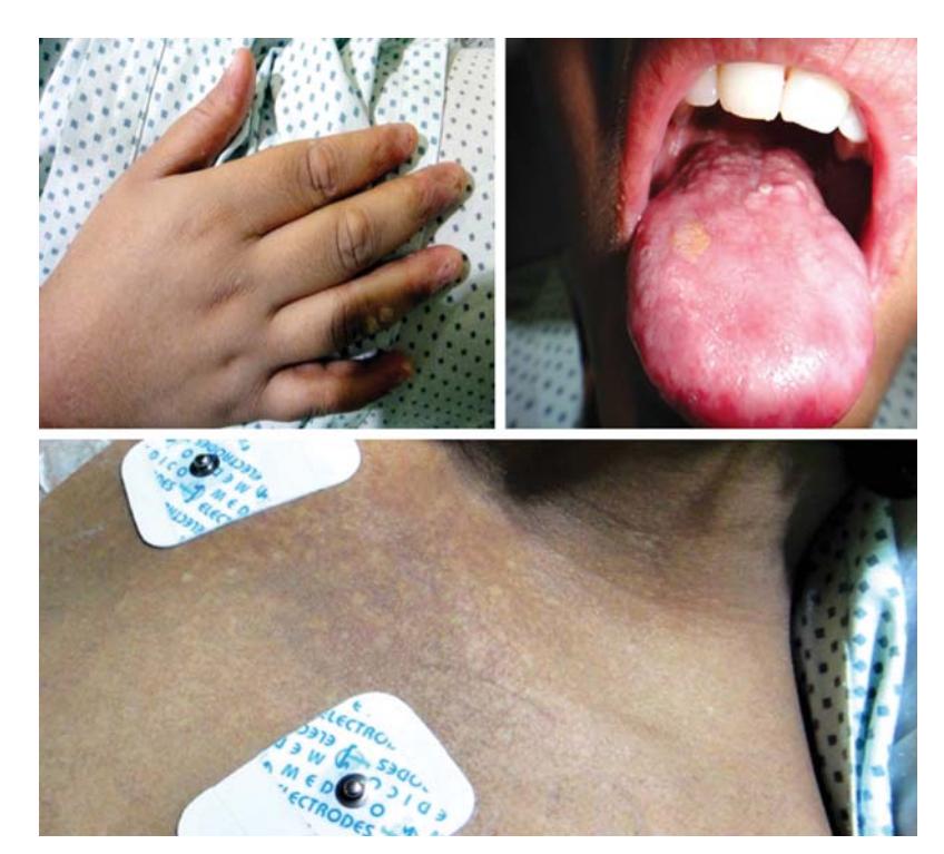
a. What is your diagnosis?
b. What is the basic pathophysiological defect in these patients?
c. How do you treat them?
d. What are other potential lethal complications?
Answer
- a. Dyskeratosis congenita (triad of reticular skin pigmentation, nail dystrophy, and oral leukoplakia).
- b. Defect in telomere biology / maintenance (short telomere length due to mutations in DKC1, TERC, TERT, or TINF2 genes).
- c. Allogeneic Bone Marrow Transplantation (BMT) with reduced-intensity conditioning, or Androgen therapy (Oxymetholone / Danazol).
- d. Progressive bone marrow failure, pulmonary fibrosis, hepatic cirrhosis, and malignancies (MDS, AML, squamous cell carcinoma of head, neck, and anogenital tract).
VVK07-011
A 7-year-old male child presents with fever, pallor and ecchymotic spots:
- O/E: Pallor, Spleen 3 cm BLCM, Liver 2 cm BRCM, Testes enlarged.
- CBC: Shows pancytopenia.
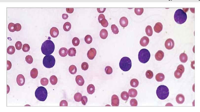
a. What is your diagnosis?
b. How to confirm the diagnosis?
c. Name 3 agents used to treat this.
d. What is the success rate of treatment with modern day regimens?
Answer
- a. Acute Lymphoblastic Leukemia (ALL) (with testicular involvement).
- b. Bone marrow aspiration and biopsy showing > 20–25% lymphoblasts, confirmed by Flow cytometry / Immunophenotyping (CD10, CD19, CD20, CD22, TdT for B-ALL; CD3, CD7 for T-ALL).
- c. Induction chemotherapy agents: Vincristine, Corticosteroids (Prednisolone/Dexamethasone), L-asparaginase, Daunorubicin. (Also Methotrexate, 6-Mercaptopurine, Cyclophosphamide).
- d. 85–90% 5-year overall cure/survival rate in children.
VVK07-012
A 15-year-old girl with history of generalised weakness and increasing pallor since one month, had history of fever 1 week back. No history of bleeding, loose stools, passage of worms:
- Afebrile, Pallor ++, Petechiae +, No icterus, No significant lymphadenopathy, No organomegaly.
- Investigations:
- Hb: 6.1 g/dL
- TLC: 5000/cumm (ANC low)
- Platelets: 27,000/cumm
- MCV: 102.8 fl, MCH: 28.6 pg, MCHC: 32.2 g/dL, RDW: 31.2%
- Reticulocyte count: 2%
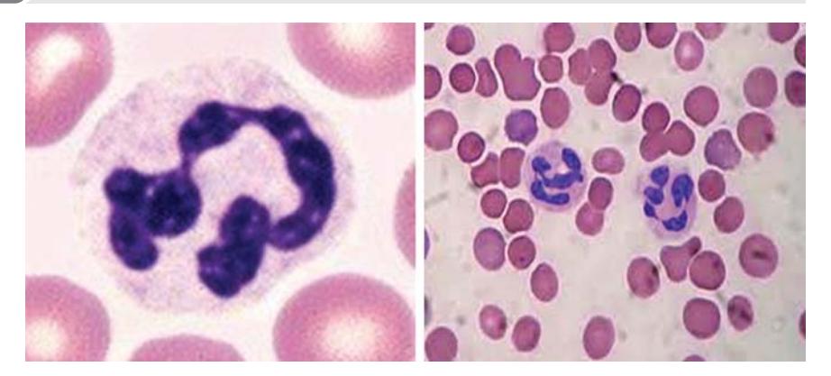
a. Name the cell seen in the smear.
b. What is your diagnosis?
c. How do you treat such a patient?
Answer
- a. Hypersegmented neutrophil (neutrophil with ≥ 6 nuclear lobes).
- b. Megaloblastic anemia (due to Vitamin B12 or folic acid deficiency).
- c. Parenteral Vitamin B12 (Cyanocobalamin / Hydroxocobalamin 1000 mcg IM) and/or Oral Folic acid (1–5 mg/day). (Always ensure B12 is given or co-administered with folate to prevent subacute combined degeneration of spinal cord).
VVK07-013
A 6-year-old boy being looked after by a single mother as father died a year back of a chronic illness, presents with excessive bruising for past 9 months. Platelets documented to be < 20,000/cumm on many occasions. Bone marrow showed megakaryocytic thrombocytopenia. Initially responded to steroids but thrombocytopenia recurs on tapering of steroids. Significant weight loss in last 6 months.
Examination: Bruises on both legs, mild pallor, bilateral cervical lymph nodes, oral thrush, spleen 3 cm palpable.
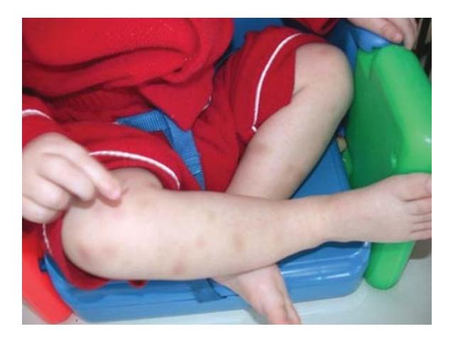
a. What is the most likely diagnosis?
b. What is the investigation of choice?
c. What agents are used to treat this condition?
Answer
- a. HIV-associated immune thrombocytopenia (HIV-ITP / pediatric HIV infection).
- b. HIV ELISA / Western Blot (or HIV RNA PCR / DNA PCR if < 18 months).
- c. Combination Antiretroviral Therapy (cART). For refractory ITP: IVIG, Anti-D immunoglobulin (in Rh+ non-splenectomized), short-course corticosteroids, Dapsone, or Thrombopoietin Receptor Agonists (Eltrombopag/Romiplostim).
VVK07-014
A 4-year-old boy is under evaluation for mild anemia:
- CBC: Hb 9.5 g/dL, TLC 6500/cumm, Platelet count 3.5 lakhs/cumm
- MCV: 68 fl, RBC: 5.8 million/cumm, RDW: 13.5%
- Mentzer index:
(< 13) - Peripheral smear shows microcytic hypochromic anemia.
a. What is the likely diagnosis?
b. What will be the test of choice and the expected results?
c. What treatment will be given to this child?
Answer
- a. Thalassemia minor / Beta-thalassemia trait.
- b. Hemoglobin High-Performance Liquid Chromatography (HPLC) / Hemoglobin Electrophoresis showing elevated
(normal , rest HbA). - c. No active medical treatment or iron therapy required (iron is contraindicated unless coexisting iron deficiency is documented). Provide genetic counselling and screening for parents/family.
VVK07-015
A 2-year-old girl presented with fever of 1 month duration, difficulty in walking because of bone pain, multiple bony swellings on the skull and periorbital ecchymosis with proptosis ("raccoon eyes").
On examination, she is pale, hypertensive, and a diffuse non-tender abdominal mass crossing the midline is palpable.
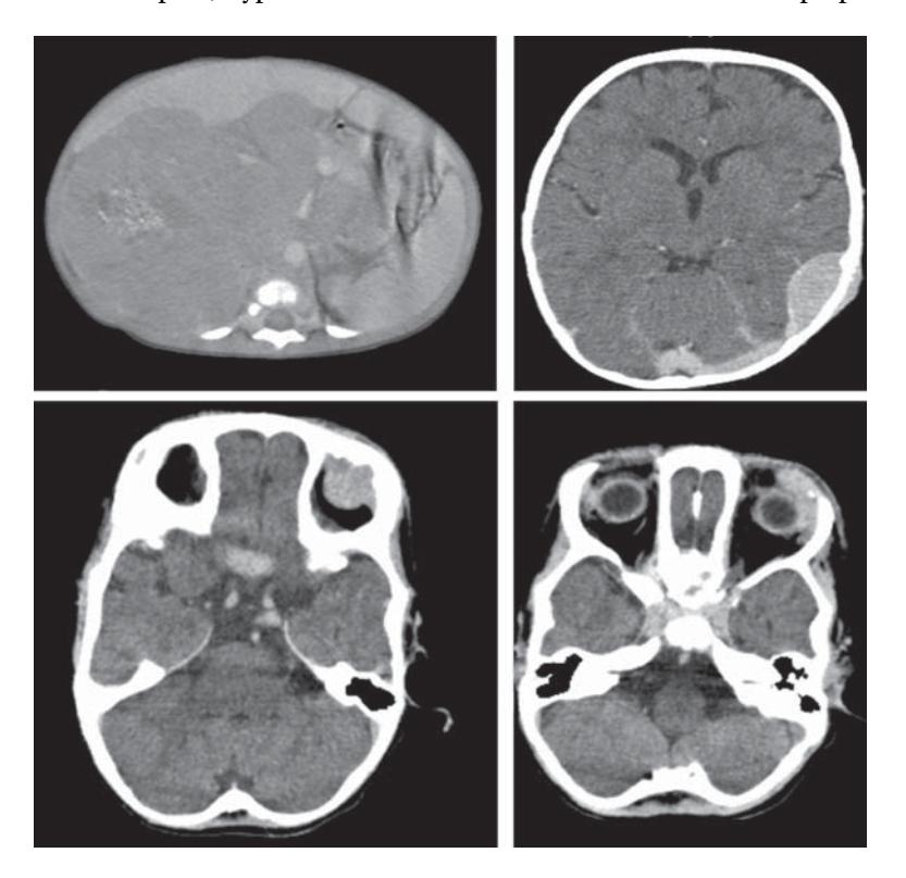
a. What is the likely diagnosis?
b. Name a non-invasive and an invasive test that can lead to a diagnosis.
c. What is the most likely site of origin of the disease?
Answer
- a. Neuroblastoma (Stage IV / Metastatic).
- b.
- Non-invasive test: 24-hour urinary catecholamine metabolites — Vanillylmandelic acid (VMA) and Homovanillic acid (HVA); MIBG scan (Metaiodobenzylguanidine).
- Invasive test: Core needle/open biopsy of the mass with histopathology (Homer-Wright pseudorosettes, synaptophysin/chromogranin) or Bone marrow aspirate/biopsy.
- c. Adrenal medulla (most common, ~40–50%) or sympathetic chain / retroperitoneal paravertebral sympathetic ganglia.
VVK07-016
A 2-year-old boy presents with a new onset unsteady gait. Mother has noticed chaotic, dancing rapid eye movements (roving movements in eyes) and jerky body movements. Local doctor has done a chest X-ray along with other routine workup which reveals a significant posterior mediastinal shadow.
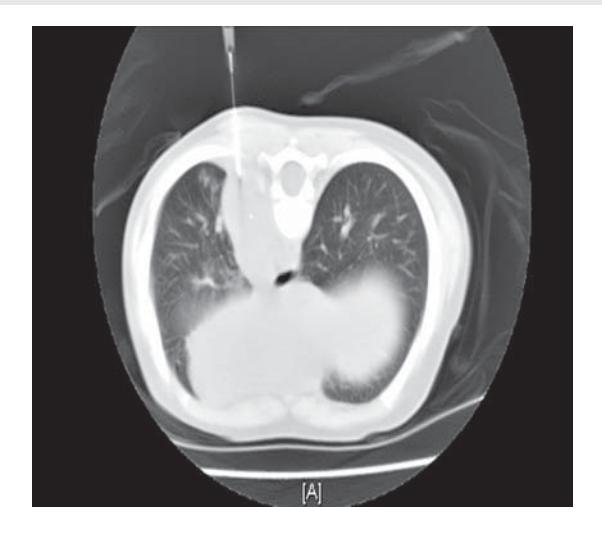
a. What is the likely diagnosis?
b. What is the treatment?
c. What is the prognosis of this condition?
Answer
- a. Paraneoplastic Opsoclonus-Myoclonus-Ataxia Syndrome (OMAS / "Dancing Eyes-Dancing Feet") secondary to Mediastinal Neuroblastoma.
- b. Surgical excision of the neuroblastoma mass combined with immunomodulatory therapy: High-dose Corticosteroids / ACTH, Intravenous Immunoglobulin (IVIG), and Rituximab.
- c. Excellent oncological survival (> 90%), though neurocognitive and behavioral sequelae may persist in some children.
VVK07-017
A 14-year-old boy presents with cough for 1 month and difficulty in breathing for last 2 weeks. He was given oral antibiotics after 1 week of illness. A local doctor did a Chest X-ray which revealed a significant pleural effusion on the left side and empirically started him on ATT. However his condition worsened and he presented in the emergency with severe respiratory distress, orthopnea and stridor. Chest X-ray reveals complete opacification of left hemithorax. CBC is normal. Pleural fluid analysis reveals abnormal blast cells. CT scan done after pleural tap reveals an anterior mediastinal mass compressing the trachea (Superior Mediastinal Syndrome).
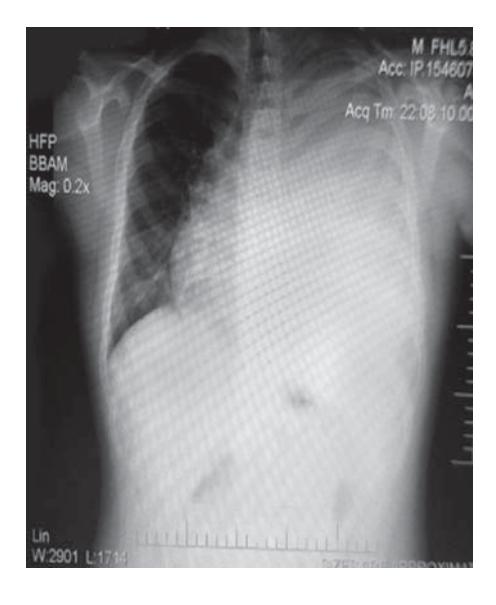
a. What is the most likely diagnosis?
b. What test will most easily clinch the diagnosis?
c. What are the precautions to be taken in this condition?
d. What is the life saving therapy in this condition?
Answer
- a. T-cell Acute Lymphoblastic Lymphoma / Leukemia (T-ALL / T-LBL) presenting with Superior Vena Cava / Superior Mediastinal Syndrome.
- b. Pleural fluid cytopathology and Flow cytometry / Immunophenotyping.
- c.
- Avoid general anesthesia, sedation, and muscle relaxants (high risk of total airway collapse / cardiopulmonary arrest).
- Avoid invasive mediastinal biopsy in acute phase.
- Keep patient in upright / propped-up position.
- d. Immediate empiric IV Corticosteroids (e.g. Methylprednisolone or Dexamethasone), emergency mediastinal radiation therapy if refractory, and supplemental oxygen.
VVK07-018
A 12-year-old boy presents with fever of 2 weeks duration with massive swellings in the neck. His CBC shows a TLC of 2.9 Lakh/cumm. His serum potassium is 6.0 mEq/L and serum creatinine is 1.2 mg/dL.
a. What is the likely diagnosis?
b. What biochemical abnormalities are expected?
c. What management needs to be done before starting definitive therapy?
Answer
- a. Acute Leukemia with Tumor Lysis Syndrome (TLS).
- b. The TLS tetrad:
- Hyperkalemia (
) - Hyperuricemia (
) - Hyperphosphatemia (
) - Hypocalcemia (
) - Deranged renal function (elevated BUN and creatinine)
- Hyperkalemia (
- c.
- Hyperhydration (2–3 times maintenance IV fluids without potassium).
- Allopurinol or Rasburicase (urate oxidase) to reduce uric acid.
- Oral phosphate binders (e.g. sevelamer, aluminium hydroxide) for hyperphosphatemia.
- Management of hyperkalemia (calcium gluconate, insulin + dextrose, salbutamol nebulization, potassium binders).
- Avoid routine IV calcium unless life-threatening symptomatic hypocalcemia is present (risk of calcium phosphate precipitation).
VVK07-019
A 5-year-old boy presents with pallor and passing dark-colored urine. Three days prior the patient had low-grade fever and dry cough. Initial CBC revealed a hemoglobin level of 4.5 g/dL, a HCT of 14%, a WBC count of 9,100/mm3 (normal DLC) and a platelet count of 3,72,000/mm3. In the past he has received 2 blood transfusions for fever and acute anemia at 18 months of age. Both parents are thalassemia carriers. Physical examination: Temperature 38.3°C, marked pallor. CVS: Systolic ejection murmur grade II/VI at left upper sternal border. Abdomen: Splenohepatomegaly is present. The Hb HPLC report is shown:
| ANALYTE | ID | % | TIME | AREA |
|---|---|---|---|---|
| Unknown / P1 / F / P2 / P3 / Ao / C-WINDOW | 1, 2, 3 | 11.9, 1.5, 5.7, 2.1, 3.5, 3.7, 3.4, 67.2, 0.9 | 0.013–5.07 | 4458732 |
| Hb F | 3.5% | Hb A2 | 0.0% |
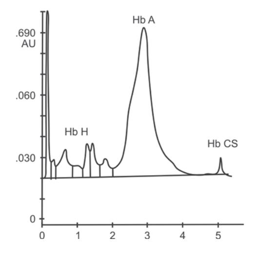
a. What is your diagnosis?
b. What may be the trigger for this crisis?
Answer
- a. Hemoglobin H (Hb H) disease / Hb H-Constant Spring with acute hemolytic crisis.
- b. Preceding acute viral/bacterial infection or oxidant stress / antipyretic drugs.
VVK07-020
A 7-year-old child is admitted for LRTI. This is his 5th episode of severe pneumonia apart from 4 episodes of severe diarrhea in past. On transfusing blood in view of anemia (Hb 5.5 g/dL), he develops a severe anaphylactic transfusion reaction. He has never received a transfusion in the past and clerical errors of a blood group mismatch are excluded.
a. What immunological defect is most likely present?
b. What would you advise the parents of this child to prevent a recurrence of reaction?
Answer
- a. Selective IgA Deficiency (patient develops anti-IgA antibodies which react with IgA in donor blood).
- b.
- Avoid unwashed donor blood products.
- If transfusion is necessary, use extensively washed RBCs (depleted of plasma/IgA) or blood products from known IgA-deficient donors.
- Wear a medical alert bracelet indicating IgA deficiency.
VVK07-021
A 5-year-old male child is recently diagnosed as a case of acute myeloid leukemia. He is clinically stable with a RR of 20/min, normal blood pressure and SpO2 of 95% in room air. His blood gas is as follows: pH 7.43; PaCO2 34 mmHg; PaO2 47.6 mmHg; HCO3– 24 mEq/L.
Further investigations show leucocytosis (TLC 60,000/cu mm) and a normal chest X-ray.
a. Analyze the blood gas report.
b. What step will you take to improve PaO2 in this patient?
Answer
- a. Pseudohypoxemia (Leukocyte Larceny) due to rapid in vitro oxygen consumption by the large number of metabolically active leukocytes in the uncooled blood sample.
- b. Analyze the arterial blood sample immediately after drawing, transport the heparinized syringe plunged on ice, or immediately separate plasma/cells. (Supplemental oxygen is not required as the patient's true arterial PaO2 is normal).
VVK07-022
Study the photograph of the peripheral smear provided and answer the questions:
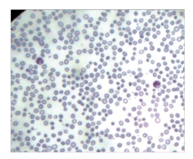
a. What is your diagnosis?
b. What is the drug used to treat this condition?
c. What is the first clinical evidence of improvement after starting the therapy?
Answer
- a. Iron Deficiency Anemia (marked microcytic, hypochromic RBCs with anisopoikilocytosis, pencil cells, and central pallor > 1/3 of cell diameter).
- b. Oral elemental iron at 3–6 mg/kg/day in 2–3 divided doses.
- c. Subjective clinical improvement: Decreased irritability, improved alertness, and increased appetite within 12–24 hours (before reticulocyte count rises at 3–7 days).
VVK07-023
Answer the given questions in relation to methemoglobinemia:
a. At what level of Met-Hb is cyanosis visible?
b. What is the expected pulse oximetry saturation in such a patient?
c. Name the drug used in the treatment of this condition and give its dose.
d. In which scenario is this drug contraindicated?
e. Acute methemoglobinemia is characterized by all except:
- i. Cyanosis not responding to oxygen
- ii. Normal cardiopulmonary examination
- iii. Chocolate brown colored arterial blood sample
- iv. Pulse oximetry SpO2 low with low PaO2
Answer
- a. Methemoglobin level > 1.5 g/dL (or > 10–15% of total hemoglobin).
- b. Characteristically plateaus at approximately 85% on standard two-wavelength pulse oximeters.
- c. Intravenous Methylene Blue 1–2 mg/kg (0.1–0.2 mL/kg of 1% solution) given over 5 minutes. Oral/IV Ascorbic acid (Vitamin C) 200–500 mg/day can also be used as adjunctive therapy.
- d. Glucose-6-Phosphate Dehydrogenase (G6PD) deficiency (risk of severe acute hemolysis and ineffective methylene blue reduction).
- e. iv (Pulse oximetry SpO2 is low, but the arterial PaO2 measured on ABG is normal because dissolved oxygen in plasma is unaffected).
VVK07-024
A 1½-year-old male child presented with episodes of on and off fast breathing for the past 5 months, occasionally associated with cough. He is also having high-grade intermittent fever for last 2 weeks. Empirical antitubercular treatment for past 3 months has had no benefit. There is a past history of chronic otitis media with perforation and the child has been taken to the dentist twice for loose teeth and swollen gums. On examination, seborrheic dermatitis, pallor and hepatomegaly (5 cm below costal margin) are the salient positive findings.
a. What is your most probable diagnosis?
b. What is the characteristic lesion found on skeletal survey?
c. What do you expect to find in the bone marrow / lesion biopsy?
d. What is the characteristic electron microscopy finding?
Answer
- a. Langerhans Cell Histiocytosis (LCH / Letterer-Siwe disease / Hand-Schüller-Christian triad).
- b. Punched-out osteolytic lesions ("floating teeth" appearance in mandible; geographic skull defects) with no surrounding sclerosis.
- c. Clonal proliferation of CD1a+ / CD207+ (Langerin+) Langerhans histiocytes with folded/grooved nuclei, accompanied by eosinophils, lymphocytes, and giant cells.
- d. Birbeck granules ("tennis-racket" shaped cytoplasmic pentalaminar organelles).
VVK07-025
Study the photomicrograph and answer the question:
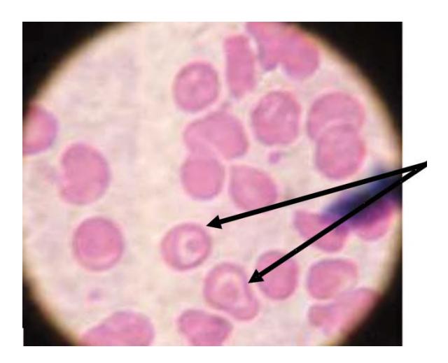
a. Identify the abnormality on peripheral smear.
b. Name 4 conditions where these cells can be seen.
Answer
- a. Target cells (Codocytes / "Mexican hat" cells).
- b. Conditions with target cells:
- Thalassemia syndromes (Alpha and Beta thalassemia)
- Hemoglobinopathies (HbC, HbS, HbE, HbSC disease)
- Obstructive liver disease / Chronic liver disease
- Post-splenectomy / Hyposplenism
- Severe iron deficiency anemia
- LCAT (Lecithin-cholesterol acyltransferase) deficiency
- Abetalipoproteinemia
VVK07-026
A 10-year-old girl, with ITP diagnosed since 6 months of age presents with ecchymotic patch on her waist (shown in the photograph) and has uncontrollable epistaxis. Her platelet count is 4000/mm3. Answer the following questions:
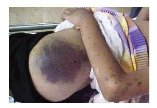
a. Which investigations would you advise in her?
b. What measures either singly or in combination can be used to treat life-threatening hemorrhage in ITP?
c. What is the indication of doing bone marrow examination in a suspected case of ITP?
d. When do you call ITP to be chronic?
e. Give drugs that cause thrombocytopenia.
Answer
- a. Antinuclear antibody (ANA), anti-dsDNA, HIV serology, HCV serology, serum immunoglobulin levels (IgG, IgA, IgM), Direct Antiglobulin Test (Coombs).
- b.
- High-dose IV Methylprednisolone (30 mg/kg/day or 500–1000 mg/m2/day for 3 days).
- IVIG (0.8–1.0 g/kg).
- Platelet transfusion (given in higher amounts together with IVIG/steroids).
- Anti-D immunoglobulin (in Rh-positive, non-splenectomized patients).
- Emergency splenectomy / Tranexamic acid / recombinant Factor VIIa.
- c. Indications for bone marrow aspiration: Unexplained cytopenias in other cell lines (anemia, leukopenia), abnormal smear with blasts/dysplasia, organomegaly, bone pain, lack of response to first-line therapy, or prior to starting corticosteroid therapy.
- d. Chronic ITP: Thrombocytopenia persisting for > 12 months from diagnosis.
- e. Valproic acid, Carbamazepine, Phenytoin, Sulfonamides, Cotrimoxazole, Vancomycin, Linezolid, Heparin (HIT), Quinine, Quinidine.
VVK07-027
This 3-year-old boy develops easy bruising over the body with hematoma formation even with minor trauma.
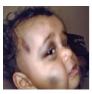
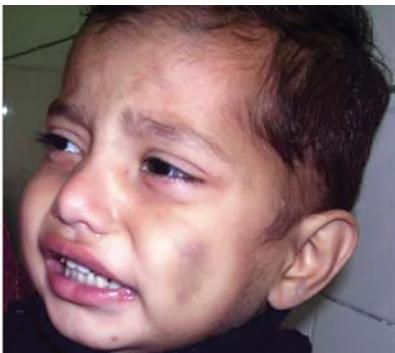
a. What is the most likely diagnosis?
b. Which one screening investigation would you advise?
c. What is the mode of inheritance?
d. Which type / severity category would he be categorized as?
Answer
- a. Hemophilia A (Factor VIII deficiency) or Hemophilia B (Factor IX deficiency).
- b. Activated Partial Thromboplastin Time (aPTT) (which will be prolonged; followed by specific factor assay).
- c. X-linked recessive.
- d. Severity classification of Hemophilia:
- Severe: < 1% factor activity (frequent spontaneous joint/muscle bleeds).
- Moderate: 1–5% factor activity (bleeds with mild trauma / minor surgery).
- Mild: 5–40% factor activity (bleeds only with major trauma or major surgery).
VVK07-028
A 12-year-old boy is tested for sickle cell disease. His father is known to have sickle cell trait. His lab results are as follows: Hb 10.1 g/dL, RBC 6.0 million/cumm, MCV 65 fl, MCH 21.1 pg, MCHC 30 g/dL, sickle test positive, HbS 71%, HbA 21.5%, HbA2 4.5%, HbF 3%, Serum ferritin 19 mcg/L.
a. What hemoglobinopathy does the boy have?
b. What hemoglobinopathy do you expect on testing the mother?
c. Which bacterial infections are more common in such patients?
Answer
- a. Sickle-
thalassemia ( ). - b. Beta-thalassemia trait / carrier (
). - c. Encapsulated organisms: Streptococcus pneumoniae, Haemophilus influenzae type b, Neisseria meningitidis, Salmonella species (osteomyelitis).
VVK07-029
Match the clinical diagnosis with the hematological findings:
| Clinical Diagnosis | Hematological Findings |
|---|---|
| a. Kawasaki disease | 1. Thrombocytosis |
| b. Alpha thalassemia major | 2. Hydrops fetalis |
| c. Beta thalassemia major | 3. Increased fetal Hb |
| d. Sickle cell disease | 4. Hyposplenism in a 6-year-old |
| e. G6PD deficiency | 5. Oxidant induced red cell damage |
| f. Hereditary spherocytosis | 6. Splenomegaly in a 6-year-old |
Answer
- a. Kawasaki disease — 1. Thrombocytosis
- b. Alpha thalassemia major — 2. Hydrops fetalis (Hb Barts)
- c. Beta thalassemia major — 3. Increased fetal Hb (HbF)
- d. Sickle cell disease — 4. Hyposplenism in a 6-year-old (autosplenectomy)
- e. G6PD deficiency — 5. Oxidant-induced red cell damage (Heinz bodies, bite cells)
- f. Hereditary spherocytosis — 6. Splenomegaly in a 6-year-old
VVK07-030
A 4-year-old boy presents with severe pain in both of his legs. On physical examination, his temperature is 37.7°C (99.8°F), blood pressure is 108/68 mmHg, pulse rate is 96/min, and respirations are 22/min. He is noted to have marked pallor on his lips and palpebral conjunctiva. Numerous purpura and petechiae are noted on his skin. There is bilateral cervical lymphadenopathy. His spleen is palpable 3 cm below his left costal margin. Laboratory evaluation reveals a white blood cell count of 1600/mm3; hemoglobin 6.1 g/dL; and platelets 36,000/mm3.
a. What is your most probable diagnosis?
b. What is the peak age incidence of this disease and in which sex is it more common?
c. What is the diagnostic investigation and what do you find?
d. Mention poor prognostic factors for this disease.
Answer
- a. Acute Lymphoblastic Leukemia (ALL).
- b. Peak age incidence: 2–5 years; more common in males than females.
- c. Bone marrow aspiration/biopsy showing
lymphoblasts, with characteristic immunophenotyping on flow cytometry. - d. Poor prognostic factors in ALL:
- Age < 1 year (infants) or
years - Initial WBC count
(or > 100,000/cumm) - Cytogenetics: Hypodiploidy (< 44 chromosomes), Philadelphia chromosome positive
BCR-ABL1, (MLL) gene rearrangements , - CNS or testicular disease at presentation
- Slow early response to induction chemotherapy (Minimal Residual Disease / MRD
at Day 29).
- Age < 1 year (infants) or
VVK07-031
On routine screening of a 12-month-old child, the hemoglobin was found to be 8 g/dL:
a. What details in a nutritional history would be important?
b. What nutritional intervention might you suggest besides or in addition to iron supplementation?
c. What specific instructions and plans for follow-up would you give the parents in order to optimize iron supplementation?
Answer
- a. Detailed dietary intake (excessive unfortified cow's milk intake > 500 mL/day, late introduction of solid complementary feeds, lack of iron-rich foods), birth history (prematurity, low birth weight, early cord clamping, multiple gestations).
- b. Limit cow's milk intake to < 500 mL/day (16–24 oz/day); introduce iron-rich foods (iron-fortified cereals, pureed meats, legumes, dark green leafy vegetables); encourage vitamin C-rich foods (citrus fruits) to enhance absorption; avoid tea/tannins with meals.
- c.
- Prescribe oral elemental iron (3 mg/kg/day) once or twice daily between meals with citrus juice; do not give with milk.
- Warn about harmless black stools, temporary teeth staining (use dropper/straw), and transient constipation.
- Recheck Hb and reticulocyte count after 4 weeks (expect Hb increase of
). Continue iron therapy for 2–3 months after Hb normalizes to replenish marrow iron stores.
VVK07-032
Study the peripheral smear shown below and answer the following questions:
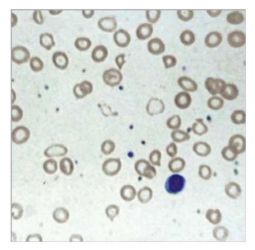
a. Comment on this smear on all the cell lines.
b. Mention at least 4 differentials for this smear.
c. What is the diagnostic test for confirmation?
Answer
- a.
- RBCs: Microcytic hypochromic RBCs with marked anisopoikilocytosis, target cells, pencil cells, and tear drop cells.
- WBCs: Normal in number and morphology.
- Platelets: Normal to mildly thrombocytosis.
- b. Differentials:
- Iron deficiency anemia
- Beta-thalassemia trait
- Anemia of chronic disease
- Sideroblastic anemia
- Lead poisoning
- c. Serum Iron, Total Iron Binding Capacity (TIBC), Serum Ferritin, and Hemoglobin HPLC / Electrophoresis (or bone marrow iron / Perls' Prussian blue stain).
VVK07-033
Answer the following questions regarding histiocytosis syndromes in children:
a. What are the classes of histiocytosis syndromes in children?
b. Name features of each in Class I histiocytosis:
- i. Dermal manifestations
- ii. Endocrine problems
- iii. X-ray skeletal findings
c. Name three biochemical abnormalities seen in Class II histiocytosis (HLH).
Answer
- a. Classification:
- Class I: Langerhans Cell Histiocytosis (LCH).
- Class II: Histiocytosis of Mononuclear Phagocytes other than Langerhans cells (Hemophagocytic Lymphohistiocytosis / HLH and infection-associated hemophagocytic syndromes).
- Class III: Malignant Histiocytosis / Monocytic Leukemia.
- b. Class I (LCH) features:
- i. Dermal: Seborrheic dermatitis-like greasy scaling rash on scalp, trunk, groin, and retroauricular areas; purpuric petechiae.
- ii. Endocrine: Central diabetes insipidus (polydipsia, polyuria) due to posterior pituitary/hypothalamic infiltration; anterior pituitary hormone deficiencies (growth failure).
- iii. Skeletal: Punched-out osteolytic bone lesions without reactive sclerosis ("floating teeth" in mandible, vertebra plana, calvarial lesions).
- c. Class II (HLH) biochemical abnormalities:
- Markedly elevated serum ferritin
- Hypertriglyceridemia
- Hypofibrinogenemia
- Elevated transaminases and hyperbilirubinemia
- Elevated soluble CD25 (sIL-2R) levels
VVK07-034
Study the smear shown below and answer the following questions:
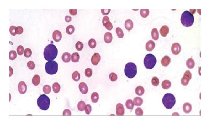
a. What is your diagnosis based on the smear shown above?
b. What other test would you like to do to confirm your diagnosis?
c. What possible differential diagnosis can you think of?
d. Do you need to screen the family if your suspicion comes out positive?
e. Can there be skin involvement in this condition? What is it known as?
Answer
- a. Acute Leukemia (presence of large blast cells with high N:C ratio).
- b. Bone marrow aspirate and biopsy with cytochemistry (MPO, PAS, NSE) and Flow Cytometry / Immunophenotyping.
- c. Acute Lymphoblastic Leukemia (ALL) vs Acute Myeloid Leukemia (AML).
- d. No (most childhood acute leukemias are sporadic, unless there is an underlying cancer predisposition syndrome like Li-Fraumeni or Fanconi anemia).
- e. Yes; Leukemia cutis / chloroma (granulocytic sarcoma).
| Characteristic | Myeloblast | Lymphoblast |
|---|---|---|
| Size | Larger | Smaller |
| Cytoplasm | Moderate, may contain Auer rods | Scanty, no Auer rods |
| Nuclear chromatin | Fine, delicate | Coarse, clumped |
| Nucleoli | Prominent (2–5) | Indistinct / 1–2 |
| Myeloperoxidase (MPO) | Positive | Negative |
VVK07-035
Study the smear shown below and answer the following questions:
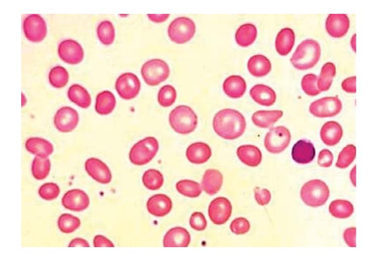
a. Identify the abnormality shown on the peripheral smear above.
b. Name four causes for this condition?
c. Name one drug which can cause this condition?
Answer
- a. Macrocytic anemia / Megaloblastic anemia (macro-ovalocytes and hypersegmented neutrophils).
- b. Causes:
- Folic acid deficiency (nutritional, goat milk feeding)
- Vitamin B12 deficiency (maternal B12 deficiency in breastfed infants, strict vegan diet)
- Malabsorption syndromes (Celiac disease, Crohn disease)
- Congenital pernicious anemia / Imerslund-Gräsbeck syndrome
- Surgical resection of ileum/stomach
- c. Antifolate drugs (Methotrexate, Trimethoprim, Pyrimethamine), Anticonvulsants (Phenytoin, Phenobarbital), Hydroxyurea, Zidovudine (AZT).
VVK07-036
Study the smear shown below and answer the given questions:
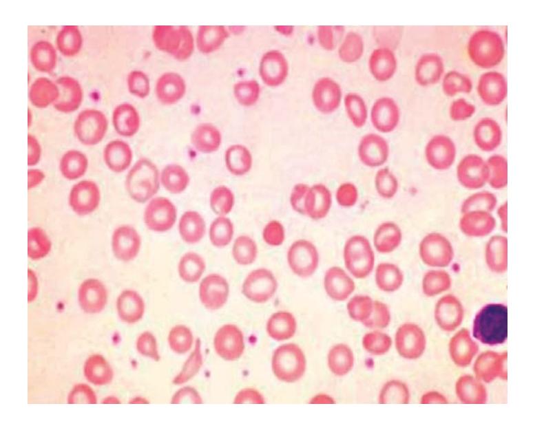
a. Name the type of anemia seen on this peripheral smear.
b. Enumerate causes of this type of anemia.
c. What is RDW and in which of these anemias is RDW increased?
Answer
- a. Microcytic hypochromic anemia.
- b. Causes:
- Iron deficiency anemia
- Thalassemia trait / minor
- Anemia of chronic disease / inflammation
- Sideroblastic anemia
- Lead poisoning
- c. RDW (Red Cell Distribution Width) is a statistical measure of the variation in red blood cell volume/size (anisocytosis). RDW is increased in Iron deficiency anemia (and sideroblastic anemia), whereas it is typically normal in uncomplicated Thalassemia minor.
VVK07-037
Study the smear below and answer the given questions:
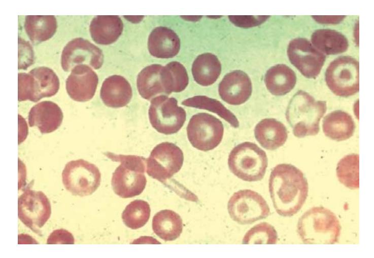
a. What are your findings on this smear?
b. What is the mutational change that leads to this?
c. Which viral infection possesses a unique threat for patients with this disease?
Answer
- a. Sickle cell anemia (showing elongated, crescent/sickle-shaped RBCs, target cells, and Howell-Jolly bodies).
- b. Point mutation in the
-globin gene (substitution of thymine for adenine, GAG to GTG), resulting in the replacement of glutamic acid by valine at position 6 of the -globin polypeptide chain. - c. Human Parvovirus B19 (causes transient pure red cell aplasia / acute aplastic crisis).
VVK07-038
Study the smear below and answer the given questions:
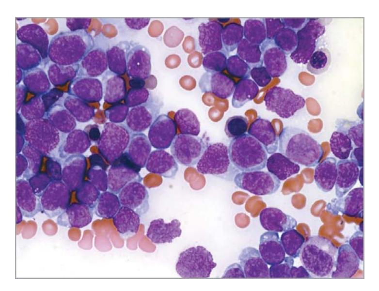
a. What are your findings on this smear?
b. What is your most probable diagnosis?
c. Name four diseases/genetic syndromes in which this condition can occur with increased frequency.
d. An increased association with which viral infection has been associated with this condition?
Answer
- a. Numerous circulating lymphoblasts (high N:C ratio, immature chromatin).
- b. Acute Lymphoblastic Leukemia (ALL).
- c.
- Down syndrome (Trisomy 21)
- Bloom syndrome
- Ataxia-telangiectasia
- Fanconi anemia
- Neurofibromatosis type 1
- d. Epstein-Barr Virus (EBV) (especially associated with Burkitt ALL / Mature B-ALL).
VVK07-039
Study the smear below and answer the given questions:
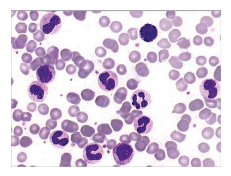
a. What are your findings on this smear?
b. What is the average half-life of a neutrophil in circulation?
c. Mention drugs that can cause neutropenia.
Answer
- a. Band cell (immature neutrophil / left shift).
- b. Approximately 6 to 8 hours in circulating blood (survives 1–2 days in tissues).
- c. Chemotherapeutic agents, Antithyroid drugs (Methimazole, Carbimazole, Propylthiouracil), Antibiotics/Antimicrobials (Sulfonamides, Cotrimoxazole, Chloramphenicol, Vancomycin), Anticonvulsants (Carbamazepine, Valproate, Phenytoin), Antipsychotics/NSAIDs (Clozapine, Aminopyrine).
VVK07-040
A 3-month-old male child presented with diarrhea and ear discharge from both ears for the last 15 days. Child is on exclusive breast milk. Since diarrhea he has lost 1 kg weight and his current weight is 3.5 kg. He was given BCG and hepatitis B at birth; BCG left no scar mark. Rest of the history including birth history is normal. On examination, he has persistent oral candidiasis (thrush) and multiple pyemic abscesses all over the body along with an eczematous rash on the abdomen. There is mild hepatosplenomegaly and absent tonsillar tissue. Initial investigations revealed severe absolute lymphopenia (< 2000/cumm) and low IgA levels. Antibody levels against HBsAg (after 2 doses) are absent. On CT scan, he was found to have a rudimentary/absent thymus (< 1 g) and very underdeveloped tonsils and adenoid tissue.
a. What is the most probable diagnosis?
b. What is the specific definitive treatment?
c. What special precaution should be taken if the infant requires transfusion of a blood product and why?
Answer
- a. Severe Combined Immunodeficiency (SCID).
- b. Allogeneic Hematopoietic Stem Cell Transplantation (HSCT) ideally before 3.5 months of life (or gene therapy for ADA-SCID/X-SCID, PEG-ADA enzyme replacement).
- c. Transfuse only irradiated, leukodepleted, and CMV-negative blood products to prevent fatal Transfusion-Associated Graft-versus-Host Disease (TA-GvHD) and CMV transmission.
VVK07-041
A 2-month-old previously healthy full term female infant presents with fever, lethargy and poor feeding for 12 hours. Family history reveals that a male sibling died suddenly at the age of two months, prior to the birth of this child. There is history of delayed umbilical cord detachment (> 3 weeks). Physical examination reveals a lethargic child with RR 70/min, HR 185/min, mean BP 25 mmHg, temperature 39.5°C and poor peripheral perfusion with absent pus in necrotic skin lesions. Lab results reveal Hb 9.8 g/dL, TLC 65,000/cumm (90% polymorphs) and platelet count 1.4 lakhs/cumm. Peripheral smear shows marked leukocytosis without blasts and no organomegaly.
a. What is the most likely diagnosis?
b. What common infections are expected in these cases?
c. What is the treatment of choice?
Answer
- a. Leukocyte Adhesion Deficiency type 1 (LAD-1) (caused by CD18 /
-integrin deficiency). - b. Recurrent severe bacterial and fungal infections without pus formation: Staphylococcus aureus, Pseudomonas aeruginosa, Escherichia coli, Candida species, and Aspergillus species.
- c. Allogeneic Hematopoietic Stem Cell Transplantation (HSCT).
VVK07-042
Match the clinical disorder with its corresponding complement deficiency:
| Disorder | Complement Deficiency |
|---|---|
| a. Lupus-like syndrome | 1. C1q, C1r, C1s, C4, C2 |
| b. Recurrent neisserial infections | 2. C5, C6, C7, C8, C9 |
| c. Severe recurrent pneumococcal infection | 3. C3 |
| d. Fatal meningococcal infections in males | 4. Properdin (X-linked) |
| e. Non-pitting angioedema | 5. C1 esterase inhibitor deficiency |
Answer
- a. Lupus-like syndrome — 1. C1q, C1r, C1s, C4, C2 (Early classic pathway components)
- b. Recurrent neisserial infections — 2. C5, C6, C7, C8, C9 (Terminal membrane attack complex / MAC)
- c. Severe recurrent pneumococcal infection — 3. C3 deficiency
- d. Fatal meningococcal infections in males — 4. Properdin deficiency (X-linked)
- e. Non-pitting angioedema — 5. C1 esterase inhibitor deficiency (Hereditary Angioedema)
VVK07-043
Answer the following questions on Thrombotic Thrombocytopenic Purpura (TTP):
a. Give the characteristic pentad of TTP.
b. What is the etiology of familial TTP (Upshaw-Schulman syndrome)?
c. What treatment strategy is most suitable for TTP?
d. What cells do you see on peripheral smear in this case?
Answer
- a. Classic Pentad of TTP:
-
- Microangiopathic hemolytic anemia (MAHA)
-
- Thrombocytopenia
-
- Neurological manifestations (headache, confusion, seizures, focal deficits)
-
- Renal dysfunction (mild/moderate)
-
- Fever
-
- b. Congenital deficiency of ADAMTS13 (vWF-cleaving metalloproteinase) due to autosomal recessive mutations.
- c. Therapeutic Plasma Exchange (PEX) with FFP. Corticosteroids, Rituximab, and Caplacizumab (anti-vWF nanobody) in acquired autoimmune TTP. (Platelet transfusions are contraindicated).
- d. Schistocytes (helmet cells, fragmented red cells), microspherocytes, polychromasia, and nucleated red cells with marked thrombocytopenia.
VVK07-044
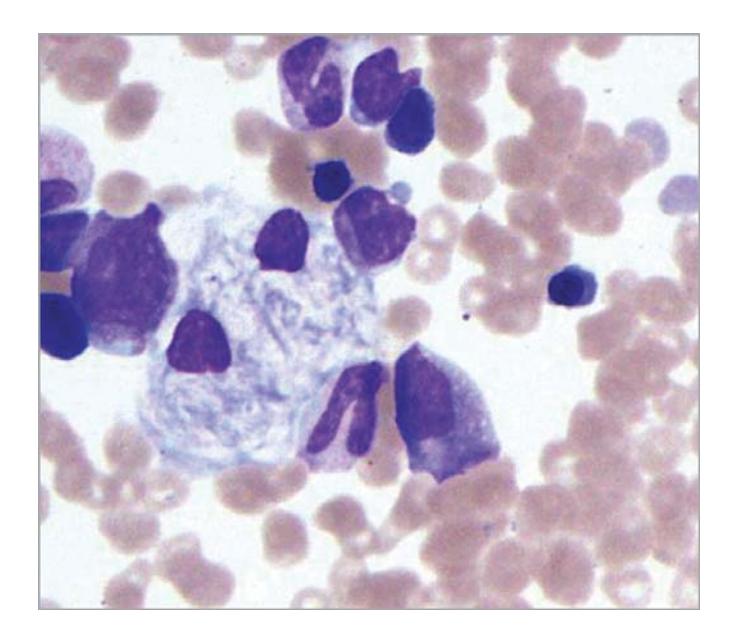
A bone marrow aspirate of an 18-month-old child with features of delayed milestones, muscle hypertonia, recurrent aspiration pneumonia and hepatosplenomegaly is shown below:
a. Describe the findings seen on the smear.
b. Give clinical differential diagnoses for this condition.
c. What management strategy is advised in such a case?
Answer
- a. Gaucher cells: Large lipid-laden macrophages with eccentric small nuclei and voluminous cytoplasm having a "wrinkled tissue paper" or fibrillar appearance.
- b. Niemann-Pick disease, Mucopolysaccharidoses (MPS), GM1 gangliosidosis, Chronic myelogenous leukemia (pseudo-Gaucher cells).
- c.
- Enzyme Replacement Therapy (ERT) with recombinant glucocerebrosidase (Imiglucerase / Velaglucerase alfa 60 U/kg IV every 2 weeks).
- Substrate reduction therapy (Miglustat, Eliglustat).
- Hematopoietic stem cell transplant in selected cases.
VVK07-045
Study the smear shown below and answer the given questions:
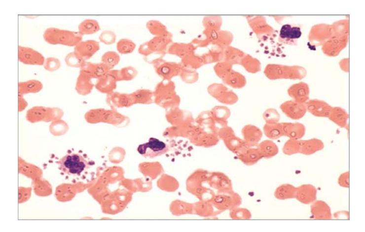
a. Describe platelet satellitism.
b. What is its clinical significance?
c. How do you take care of such a situation?
Answer
- a. Platelet satellitism: In vitro phenomenon where platelets rosette and adhere around the periphery of mature neutrophils (PMNs) in blood collected in EDTA anticoagulant.
- b. Causes spurious/factitious thrombocytopenia (pseudothrombocytopenia) on automated hematology analyzers.
- c. Recollect blood sample in a sodium citrate or heparin tube (or perform a direct fingerstick smear) to obtain an accurate platelet count.
VVK07-046
Answer the following questions:
a. What is the effect on PT and PTT in hemophilia A?
b. Calculate the dose of factor VIII required to increase factor VIII levels by 50% in a child weighing 20 kg.
c. Give the other name for hemophilia C.
d. Give examples of gene therapy being tried for hemophilia.
Answer
- a. PT is normal; aPTT is significantly prolonged.
- b. Calculation of Factor VIII dose:
(Note: 1 Unit/kg of Factor VIII raises plasma activity by 2%). - c. Factor XI deficiency (Rosenthal syndrome).
- d. Adeno-Associated Virus (AAV) vector-mediated liver-directed gene transfer (e.g. Valoctocogene roxaparvovec for Hemophilia A, Etranacogene dezaparvovec for Hemophilia B), lentiviral and retroviral vector delivery.
VVK07-047
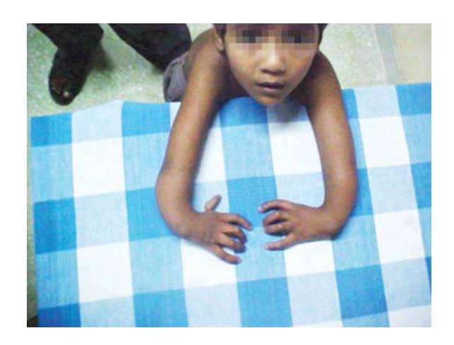
A 5-year-old child presented with pallor and cutaneous markers at the back with an abnormal skeletal profile (hypoplastic/absent thumbs, short stature). Peripheral smear was consistent with macrocytic anemia and pancytopenia.
a. What is the most probable diagnosis?
b. Name the cutaneous marker.
c. What malignancies are expected in this patient?
Answer
- a. Fanconi Anemia (inherited bone marrow failure syndrome).
- b. Café-au-lait spots and areas of cutaneous hyperpigmentation/hypopigmentation.
- c. Acute Myeloid Leukemia (AML / MDS) and solid tumors (squamous cell carcinomas of head, neck, esophagus, and anogenital tract).
VVK07-048
A previously asymptomatic 5-year-old girl is referred with an abdominal mass found by her mother as she was undressing her. On examination, a mass arising from right renal angle is noted which extends medially almost up to midline and inferiorly 10 cm. Her BP is 160/110 mmHg. Investigations show normal plasma urea, creatinine and electrolytes. Urine examination reveals microscopic hematuria but culture is sterile.
a. Give the most likely differential diagnoses.
b. What is the mechanism of raised blood pressure?
c. For each diagnosis give one site where metastases might be commonly found.
Answer
- a. Wilms tumor (Nephroblastoma) and Neuroblastoma.
- b.
- In Wilms tumor: Renal parenchymal compression causing renal ischemia and activation of the Renin-Angiotensin-Aldosterone System (RAAS), or direct renin secretion by tumor cells.
- In Neuroblastoma: Secretion of excessive catecholamines (epinephrine, norepinephrine).
- c. Common metastatic sites:
- Wilms tumor: Lungs (most common), liver.
- Neuroblastoma: Long bones, bone marrow, orbit/skull, liver, lymph nodes.