MNJ07-001
A six-year-old child investigated for increasing pallor, peripheral blood examination was done that is shown below:
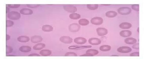
- Name the intracellular inclusion seen in this blood film.
- Name the conditions in which it is seen (Write 3 at least).
Answer
- Cabot ring
- Seen in:
- Lead poisoning
- Pernicious anemia (vitamin B12 deficiency)
- Hemolytic anemias
MNJ07-002
(2014 Exam):
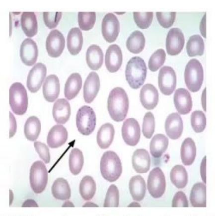
- Identify the abnormality pointed in the given PBF (Peripheral blood film).
- Give 2 differential diagnoses.
Answer
- Basophilic stippling (*can be asked along with X-ray knee of lead poisoning).
- Differential diagnoses:
- Lead poisoning
- Thalassemia
- Vitamin B12 deficiency
Note: The Desferal therapeutic index or Porter index is defined as mean daily dose of desferrioxamine in mg/kg, divided by serum ferritin (useful in thalassemia patients; calculated every 6 months in patients receiving desferrioxamine; should not exceed 0.025 to minimize sensorineural hearing loss).
MNJ07-003
PBF of an 8-year-old child was investigated and pathologist informed you about the satellitism of platelets in PBF:
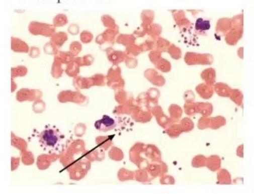
- Describe platelet satellitism.
- What is its clinical significance?
- Next step in evaluation in these patients.
Answer
- Platelet satellitism: Platelet clustering around neutrophils (and rarely monocytes) in the presence of EDTA.
- Clinical significance: Causes pseudothrombocytopenia (spurious thrombocytopenia because cellular aggregates are counted as leukocytes rather than platelets).
- Next step: Repeat platelet count in a heparinized or citrate blood sample.
MNJ07-004
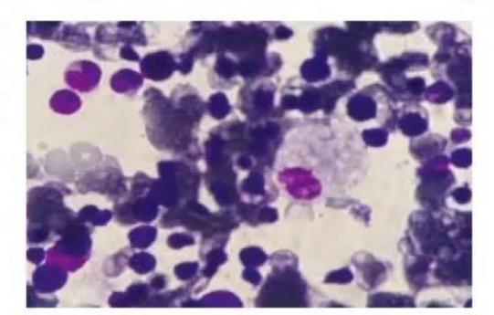
Bone marrow aspirate of an 18-month-old child with a history of hypertonicity, aspiration pneumonia and hepatosplenomegaly: See the film and answer the following questions:
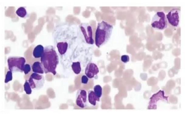
- Describe the findings and write the diagnosis.
- Give 2 clinical differential diagnoses.
- Management.
Answer
- Two macrophages are shown which have a fibrillar, crumpled tissue paper-appearing cytoplasm and eccentric nuclei, consistent with Gaucher cells. Diagnosis: Gaucher disease.
- Differential diagnoses: Niemann-Pick disease, Mucopolysaccharidosis (MPS).
- Management: Enzyme replacement therapy (ERT) with Cerezyme (imiglucerase for injection) at
.
MNJ07-005
A 4-year-old male child presents with fever, hepatosplenomegaly, lymphadenopathy and petechial rash over body, bone marrow was done:
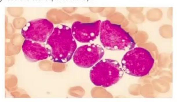
- What is this cell and type of malignancy?
- Write down 4 prognostic factors (favorable).
Answer
- Blast cell: L3 type lymphoblast — Burkitt lymphoma / B-cell ALL.
- Favorable prognostic factors:
- Age between 1 and 10 years
- Hyperdiploidy (>50 chromosomes)
- Presence of t(12;21) [TEL-AML1] translocation
- Low TLC / WBC count at diagnosis (<50,000/μL)
- Rapid early response to therapy
MNJ07-006
A 10-year-old male boy from Bihar, presents with complaints of prolonged fever, weight loss and hepatosplenomegaly. Bone marrow was done in film below:
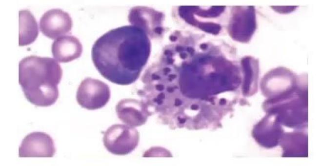
- Identify the bone marrow abnormality and write the possible diagnosis.
- What are diagnostic tests for this disease?
- Write treatment with two new drugs.
Answer
- Leishman-Donovan (LD) bodies (amastigotes) in bone marrow macrophage — Kala-azar (Visceral Leishmaniasis).
- Diagnostic tests:
- Bone marrow / splenic aspirate smear (LD bodies)
- NNN media culture (Novy-MacNeal-Nicolle medium)
- rK39 immunochromatographic rapid dipstick test
- Direct Agglutination Test (DAT) / PCR
- Treatment:
- Standard: Sodium stibogluconate (20 mg/kg/day for 28–30 days) or Amphotericin B (1 mg/kg/day alternate days for 14–20 days) / Liposomal Amphotericin B.
- Newer oral/parenteral drugs: Miltefosine, Paromomycin, Pentamidine.
MNJ07-007
A 7-month-old male child was brought from Abbottabad with complaints of loose stools and fever off and on for the last 6 months. On examination, he had hepatomegaly 10 cm, splenomegaly 5 cm, delayed milestones, and fundoscopy showed cherry-red spots:
- What is the diagnosis of this bone marrow and clinical scenario?
- Write the inheritance of this disease.
- Enzyme defect responsible for this.
Answer
- Diagnosis: Niemann-Pick disease (Type A) showing foamy histiocytes / lipid-laden macrophages ("foam cells").
- Inheritance: Autosomal Recessive (AR).
- Enzyme defect: Acid sphingomyelinase deficiency.
MNJ07-008
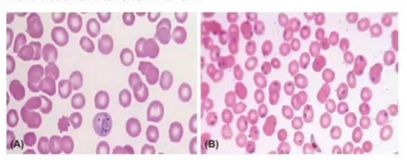
- Identify slide A and B.
- Write down the treatment for both A and B.
Answer
- Identification:
- Slide A: Plasmodium vivax (trophozoite / ring form with Schüffner dots / enlarged RBC).
- Slide B: Plasmodium falciparum (multiple delicate ring forms / crescent-shaped gametocyte, high parasitemia >80% RBCs infected).
- Treatment:
- A (P. vivax): Chloroquine for 3 days followed by Primaquine (0.25–0.5 mg/kg/day) for 14 days (after ruling out G6PD deficiency).
- B (P. falciparum): Artemisinin-based combination therapy (ACT, e.g., Artesunate + Sulfadoxine-Pyrimethamine or Artemether-Lumefantrine) for 3 days plus single dose Primaquine on day 2.
MNJ07-009
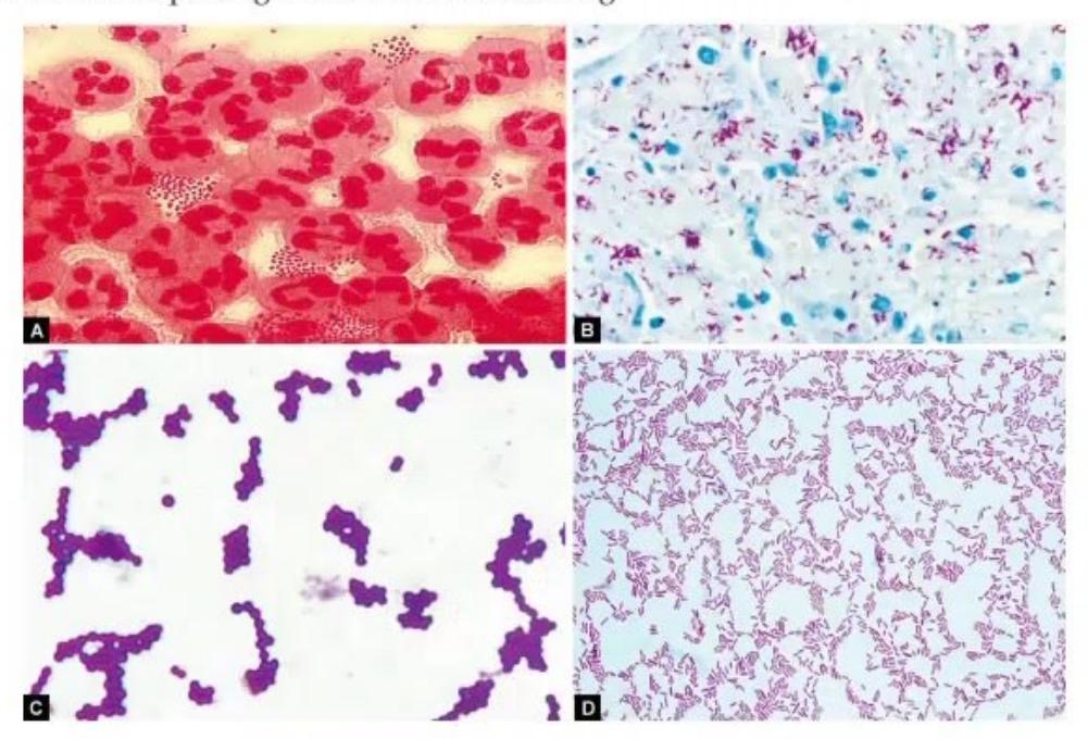

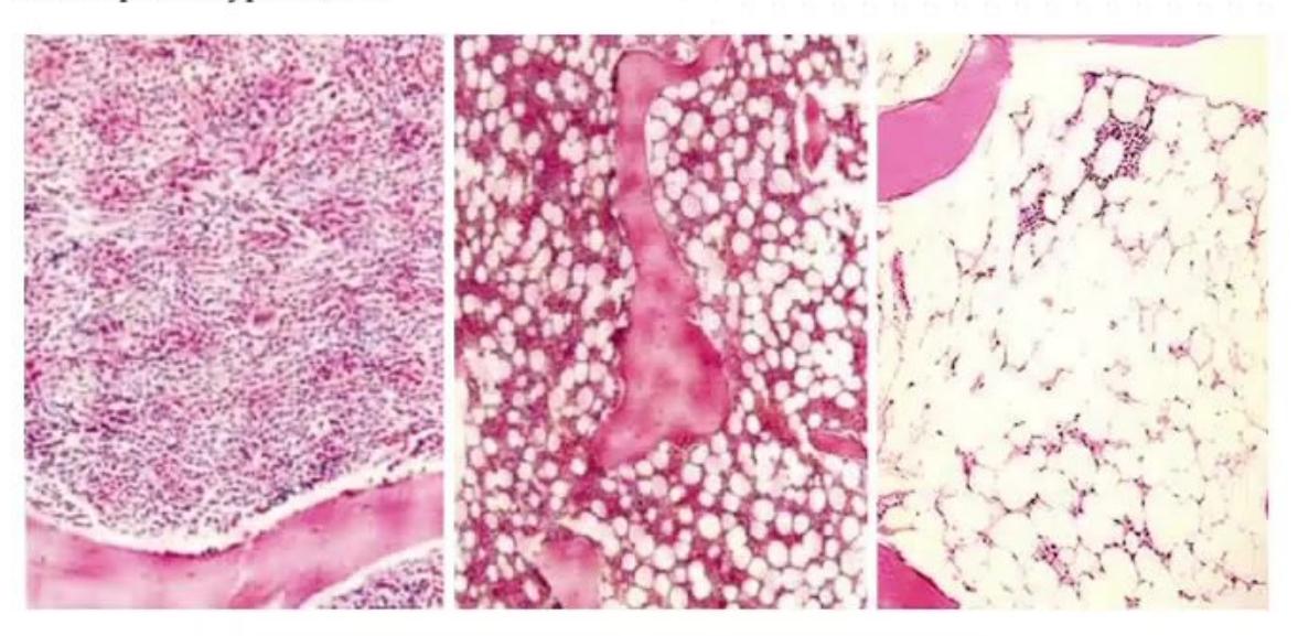
- Identify slides A, B, C, and D.
- Write down the steps for Gram staining and Acid-Fast (Ziehl-Neelsen) staining.
Answer
-
Identification:
- A: Gram-negative bacilli / cocci
- B: Acid-Fast Bacilli (AFB)
- C: Gram-positive cocci
- D: Gram-negative bacilli
-
Staining Procedures:
-
Steps of Gram Staining:
- Heat fixation of smear
- Primary stain: Apply Crystal Violet for 1 minute
- Mordant: Flood with Gram's Iodine for 1 minute (or 10–30 sec)
- Decolorization: Decolorize with 95% ethyl alcohol / acetone-alcohol rapidly (10–15 sec) and wash with water immediately
- Counterstain: Apply Safranin / Basic Fuchsin for 30–60 seconds, wash with water and dry
-
Steps of Acid-Fast (Ziehl-Neelsen) Staining:
- Heat and dry-fix the smear
- Primary stain: Add Strong Carbol Fuchsin and heat gently until steaming (do not boil) for 5 minutes
- Decolorization: Decolorize with 20% sulfuric acid (or acid-alcohol) and wash with water
- Counterstain: Apply Methylene Blue for 1–2 minutes, wash with water, air dry and examine
-
MNJ07-010
(Exam 2014) Match the following red blood cell abnormalities with their associated condition:
| RBC Morphology | Condition |
|---|---|
| 1. Spur cell (Acanthocyte) | A. Renal disease |
| 2. Burr cell (Echinocyte) | B. Thalassemia |
| 3. Target cell (Codocyte) | C. Pernicious anemia |
| 4. Cabot ring | D. G6PD deficiency |
| 5. Bite cell (Degmacyte) | E. Myelofibrosis |
| 6. Teardrop cell (Dacrocyte) | F. Liver disease |
Answer
- 1 – F: Spur cell (Acanthocyte)
Liver disease / Abetalipoproteinemia - 2 – A: Burr cell (Echinocyte)
Renal disease (Uremia) - 3 – B: Target cell (Codocyte)
Thalassemia / Hemoglobinopathies - 4 – C: Cabot ring
Pernicious anemia (Megablastic anemia / Lead poisoning) - 5 – D: Bite cell
G6PD deficiency - 6 – E: Teardrop cell (Dacrocyte)
Myelofibrosis
MNJ07-011
Peripheral smear:
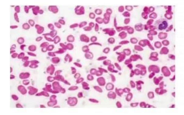
- What is the diagnosis?
- What is the drug of choice for the prevention of painful vaso-occlusive episodes?
- What are measures for the primary prevention of stroke in these children?
- What are the antibiotics of choice in acute chest syndrome?
- What type of renal malignancy is common in this condition?
Answer
- Diagnosis: Sickle Cell Anemia (showing sickle cells / drepanocytes and target cells).
- Drug of choice for prevention of painful episodes: Hydroxyurea.
- Primary prevention of stroke:
- Screening with Transcranial Doppler (TCD) ultrasonography annually (evaluating time-averaged mean maximum velocity of internal carotid/middle cerebral artery; if velocity
, high risk). - Chronic transfusion therapy (to maintain
).
- Screening with Transcranial Doppler (TCD) ultrasonography annually (evaluating time-averaged mean maximum velocity of internal carotid/middle cerebral artery; if velocity
- Antibiotics of choice in acute chest syndrome: Third-generation cephalosporin (e.g., Cefotaxime/Ceftriaxone) plus a Macrolide (e.g., Azithromycin) to cover atypical pathogens (Mycoplasma, Chlamydia).
- Renal malignancy: Renal medullary carcinoma.
MNJ07-012
A 1-year-old child with high fever and convulsions:
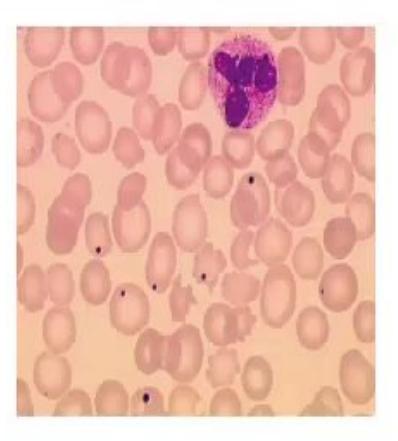
- What does the smear show?
- What association will you suspect on clinical examination with this blood film?
- What is the likely cause of the symptoms?
- Which organism is responsible?
- How could you have prevented this infection?
Answer
- Smear finding: Howell-Jolly bodies (small, round, purple nuclear remnants of condensed DNA in RBCs).
- Clinical association: Asplenia / Functional asplenia (or post-splenectomy).
- Likely cause of symptoms: Overwhelming post-splenectomy infection (OPSI) / Bacterial meningitis / Sepsis.
- Responsible organism: Streptococcus pneumoniae (encapsulated organisms).
- Prevention:
- Immunization: Pneumococcal conjugate/polysaccharide vaccine, Meningococcal vaccine, Haemophilus influenzae type b (Hib) vaccine.
- Prophylactic oral penicillin.
MNJ07-013
(Exam 2013, 2014):
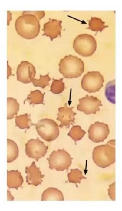
- Describe the cells seen.
- Name one condition each from following category in which these cells are seen:
- Neurological
- Metabolic
- Hepatic
- Endocrinal
Answer
- Description: Acanthocytes (spur cells) — RBCs lacking central pallor with multiple thorn-like, spiked projections of variable length located at irregular intervals.
- Conditions:
- Neurological: Neuroacanthocytosis (e.g., Chorea-acanthocytosis, McLeod syndrome)
- Metabolic: Abetalipoproteinemia (Bassen-Kornzweig syndrome)
- Hepatic: Severe liver dysfunction / Cirrhosis (spur cell anemia)
- Endocrinal: Hypothyroidism
MNJ07-014
A 10-year-old girl presents with fatigue and tingling sensation for the last one month. On examination, pallor was present with a murmur; all other examination was normal. PBF is given below:
- Describe this peripheral smear.
- Diagnose the condition and give 6 differential diagnoses of macrocytes.
- Define this abnormality of neutrophil and write down 4 causes of it.
Answer
- Description: Macro-ovalocytes (large oval RBCs) and hypersegmented neutrophils.
- Diagnosis: Megaloblastic anemia (Vitamin B12 / Folate deficiency).
- Differential diagnoses of Macrocytosis:
- Vitamin B12 deficiency
- Folate deficiency
- Chronic liver disease
- Hypothyroidism
- Aplastic anemia
- Diamond-Blackfan anemia
- Orotic aciduria / Drugs (hydroxyurea, methotrexate, AZT)
- Differential diagnoses of Macrocytosis:
- Hypersegmented neutrophil:
- Definition: Presence of
neutrophils with 5 lobes or neutrophil with 6 or more lobes on a 100-neutrophil differential count. - Causes:
- Vitamin B12 deficiency
- Folate deficiency
- Iron deficiency anemia (rare/mixed)
- Uremia
- Hydroxyurea / Chemotherapeutic drug therapy
- Definition: Presence of
MNJ07-015
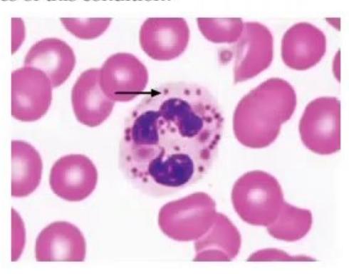
- Identify the cell and write the condition it is seen in.
- What are the clinical features of this condition?
Answer
- Identification: Giant lysosomal granule within leukocyte (granulocyte/neutrophil), seen in Chédiak-Higashi syndrome.
- Clinical features:
- Partial oculocutaneous albinism (silvery hair, photophobia, nystagmus)
- Recurrent severe pyogenic infections
- Peripheral neuropathy
- Bleeding diathesis (platelet dense granule defect)
- Accelerated phase (hemophagocytic lymphohistiocytosis / pancytopenia)
MNJ07-016
An 8-year-old boy presented with pain in throat with difficulty in respiration for 1 day. Throat examination showed palatal weakness with a white patch on the tonsil:
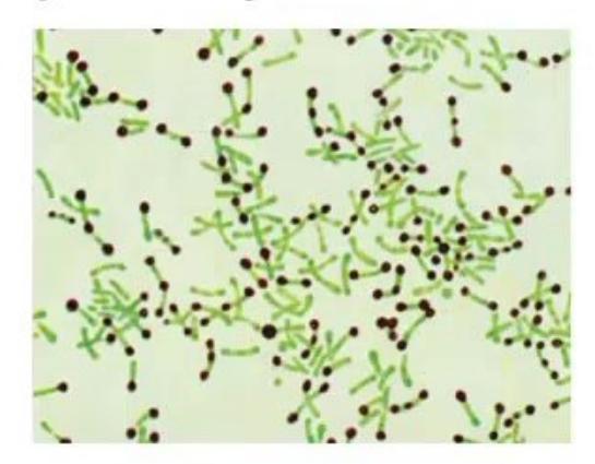
- Name two common complications in this situation.
- What is the name of the stain in the slide?
- Write differential diagnoses of white patch on tonsil.
Answer
- Complications (of Diphtheria):
- Toxic myocarditis / Arrhythmias / Heart failure
- Neurological complications / Cranial neuropathies (palatal weakness, ciliary paralysis) and peripheral neuropathy
- Airway obstruction due to pseudomembrane
- Stain: Albert stain (shows metachromatic granules of Corynebacterium diphtheriae).
- Differential diagnoses of white patch / membrane on tonsil:
- Streptococcal tonsillitis / pharyngitis
- Infectious mononucleosis (EBV)
- Candidiasis (Oral thrush)
- Vincent angina (acute necrotizing ulcerative gingivostomatitis)
- Agranulocytosis
MNJ07-017
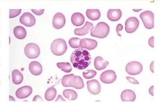
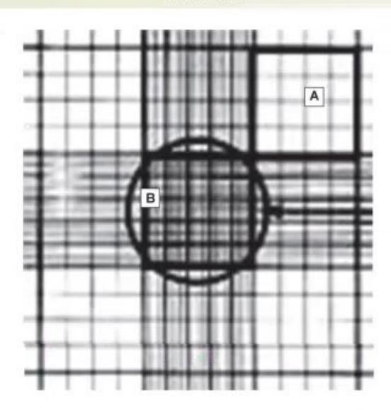
- Name the spot.
- What are these chambers used for (A and B)?
- If doing CSF cytology for WBCs, which chambers are used for this purpose?
Answer
- Spot: Improved Neubauer hemocytometer counting chamber.
- Chambers used:
- A (Large 4 corner squares): Used for White Blood Cell (WBC) counting.
- B (Central large square / 5 smaller squares within): Used for Red Blood Cell (RBC) and Platelet counting.
- CSF cytology for WBCs: All 9 large squares (including 4 corners and central squares) are counted.
MNJ07-018
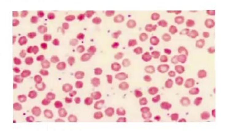
- Identify the abnormality in RBC.
- This child has chronic diarrhea with foul smelling stool. What is the possible diagnosis?
- Which vitamin deficiency in these children is associated with neurological symptoms?
- Which lipid abnormalities are characteristic?
- What is the fundus finding?
- What is the mode of inheritance?
Answer
- Abnormality: Acanthocytes (spur cells).
- Diagnosis: Abetalipoproteinemia (Bassen-Kornzweig syndrome).
- Vitamin deficiency: Vitamin E deficiency (causing ataxia, neuropathy, loss of proprioception).
- Lipid abnormalities:
- Markedly decreased / absent Serum triglycerides
- Markedly decreased Serum cholesterol
- Absence of ApoB-containing lipoproteins (chylomicrons, VLDL, LDL).
- Fundus finding: Retinitis pigmentosa (atypical).
- Mode of inheritance: Autosomal recessive.