MNJ32-001
A 6-year-old child presents with fever and toxemia:
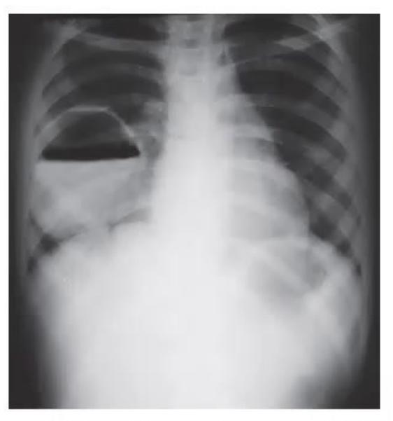
- Give three differential diagnoses for this X-ray picture.
- Give three modalities of management.
Answer
- Differential diagnoses:
- Lung abscess
- Infected bronchogenic cyst
- Infected hydatid cyst
- Modalities of management:
- Antibiotics (anaerobic + aerobic coverage)
- Chest physiotherapy
- Percutaneous CT-guided aspiration
MNJ32-002
See the X-ray image below and answer the following questions:
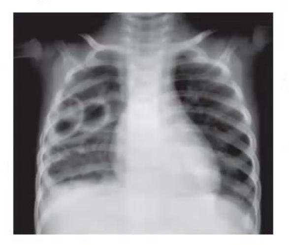
- What is the diagnosis?
- What are the likely two organisms?
Answer
- Pneumatoceles
- Staphylococcus, Klebsiella
MNJ32-003
See the X-ray image below:
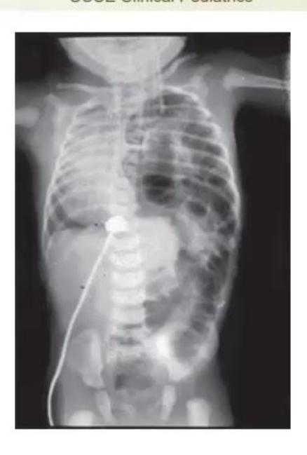
- What is the abnormality?
- What is the diagnosis?
- How is it suspected clinically?
- What is the management?
Answer
- X-ray abnormality: Bowel loops in left hemithorax, mediastinal shift to right
- Diagnosis: Congenital diaphragmatic hernia (CDH)
- Clinical suspicion:
- Respiratory distress
- Mediastinal shift (dextroposition of heart sounds)
- Bowel sounds in the thorax
- Scaphoid abdomen
- Management:
- Avoid bag and mask (B&M) ventilation; immediate endotracheal intubation
- Prop up head end and decompress stomach with nasogastric tube
- Mechanical ventilation
- Treat PPHN (pulmonary hypertension)
- Surgical correction (delayed until hemodynamically stable)
- Antenatal intervention: Tracheal occlusion / ligation (FETO)
MNJ32-004
See the X-ray image below:
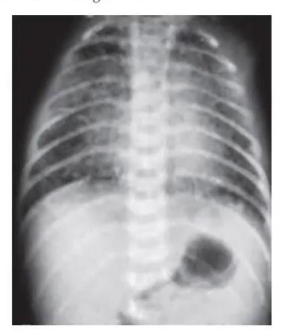
- Describe the X-ray in a neonate with difficult labour.
- What is the diagnosis?
- Give three recent advances in management.
Answer
- X-ray findings: Hyperinflated lung fields with areas of atelectasis interspersed with areas of overinflation / patchy infiltrates.
- Diagnosis: MAS (Meconium Aspiration Syndrome)
- Recent advances in management:
- Lung lavage
- Surfactant instillation
- HFOV (High Frequency Oscillatory Ventilation)
- Inhaled Nitric Oxide (iNO for PPHN)
- Liquid ventilation
MNJ32-005
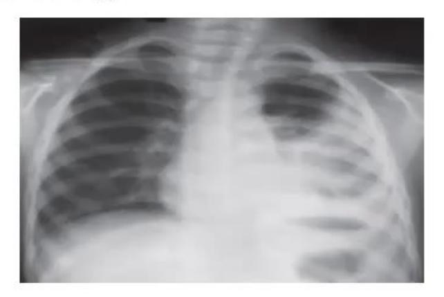
See the X-ray images below:
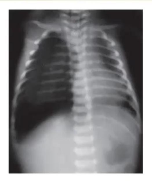
- What is the abnormality?
- Three high risk situations when this condition is imminent.
- What is the management?
Answer
- Abnormality: Tension pneumothorax
- High risk situations:
- Congenital diaphragmatic hernia (CDH)
- Positive pressure mechanical ventilation
- Post-surfactant ventilation (due to sudden compliance change)
- Meconium aspiration syndrome
- Management:
- Immediate needle thoracocentesis (decompression)
- Chest tube / intercostal drainage (ICD)
MNJ32-006
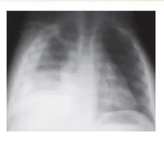
See the X-ray image below:
- What is the X-ray suggestive of?
- What is the likely organism?
- What are the complications?
- What is the drug of choice?
- What is the duration of therapy?
Answer
- Lobar pneumonia (Right upper lobe consolidation)
- Streptococcus pneumoniae / Staphylococcus aureus
- Synpneumonic / parapneumonic effusion, empyema, lung abscess, pneumatocele, septicemia
- Penicillin / Ampicillin for susceptible organisms; Cefotaxime / Ceftriaxone / Vancomycin for penicillin-resistant strains
- 10–14 days
MNJ32-007
See the X-ray image below:
- What is the diagnosis?
- Fill in the blanks regarding exudative pleural fluid:
- Proteins > ........
- Pleural fluid LDH > ........
- Fluid to serum LDH ratio > ........
- Cell count > ........
- What does VATS stand for?
Answer
- Diagnosis: Pleural effusion (right-sided)
- Exudative pleural fluid criteria (Light's criteria & parameters):
- Proteins > 3.0 g/dL (or Pleural/Serum Protein ratio > 0.5)
- Pleural fluid LDH > 200 IU/L (or > 2/3 upper limit of normal serum LDH)
- Fluid to serum LDH ratio > 0.6
- Cell count > 1000 /mm³
- VATS: Video-Assisted Thoracoscopic Surgery
MNJ32-008
See the chest radiograph below:
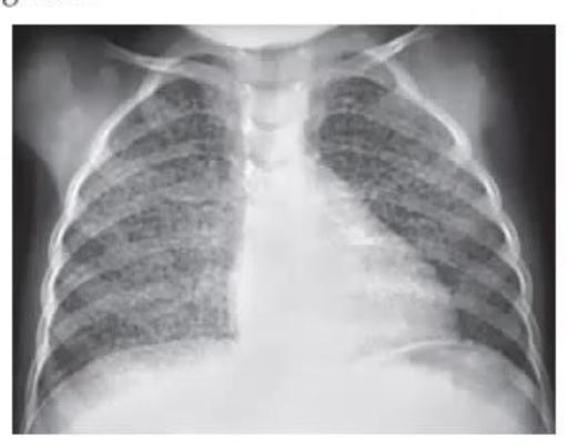
- Diagnosis.
- What is the treatment (mention complete schedule)?
- Write 4 differential diagnoses.
Answer
- Diagnosis: Miliary tuberculosis
- Treatment schedule:
- 2 months HRZES + 1 month HRZE (Intensive phase) followed by 5 months HRE (Continuation phase)
- (Or Pediatric ATT regimen: 2 months HRZE + 4 months HRE)
- Differential diagnoses:
- Chronic CHF
- Eosinophilic pneumonia (Tropical pulmonary eosinophilia)
- Staphylococcal pneumonia / septic emboli
- Hemosiderosis
- Histoplasmosis / fungal infections
MNJ32-009
See the radiographs below:
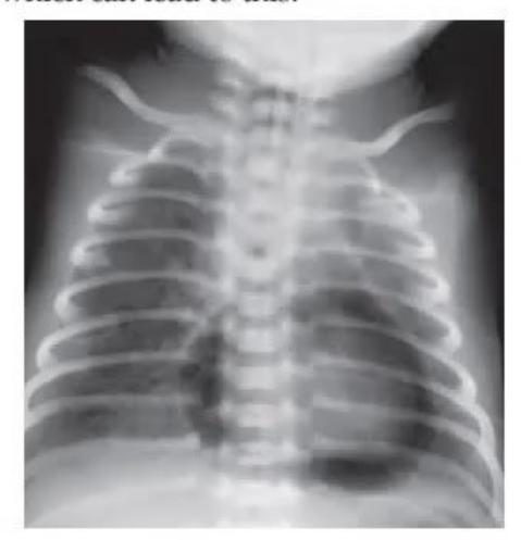
- What is the diagnosis?
- What is the clinical sign for the diagnosis called?
- Name one intervention which can lead to this.
Answer
- Diagnosis: Pneumopericardium / Pneumomediastinum
- Clinical sign: Hamman's sign (Hamman's crunch / click)
- Intervention leading to this: Invasive mechanical ventilation with high peak inspiratory pressures (barotrauma) / central line insertion / thoracic surgery
MNJ32-010
See the neck radiograph below:
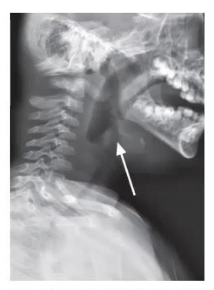
- What is the diagnosis and characteristic X-ray sign?
- Which is the commonest organism implicated in this disease?
- What antibiotics are useful in this condition?
Answer
- Diagnosis & Sign: Acute Epiglottitis — "Thumb sign" (swollen epiglottis on lateral neck X-ray). (Note: Croup presents with "Steeple sign" on AP view).
- Organisms: Haemophilus influenzae type b (Hib), Streptococcus pneumoniae, Staphylococcus aureus
- Antibiotics: 3rd generation cephalosporins (Ceftriaxone / Cefotaxime) or Ampicillin-Sulbactam
MNJ32-011
A 16-year-old boy followed up in your clinic for several years. His latest lung function tests are:
| Parameter | Value |
|---|---|
| FVC | 85% |
| FEV1 | 57% |
| PEF | 53% |
| FEV1/FVC | 67% |
- Diagnosis.
- Which of the above measurements is best for monitoring?
Answer
- Asthma (severe persistent, obstructive pattern)
- FEV1/FVC (67%) and PEF (Peak Expiratory Flow)
MNJ32-012
Write changes in RNTCP guidelines in following aspects:
- Explain categorizations
- Definition of smear +ve
- Definition of TB suspect in HIV
- Drug in HIV
- XDR-TB
Answer
- Categorizations:
- Category I: New cases (smear-positive, smear-negative, extra-pulmonary)
- Category II: Previously treated cases (relapse, failure, treatment after default)
- Definition of smear +ve: Sputum samples collected are only "2" (one spot, one early morning); if one of two samples is positive, the patient is considered smear-positive.
- TB suspect in HIV: Any duration of cough, fever, night sweats, or weight loss.
- Drug in HIV: Rifabutin (used when co-administering with certain antiretroviral protease inhibitors) / standard Rifampicin with efavirenz-based regimen.
- XDR-TB: MDR-TB (resistance to INH + Rifampicin) plus resistance to any fluoroquinolone and at least one second-line injectable agent (amikacin, kanamycin, or capreomycin).
MNJ32-013
An 11-year-old boy performed standard spirometry and produced the following results:
| Investigation | Actual | Predicted |
|---|---|---|
| FVC | 1.13 | 2.07 |
| FEV1 | 1.08 | 1.87 |
| FEV1/FVC | 95% | 87% |
| FEF 25-75% | 1.89 | 2.26 |
- What type of respiratory impairment is present?
- Give three possible causes for getting this result.
Answer
- Restrictive pattern (Reduced FVC and FEV1 with normal or elevated FEV1/FVC ratio)
- Causes:
- Neuromuscular disease (e.g., Myasthenia gravis, Duchenne muscular dystrophy)
- Interstitial lung disease / Fibrosing alveolitis
- Chest wall deformities (e.g., Kyphoscoliosis, pectus excavatum)
MNJ32-014
A spirometry is performed in an asthmatic child:
- What will be abnormalities in the following:
- FEV1
- FEV1/FVC
- Improvement in FEV1 with bronchodilator
- Exercise challenge test
- What PEFR diurnal variation is consistent with a diagnosis of asthma?
Answer
- Abnormalities:
- FEV1: Low / Reduced (<80% predicted)
- FEV1/FVC ratio: Reduced (<0.80 or <80%)
- Improvement in FEV1: Reversibility ≥12% (or ≥200 mL) post-inhaled β2 agonist
- Exercise challenge: Worsening / fall in FEV1 ≥15%
- PEFR variation: Diurnal (morning to afternoon) variation ≥20%
MNJ32-015
An infant has cough and difficult breathing. The respiratory rate is 70/min. The infant has severe respiratory distress (head nodding):
- What does the infant have as per ARI / IMNCI programme?
- What are other criteria for this status?
- Will you treat this infant as OP or IP?
- What is the antibiotic therapy regimen?
Answer
- ARI/IMNCI classification: Very severe disease / Very severe pneumonia
- Other danger signs / criteria:
- Central cyanosis
- Inability to feed / drink or vomiting everything
- Convulsions / lethargy / unconsciousness
- Severe chest indrawing / stridor in calm child
- Setting: IP (Inpatient admission)
- Antibiotic therapy:
- IV Ampicillin + Gentamicin for 5–7 days
- OR IV Ceftriaxone / Cefotaxime for 5–7 days
MNJ32-016
A 3-day-old male child born of third-degree consanguineous marriage as a full-term normal delivery, birth weight 3 kg, history of cried immediately, brought on day 3 with abdominal distension noticed since day 2 of life. Today, baby also had bilious vomiting. On examination, no congenital abnormality was detected. Vitals stable, anus patent, doughy abdomen. Pediatric surgeon advised conservative management and a barium enema. Post barium enema procedure large amount of meconium was passed. Also micro-colon was noted. Child gradually improved:
- Give diagnosis.
- Primary disease & mode of inheritance.
- Confirmatory tests (any 2).
- Neonatal screening test.
Answer
- Diagnosis: Meconium ileus (complicated by microcolon)
- Primary disease & Inheritance: Cystic Fibrosis; Autosomal Recessive
- Confirmatory tests:
- Sweat chloride test (Pilocarpine iontophoresis: >60 mEq/L)
- CFTR gene mutational analysis
- Nasal potential difference measurement
- Neonatal screening test: IRT (Immunoreactive Trypsinogen) assay on dried blood spot
MNJ32-017
A 3-year-old child with fever, irritability, poor feeding and drooling of saliva:
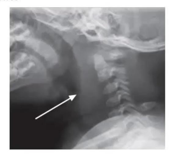
- What is the diagnosis?
- Name two most common micro-organisms associated.
- What is the drug of choice?
- Name two complications.
- What is Lemierre's disease?
Answer
- Diagnosis: Retropharyngeal abscess
- Microorganisms: Group A Streptococcus (GAS), Staphylococcus aureus, Haemophilus influenzae, anaerobes
- Drug of choice: 3rd generation cephalosporin (Ceftriaxone) + Ampicillin-Sulbactam / Clindamycin / Vancomycin
- Complications: Upper airway obstruction, aspiration pneumonia, mediastinitis, erosion into carotid artery, jugular vein thrombophlebitis
- Lemierre's disease: Parapharyngeal/retropharyngeal infection extending to cause internal jugular vein septic thrombophlebitis with metastatic septic emboli to lungs and other organs (most commonly caused by Fusobacterium necrophorum).
MNJ32-018
Full term vacuum delivery noticed to have inability to move left upper limb at all (C5-T1 injury). He was followed regularly for management of the same. Corrective neurosurgical procedure was performed at 6 months of age. Now 8 months old, brought with history of breathlessness noticed since last 6 weeks, no fever:
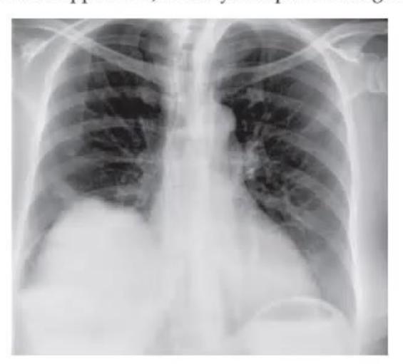
- Mention abnormalities on X-ray.
- Possible reason for this situation.
- Mention nerve roots involved in Erb's and Klumpke's palsy.
- What other signs (other than upper limb) would you expect at the age of 4 months in this child if sympathetic chain is injured?
Answer
- X-ray abnormalities:
- Eventration / elevation of left dome of diaphragm
- Shift of mediastinum and trachea to the opposite side
- Fracture / operative resection of left clavicle with callus formation
- Possible reason: Peri-operative or birth trauma damage to phrenic nerve (C3–C5) / left brachial plexus leading to paralysis of the left dome of diaphragm.
- Nerve roots:
- Erb's palsy: C5–C6 (upper trunk)
- Klumpke's palsy: C8–T1 (lower trunk)
- Other signs: Horner's syndrome (Miosis, ptosis, enophthalmos, anhidrosis) due to T1 sympathetic chain involvement.
MNJ32-019
A 6-year-old boy presents with complaints of cough, fever and dyspnoea. On examination, he is tachypneic and heart sounds are shifted to right side. Auscultation suggests decreased air entry on right side. His chest X-ray is given below:
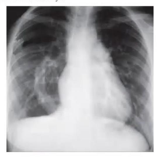
- What is the finding in this X-ray?
- What is the diagnosis?
- What is the basic vascular pathology behind this X-ray finding?
- What would you expect in antenatal history?
Answer
- X-ray findings: Opacification and low-volume right lung, mediastinum/heart shifted to the right side, compensatory hyperinflation of left lung, and curved vascular shadow (scimitar-like) descending along right cardiac border.
- Diagnosis: Scimitar syndrome (Congenital pulmonary venolobar syndrome / partial anomalous pulmonary venous return with right lung hypoplasia)
- Vascular pathology: Anomalous drainage of right pulmonary veins directly into the inferior vena cava (IVC).
- Antenatal history: Oligohydramnios / maternal polyhydramnios or fetal lung abnormalities detected on anomaly scan.
MNJ32-020
Five children were referred to OPD for hearing concerns. You performed Weber and Rinne tests. Match the test results with the correct diagnoses:
Test Findings:
- Weber localizing to right ear; Rinne left ear positive, right ear negative
- Weber localizing to right ear; Rinne left ear negative, right ear positive
- Weber equal (central); Rinne positive in both ears
- Weber localizing to left ear; Rinne left ear negative, right ear positive
- Weber localizing to left ear; Rinne left ear positive, right ear negative
Options:
- a. Conductive hearing loss of right ear
- b. Sensorineural hearing loss of left ear
- c. Normal hearing
- d. Conductive hearing loss of left ear
- e. Sensorineural hearing loss of right ear
Answer
- -> a (Conductive hearing loss of right ear)
- -> b (Sensorineural hearing loss of left ear)
- -> c (Normal hearing)
- -> d (Conductive hearing loss of left ear)
- -> e (Sensorineural hearing loss of right ear)
Principles:
- Weber: Lateralizes to the side of conductive loss or to the opposite side of sensorineural loss.
- Rinne: Positive (AC > BC) = Normal or SNHL; Negative (BC > AC) = Conductive hearing loss.
MNJ32-021
Sameer, a 12-year-old boy, has been admitted to the pediatric ward with respiratory problems. Lung function tests were performed:
| Parameter | Measured | Predicted |
|---|---|---|
| FVC | 1.3 L | 2.6 L |
| FEV1 | 1.1 L | 2.3 L |
| FEV1/FVC | 92% | 90% |
- What type of respiratory impairment does he have according to these test results?
- What are 4 causes that can present with a similar lung function picture?
- What respiratory impairment will you get in a child with cystic fibrosis?
Answer
- Respiratory impairment: Restrictive lung disease (both FVC and FEV1 reduced with normal/elevated FEV1/FVC ratio).
- 4 Causes:
- Kyphoscoliosis / chest wall deformity
- Fibrosing alveolitis / interstitial lung disease
- Duchenne muscular dystrophy
- Guillain-Barré syndrome (GBS)
- Cystic fibrosis impairment: Predominantly obstructive lung defect, progressing to a mixed (obstructive + restrictive) defect in advanced disease.
MNJ32-022
Match the vocal cord/laryngeal disorders with the clinical conditions described below:
Clinical Vignettes:
- A 14-year-old known asthmatic boy presents with complaints of recurrent wheeze during the day and intermittent shortness of breath associated with sensation of throat tightness. Sometimes when he gets upset, coughing is associated with an audible noise. These attacks are not relieved with his MDI therapy. On examination, you find inspiratory upper airway sounds on auscultation with no other anomaly.
- A 7-year-old boy with pulmonary atresia and intact interventricular septum has just been discharged from the ICU after cardiac surgery. His mother noticed his voice is hoarse and weak. He also suffers from frequent choking and coughing.
- An 11-year-old girl presents for hoarseness of voice. She is part of the school singing choir and feels throat pain while singing. Her mother notes she started strenuous rehearsals for her school programme next month.
Options:
- a. Vocal nodule
- b. Vocal cord dysfunction
- c. Congenital laryngeal web
- d. Unilateral vocal cord paralysis
- e. Bilateral vocal cord paralysis
Answer
- -> b. Vocal cord dysfunction (VCD): Presents with daytime wheeze and throat tightness, often misdiagnosed as refractory asthma.
- -> d. Unilateral vocal cord paralysis: Recurrent laryngeal nerve injury post-cardiac/thoracic surgery leading to hoarse/breathy voice and choking/aspiration risk.
- -> a. Vocal nodule (Screamer's/Singer's nodules): Most common cause of chronic hoarseness in children due to vocal misuse/overuse.
MNJ32-023
A 4-year-old boy known to be HIV-positive is admitted with complaints of respiratory distress. After 4 days of routine broad-spectrum antibiotics and supportive management, his condition deteriorated and he required mechanical ventilation. On auscultation the chest is clear, but chest X-ray showed bilateral infiltrates with ground-glass attenuation:
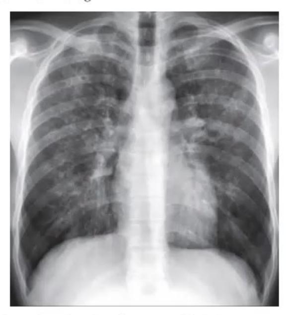
- What is the most likely diagnosis?
- Name two drugs that you will order immediately.
- Name two alternative drugs that can be used for treatment.
- What will be the typical ABG finding?
Answer
- Diagnosis: Pneumocystis jirovecii pneumonia (PCP / PJP)
- Immediate therapy: High-dose IV Co-trimoxazole (TMP-SMX) + Corticosteroids (Prednisolone)
- Alternative drugs: IV Pentamidine, Dapsone + Trimethoprim, Atovaquone, Clindamycin + Primaquine
- Typical ABG finding: Hypoxemia (low PaO2), respiratory alkalosis (early, due to hyperventilation), increased A-a gradient
MNJ32-024
Regarding cleft lip and palate, mark True (T) or False (F):
- Can be associated with Pierre Robin sequence / syndrome?
- The extent of cleft lip and palate can be readily visualized on a 20-week scan.
- Folic acid supplementation in pregnancy has been shown to reduce the risk of this condition.
- Never causes any respiratory problems.
- Common presentation in Edwards syndrome (Trisomy 18).
- Age of surgical repair is 12 weeks for both cleft lip and palate.
Answer
- True (T): Frequently seen in Pierre Robin sequence.
- False (F): Isolated cleft palate is frequently missed on routine 20-week antenatal ultrasound (only cleft lip/primary palate is readily seen).
- True (T): Periconceptional folic acid reduces the risk of orofacial clefts.
- False (F): Can cause airway obstruction, feeding difficulties, and aspiration.
- True (T): Cleft lip/palate is a known association of Edwards syndrome (Trisomy 18) and Patau syndrome (Trisomy 13).
- False (F): Rule of 10s for cleft lip repair (~3 months / 10-12 weeks), while cleft palate repair is usually performed at 9–18 months (before speech development).
MNJ32-025
A 12-year-old boy presented with complaints of high fever, cough, and desaturation. He requires oxygen to maintain saturation. He was started on IV antibiotics and later on oral oseltamivir. He improved well and maintained saturation on room air. Serial chest X-rays showed right upper lobe collapse. CT scan was performed:
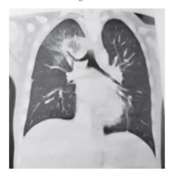
- What is the finding on CT scan with contrast?
- What are the possible treatment options at this stage?
- What are other possible associated congenital anomalies?
Answer
- CT finding: Aberrant tracheal bronchus (pig bronchus) arising directly from the lateral wall of the trachea above the carina supplying the right upper lobe.
- Treatment options:
- Conservative: Chest physiotherapy, postural drainage, inhaled bronchodilators, antibiotics for secondary infections.
- Surgical: Lobectomy / segmentectomy of the involved lobe if recurrent/refractory pneumonia or severe bronchiectasis develops.
- Associated congenital anomalies:
- Tracheoesophageal fistula (TEF) / Esophageal atresia
- VACTERL / VATER association
- Congenital heart disease
- Trisomy 21 (Down syndrome)