VVK20-001
a. What major problems can occur in using a cast?
b. What are the signs that the cast is too tight?
c. How do you prevent a tight cast?
d. How long does it take for a cast to harden?
Answer
- a. A cast that is too tight can cause serious neurovascular compromise and acute compartment syndrome.
- b.
- Pain when moving the fingers/toes (especially on passive stretch)
- Pallor, or pale/white distal extremity due to arterial insufficiency
- Paresthesias and numbness due to nerve compression and ischemia
- Paralysis / paresis due to loss of motor nerve function
- Pulselessness / delayed capillary refill
- Poikilothermia (cold extremity)
- c.
- Use sufficient padding
- Split the cast on both sides and spread the cast to allow for swelling (bivalving)
- Split the padding down to the skin
- Elevate the limb to prevent dependent edema and inflammatory swelling
- d.
- Plaster of Paris cast: 2–8 minutes to set (24–48 hours to dry completely)
- Fiberglass cast: 4–5 minutes
VVK20-002
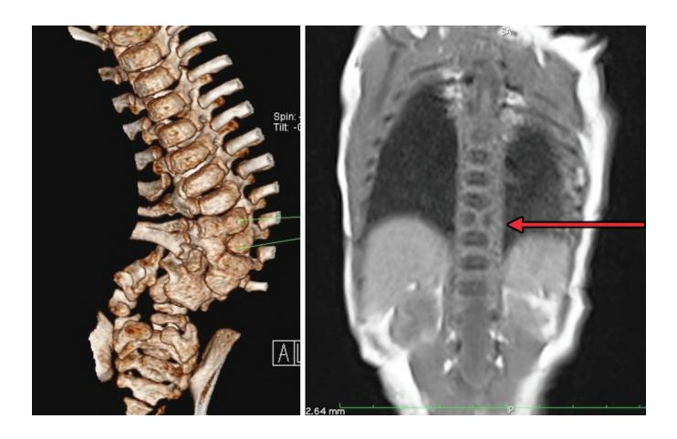
a. What is the radiological anomaly?
b. What is the incidence?
- i. 1:300
- ii. 1:3000
- iii. 1:30000
- iv. 1:300000
c. What can be the clinical abnormality associated with above abnormality?
Answer
- a. Hemivertebra (segmentation/formation failure defect of marked vertebra)
- b. ii. 1:3000
- c. Congenital scoliosis, dorsal kyphoscoliosis, lordosis
VVK20-003
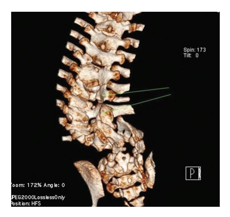
a. What is the radiological finding?
b. What is the diagnosis?
c. What are common physical signs of above diagnosis?
d. What is the single most important prenatal/antenatal factor leading into above abnormality?
Answer
- a. Unfused posterior neural elements / posterior arches of marked vertebrae (spina bifida / dysraphism)
- b. Spina bifida
- c.
- i. Lower limb weakness and paralysis
- ii. Orthopedic abnormalities (e.g., clubfoot / CTEV, hip dislocation, scoliosis)
- iii. Bladder and bowel dysfunction (neurogenic bladder/incontinence, urinary tract infections, poor renal function)
- iv. Pressure sores, trophic ulcers, and skin irritations
- v. Hydrocephalus / Chiari II malformation / abnormal eye movements
- d. Inadequate periconceptional maternal folic acid intake / folic acid deficiency during early pregnancy.
VVK20-004
A 10-year-old presents with history of weakness in the legs; low back pain; scoliosis; and incontinence.
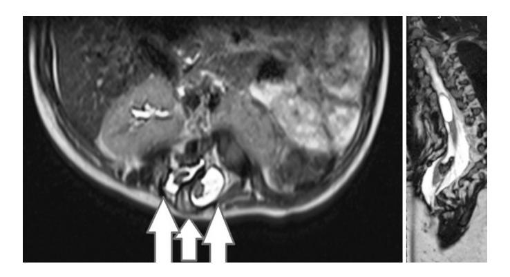
a. What is the radiological finding?
b. What is the diagnosis?
c. What is the treatment?
Answer
- a. Bony / fibrocartilaginous spur (osseous septum) noted between 2 divided hemicords (splitting of spinal cord)
- b. Diastematomyelia (split cord malformation)
- c. Surgical decompression of neural elements, resection/removal of the bony spur, and untethering of the spinal cord.
VVK20-005
Attached are pre and 2 weeks postoperative X-rays of a baby with developmental dysplasia of the hip.
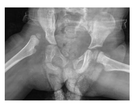
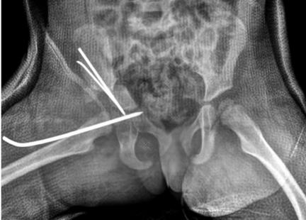
a. Which of the following is a risk factor for DDH? (True/False)
- i. First born
- ii. Male
- iii. Family history of DDH
- iv. Breech presentation
b. Enlist 2 orthopedic conditions associated with DDH.
c. Name 2 screening tests on examinations used to confirm the diagnosis.
Answer
- a.
- i. First born — True
- ii. Male — False (more common in females)
- iii. Family history of DDH — True
- iv. Breech presentation — True
- b. Any 2 of:
- Congenital muscular torticollis
- Congenital talipes equinovarus (clubfoot)
- Congenital hyperextension / dislocation of knee
- Metatarsus adductus
- Skull/facial deformities (plagiocephaly)
- c.
- Ortolani maneuver / test
- Barlow maneuver / test
(Note: Practice Ortolani and Barlow maneuvers as these can be asked for demonstration in observed OSCE stations.)
VVK20-006
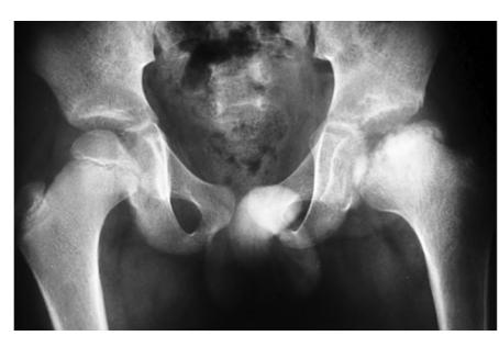
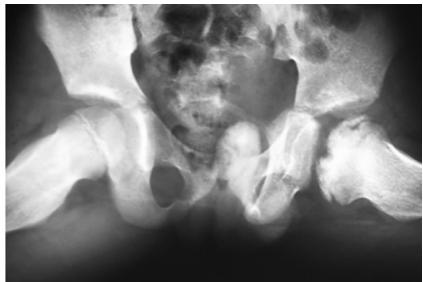
a. What are the radiological findings?
b. What is the diagnosis?
c. What is the pathological process?
d. True / False:
- i. Most commonly occur in children of 5–9 years age.
- ii. Is more common in females?
- iii. Is more common in children who have a sedentary lifestyle?
- iv. Surgical correction is most common modality of treatment.
Answer
- a. Uniformly increased density (sclerosis), flattening, fragmentation, and collapse of the left femoral capital epiphysis.
- b. Legg-Calvé-Perthes disease
- c. Idiopathic avascular necrosis (osteonecrosis) of the proximal femoral capital epiphysis.
- d.
- i. True (peak incidence 5–9 years)
- ii. False (more common in males, M:F ~ 4:1)
- iii. False (more common in active / hyperactive children)
- iv. False (conservative management / containment is the primary modality; surgery is reserved for select severe cases).
VVK20-007
Attached are MRI images of an 8-year-old female.
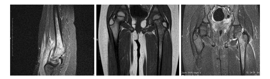
a. What are the radiological findings?
b. What is the diagnosis?
c. What is the treatment?
d. Enlist 3 complications associated with above diagnosis.
Answer
- a. Heterogeneous hyperintense signal in the neck and proximal metaphysis/upper end of the femur on T2/STIR with surrounding marrow edema.
- b. Acute osteomyelitis (proximal femur)
- c. Intravenous antimicrobial therapy (and surgical drainage/decompression if indicated).
- d. Any 3 of:
- Septic arthritis
- Distant metastatic seeding leading into pneumonia, septic pericarditis, empyema
- Pathological fracture
- Physeal bars and growth arrest / limb length discrepancy
- Chronic osteomyelitis / recurrent infections
VVK20-008
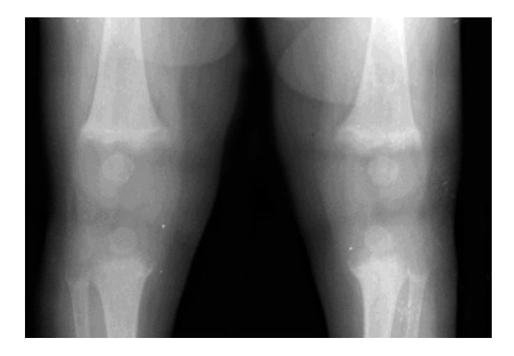
a. What is the diagnosis?
b. This infant is 10 months old, what is the most likely subtype?
c. What is the earliest sign of this disorder?
d. What is the first radiological change that occurs in response to specific therapy?
e. How could this have been prevented?
f. What are the non-specific urinary findings in this disorder? (at least 2).
Answer
- a. Active rickets (nutritional rickets)
- b. Vitamin D deficiency rickets
- c. Craniotabes
- d. Appearance of provisional zone of calcification (dense calcified white line across metaphysis)
- e. Daily supplementation of 400 IU of vitamin D from birth and adequate sunlight exposure.
- f. Any 2 of:
- Generalized aminoaciduria
- Phosphaturia
- Glycosuria
- Elevated urinary citrate
- Impaired renal acidification