VVK21-001
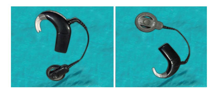
a. Name the device seen in the picture.
b. What are the essential parts of the device?
c. Enlist 3 commonest types of devices available.
Answer
- a. Hearing aid
- b.
- Microphone
- Amplifier
- Receiver / speaker
- Microchip / processor
- Battery / power source
- c.
- Invisible-in-the-canal (IIC) / Completely-in-the-canal (CIC)
- Behind-the-ear (BTE)
- In-the-canal (ITC) / In-the-ear (ITE)
VVK21-002
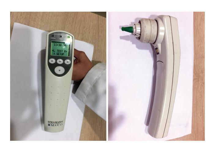
a. Name the device seen in the picture.
b. Which organ function does it detect?
c. How does this device work?
d. What are the prerequisite and limitations of working of the device?
Answer
- a. Otoacoustic emission (OAE) testing device.
- b. Inner ear (outer hair cells of the cochlea).
- c. In a healthy cochlea, vibration of the hair cells in response to sound stimulus generates acoustic energy called otoacoustic emissions. A probe placed in the ear canal delivers a click sound stimulus, and the resulting acoustic energy produced by the cochlea is detected and recorded by a microphone within the probe.
- d.
- Prerequisites: Needs a quiet baby and a clean ear canal free of vernix, fluid, or debris.
- Limitations:
- High false-positive rate if performed in the first 24 hours of birth due to vernix/debris in the canal.
- Only assesses cochlear/pre-neural ear function — does not assess the retrocochlear or central auditory neural pathway.
- It is a screening test; failed OAE must be confirmed by BERA (Brainstem Evoked Response Audiometry).
VVK21-003
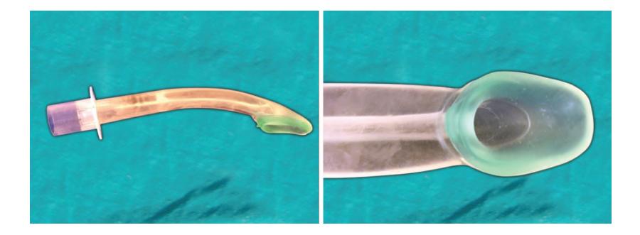
a. Name the device.
b. Indications (enlist 3).
c. Complications (enlist 3).
d. Contraindications when above device shall be avoided in a child during elective procedure.
Answer
- a. Laryngeal mask airway (LMA)
- b. Indications:
- Difficult airway management (rescue airway)
- Routine airway management during elective procedures
- Pre-hospital / emergency resuscitation airway management
- c. Complications:
- Aspiration of gastric contents
- Dislodgement / malposition
- Upper airway / pharyngeal soft tissue trauma, sore throat, or laryngospasm
- d. Contraindications:
- Complete upper airway obstruction below the glottis
- Increased risk of aspiration (non-fasted/full stomach patient, intestinal obstruction, upper gastrointestinal bleed, severe GERD)
- Need for prolonged mechanical ventilation or high peak airway pressures (>20 cm H2O)
- Suspected or known severe abnormalities in supraglottic anatomy
VVK21-004
Check functionality of self-inflating bag.
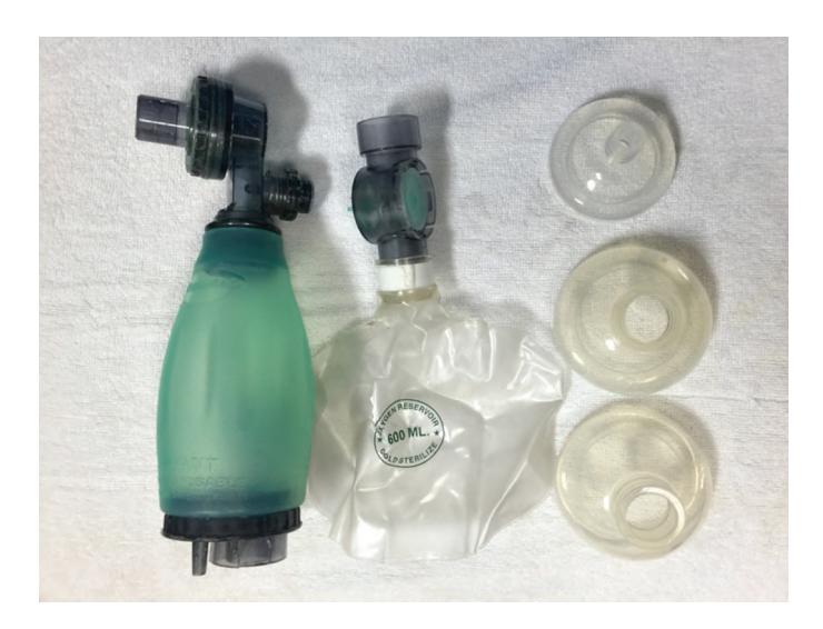
Answer
- Assemble the device correctly.
- With a self-inflating bag, occlude the patient connector / face mask against the palm of your hand and squeeze the bag.
- Look for the following 5 features as you squeeze the bag:
- a. You should feel pressure against the palm.
- b. The fish-mouth / non-rebreathing valve should open and close freely.
- c. The pop-off (pressure-relief) valve should make a hissing sound / open at preset pressure (around 35–40 cm H2O).
- d. The bag should recoil and reinflate instantly when pressure is released.
- e. If a pressure manometer is attached, it should register and display the pressure when the bag is squeezed.
- Absence of any of the above features suggests malfunction or leak.
VVK21-005
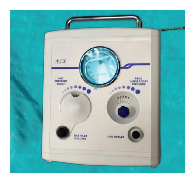
i. Name the device.
ii. Name the controls.
iii. What is the advantage and disadvantage over self inflating bag?
Answer
- i. T-piece resuscitator (e.g., Neopuff)
- ii. Controls:
- Inspiratory peak pressure control (PIP): Sets the amount of pressure delivered during a normal assisted breath.
- Maximum pressure relief control (Pop-off): Safety feature that prevents the pressure from exceeding a preset value (usually 40 cm H2O, adjustable).
- PEEP control (on the T-piece cap): Adjusts positive end-expiratory pressure.
- iii.
- Advantages:
- Consistent delivery of preset PIP and PEEP.
- Reliably provides free-flow oxygen (21–100% with blender) and continuous CPAP.
- Operator does not fatigue from prolonged hand bagging.
- Disadvantages:
- Requires a compressed gas source and blender.
- Requires pressures to be set and checked prior to use.
- Changing inflation pressure during resuscitation is more difficult.
- Risk of prolonged inspiratory time if the PEEP cap is held occluded too long.
- Advantages:
VVK21-006
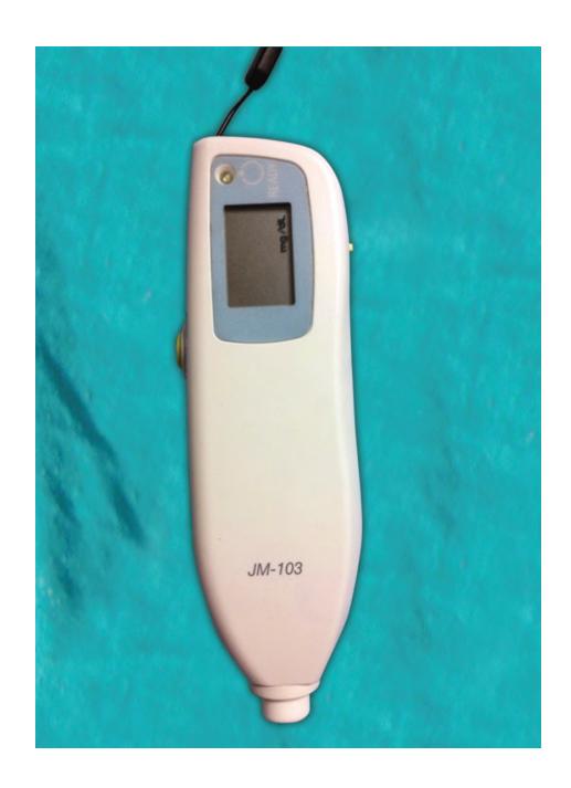
a. What is the device shown?
b. What is the site of use?
c. What are the single most important advantage and limitation of use?
Answer
- a. Transcutaneous bilirubinometer (TcB)
- b. Forehead and upper end of the sternum.
- c.
- Advantage: Non-invasive, instantaneous, and painless screening tool.
- Limitation: Inaccurate / cannot be used in babies already receiving or who have received phototherapy (unless the site was shielded during phototherapy), and unreliable at high serum bilirubin levels (>15 mg/dL).
VVK21-007
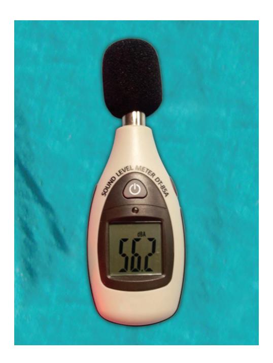
Name the device shown?
Answer
Sound level monitor / Decibel meter (used to monitor ambient noise levels in the neonatal intensive care unit).
VVK21-008
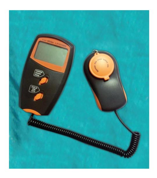
Name the device shown?
Answer
Flux meter / Phototherapy radiometer (used to measure irradiance/intensity of phototherapy units).
VVK21-009
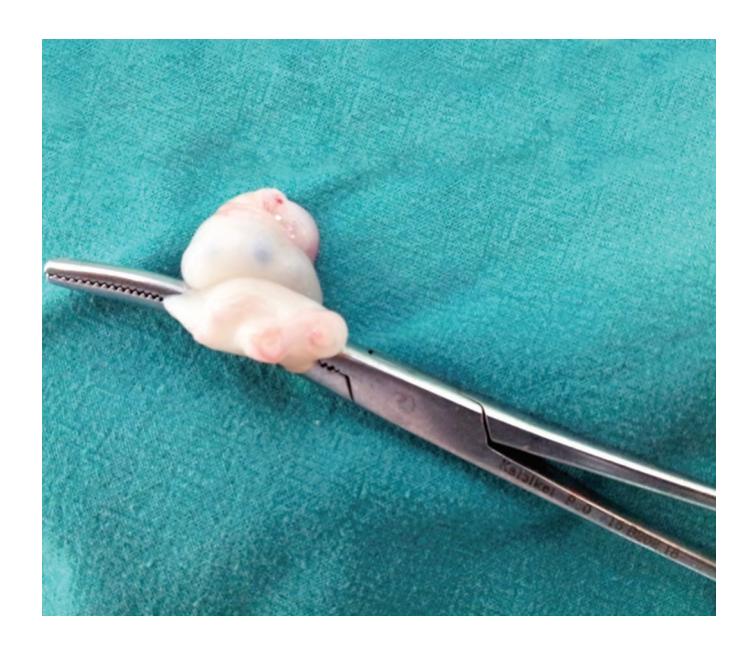
a. What is the abnormality seen?
b. What is the incidence?
- i. 1:10
- ii. 1:100
- iii. 1:1000
- iv. 1:10000
- v. 1:100000
c. It is associated with other anomalies in: - i. 1% cases
- ii. 10% cases
- iii. 25% cases
- iv. 50% cases
- v. 75% cases
d. What are the 2 most common systems involved in associated anomalies?
Answer
- a. Single umbilical artery (SUA / 2-vessel umbilical cord)
- b. ii. 1:100 (Option B)
- c. iii. 25% cases (Option C)
- d. Genitourinary system (most common) followed by Cardiovascular and Musculoskeletal systems.
VVK21-010
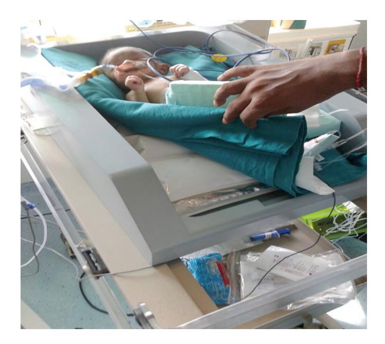
a. What procedure is this baby undergoing?
b. Name the device.
c. Enlist 5 complications associated with above procedure.
Answer
- a. Therapeutic hypothermia (targeted temperature management for neonatal hypoxic-ischemic encephalopathy).
- b. Whole-body cooling device / cooling blanket.
- c. Any 5 of:
- Sinus bradycardia and delayed intracardiac conduction
- Prolonged QT interval / ventricular arrhythmias
- Reduced cardiac output and hypotension
- Coagulopathy and thrombocytopenia (risk of thrombosis or hemorrhage)
- Pulmonary hypertension (increased PVR) and decreased surfactant production
- Leukopenia and increased risk of sepsis
- Sclerema neonatorum / subcutaneous fat necrosis
- Electrolyte disturbances (hypokalemia, hypomagnesemia, hyponatremia)
- Metabolic and lactic acidosis
- Impaired renal and liver function
VVK21-011
A lumbar puncture is performed.
a. The CSF is xanthochromic. What are the four possible causes?
b. CSF protein levels are 350 mg/dL. What are the three possible causes for the same?
c. CSF glucose is 210 mg/dL and blood glucose is 118 mg/dL (or low CSF glucose). List five causes for hypoglycorrhachia / abnormal CSF glucose.
d. CSF is cloudy, what is the likely cause?
Answer
- a. Causes of xanthochromia:
- Subarachnoid hemorrhage
- Severe hyperbilirubinemia (jaundice)
- Markedly elevated CSF protein (>150 mg/dL, Froin's syndrome / spinal block)
- Carotenemia
- Traumatic tap with delayed processing
- b. Causes of high CSF protein (>350 mg/dL):
- Tuberculous meningitis (TBM) / fungal meningitis
- Guillain-Barré syndrome (albuminocytological dissociation)
- Spinal cord tumors or brain tumors
- CNS vasculitis / neurosarcoidosis
- Multiple sclerosis
- Degenerative neurological disorders
- c. Causes of abnormal / low CSF glucose:
- Acute bacterial (pyogenic) meningitis
- Tuberculous meningitis (TBM)
- Fungal meningitis
- Neoplasms of the meninges (carcinomatous meningitis)
- Aseptic/viral meningitis (occasionally mumps, HSV, enterovirus)
- Subarachnoid hemorrhage
- d. Cloudy CSF:
- Markedly elevated cell count (pleocytosis) — high WBC count in bacterial/pyogenic meningitis or massive RBC count in intracranial hemorrhage.