MNJ09-001
A 3-month-old female child is admitted with a history of recurrent episodes of respiratory distress. On examination: Respiratory rate 70/min, suprasternal and subcostal retractions, bilateral reduced air entry, inspiratory and expiratory rhonchi. CXR shows bilateral hyperinflation:
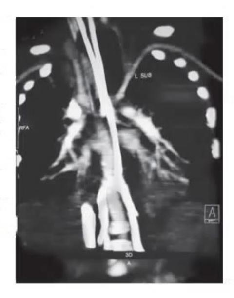
- Give your clinical diagnosis.
- What is the CT scan suggestive of?
- Enumerate initial steps of management.
- Definitive treatment.
Answer
- Diagnosis: Intrathoracic large airway obstruction / Tracheal compression.
- CT scan finding: Vascular sling (e.g., pulmonary artery sling / double aortic arch) around the trachea causing extrinsic compression and severe luminal narrowing.
- Initial management:
- Position of comfort, gentle handling, humidified oxygen.
- Minimal sedation if agitated.
- If intubation is needed, perform under controlled anesthesia using the smallest appropriate endotracheal tube with ENT/surgical backup.
- Definitive treatment: Cardiovascular surgical repair/reconstruction to relieve the vascular ring/sling compression.
MNJ09-002
In view of herniation of the brain:
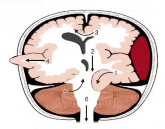
Identify the numbered types of brain herniation (1 to 6).
Answer
- 1. Cingulate (Subfalcine) herniation
- 2. Central (Transtentorial) herniation
- 3. Uncal (Transtentorial) herniation
- 4. Transcalvarial (External) herniation
- 5. Upward (Cerebellar / Transtentorial) herniation
- 6. Tonsillar herniation (Downward cerebellar / foramen magnum herniation)
MNJ09-003
A 3-year-old male child is admitted to the PICU with status epilepticus. He is on a ventilator and on midazolam infusion at
- ABG:
, , . - Laboratory tests:
, , , , Glucose ( ), ( ). - Measured Serum Osmolality:
.
- What is the metabolic disorder in this child?
- What is the cause of this metabolic disorder?
- What is the supporting evidence for this diagnosis?
- Mention other causes of a similar metabolic disorder.
Answer
- Metabolic disorder: High Anion Gap Metabolic Acidosis (HAGMA) with high osmolar gap.
(High).
- Cause: Propylene glycol toxicity (propylene glycol is used as a vehicle/solvent in parenteral midazolam, lorazepam, and phenytoin infusions).
- Supporting evidence:
. (Normal ).
- Other causes of high osmolar gap metabolic acidosis:
- Toxic alcohols: Methanol, Ethylene glycol, Isopropanol
- Drugs/Solvents: Salicylates, IV Phenytoin, IV Melphalan, IV Lorazepam
- Alcoholic or diabetic ketoacidosis, severe lactic acidosis
MNJ09-004
State which of the following situations would be expected to lower a patient's arterial
- a. Anemia
- b. Carbon monoxide poisoning
- c. An abnormal hemoglobin that holds oxygen with half the affinity of normal hemoglobin
- d. An abnormal hemoglobin that holds oxygen with twice the affinity of normal hemoglobin
- e. Lung disease with intra-pulmonary shunting
Answer
Correct Answer: Only (e) — Lung disease with intra-pulmonary shunting.
Explanation:
- (a) Anemia: Lowers total oxygen content (
), but arterial (dissolved oxygen) and are normal. - (b) Carbon monoxide poisoning: Decreases
and shifts curve to the left, but dissolved remains normal. - (c) and (d) Abnormal hemoglobins (affinity shifts): Shift the oxyhemoglobin dissociation curve affecting
and , but do not alter dissolved arterial . - (e) Intra-pulmonary shunting: Directly results in venous admixture and decreased arterial
.
MNJ09-005
Which patient is more hypoxemic, and why?
| Parameter | Patient A | Patient B |
|---|---|---|
| pH | 7.48 | 7.32 |
| 95% | 85% | |
| Hemoglobin |
Answer
Patient A is more hypoxemic.
Reasoning:
Hypoxemia reflects inadequate oxygen carriage in blood. Total arterial oxygen content (
- Patient A:
- Patient B:
Although Patient A has a higher
MNJ09-006
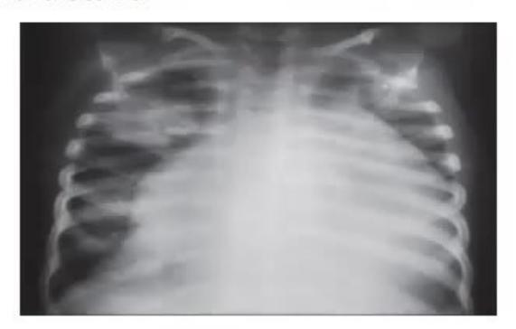
- Identify the chest X-ray.
- What is the acute emergency therapy? Mention the technique.
- Give the indications for the same.
Answer
- CXR finding: Massive cardiomegaly ("water-bottle" heart / globular cardiac shadow) suggestive of massive pericardial effusion with right mid-zone consolidation.
- Acute emergency therapy: Pericardiocentesis.
- Technique: Subxiphoid approach — insert needle at a
angle between the xiphoid process and left costal margin aiming towards the left shoulder under continuous aspiration and echocardiographic / ECG guidance.
- Technique: Subxiphoid approach — insert needle at a
- Indications:
- Clinical features of cardiac tamponade / obstructive cardiogenic shock (Beck's triad, pulsus paradoxus).
- Echocardiographic signs of tamponade (diastolic collapse/buckling of right atrium or right ventricle).
- Diagnostic aspiration in purulent/suspected tubercular pericarditis.
MNJ09-007
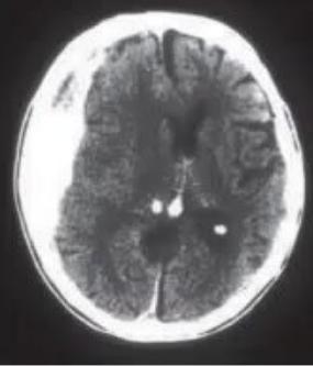
A 12-year-old female presents after head injury with low GCS, HR 60/min, irregular respiration, and is normotensive with the CT scan shown:
- Identify the CT scan abnormality.
- Immediate management.
- Further management.
- What is the most common electrolyte disturbance associated with this patient?
Answer
- CT scan finding: Acute right-sided crescentic subdural hematoma with significant midline shift and mass effect.
- Immediate management:
- Airway, Breathing, Circulation (ABC): Endotracheal intubation with in-line cervical stabilization.
- Controlled ventilation to prevent hypoxia and hypercapnia (target
35–38 mmHg). - Acute neuroprotective measures: Head end elevation
, neutral neck position, analgesia/sedation.
- Further management:
- Urgent neurosurgical consultation for craniotomy and surgical evacuation of the hematoma.
- Osmotherapy: Mannitol (
) or 3% Hypertonic Saline ( ). - Maintain Cerebral Perfusion Pressure (
).
- Electrolyte disturbance: Hyponatremia (due to SIADH or Cerebral Salt Wasting syndrome).
MNJ09-008
A 6-year-old female child comes with a history of progressive weight loss over the last several months, polydipsia, and oral thrush. Now she presents with severe breathlessness (Kussmaul breathing), abdominal pain, and vomiting.
On examination: Dehydrated, acidotic, urine sugar 4+, and urine ketones 4+.
- What is your diagnosis?
- What is the therapy in the first hour?
- After 8 hours of treatment, her
report comes as . What actions will you take? - What are her risk factors for developing cerebral edema?
- What are the major mortality predictors in this condition?
Answer
- Diagnosis: Diabetic Ketoacidosis (DKA) in newly presenting Type 1 Diabetes Mellitus.
- First hour therapy: Fluid resuscitation with Normal Saline (
) at over 1 hour (no insulin bolus in the first hour). - Actions for
: - Temporarily pause or reduce regular insulin infusion (to
). - Increase potassium replacement in IV fluids up to
(as KCl/KPO4). - Monitor continuous ECG and recheck serum potassium hourly.
- Temporarily pause or reduce regular insulin infusion (to
- Risk factors for cerebral edema:
- Younger age (
), new-onset T1DM, longer duration of symptoms. - Severe acidosis (
), lower , elevated initial BUN. - Administration of sodium bicarbonate.
- Rapid infusion of hypotonic fluids or excessive fluid volume (
). - Attenuated or failing rise in serum sodium during therapy.
- Younger age (
- Mortality predictors: Cerebral edema and severe refractory hypokalemia / cardiac arrhythmias.
MNJ09-009
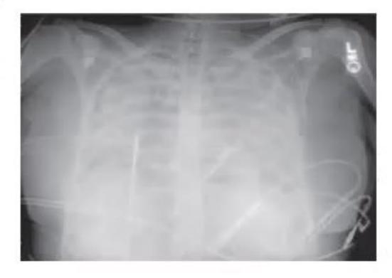

- Identify the condition shown on CXR.
- Give the diagnostic criteria for this condition.
Answer
- Condition: Acute Respiratory Distress Syndrome (ARDS) / Acute Lung Injury (ALI).
- Diagnostic Criteria (AECC / Berlin Criteria):
- Timing: Acute onset within 1 week of a known clinical insult or new/worsening respiratory symptoms.
- Chest Imaging: Bilateral opacities/pulmonary infiltrates on CXR or CT not fully explained by effusions, atelectasis, or nodules.
- Origin of Edema: Respiratory failure not fully explained by cardiac failure or fluid overload (PAOP
or no left atrial hypertension). - Oxygenation (on PEEP
): - Mild ARDS:
(formerly ALI) - Moderate ARDS:
- Severe ARDS:
- Mild ARDS:
MNJ09-010
- Identify the diagram / cardiac cycle event shown.
- What do the X-axis and Y-axis stand for?
- What do the numbers / phases denote?
Answer
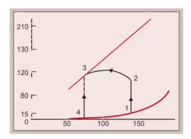
- Diagram: Left Ventricular Pressure-Volume Loop (or Wiggers diagram of cardiac cycle).
- Axes:
- X-axis: Left Ventricular Volume (
) - Y-axis: Left Ventricular Pressure (
)
- X-axis: Left Ventricular Volume (
- Phases:
- Isovolumetric contraction
- Rapid ventricular ejection
- Isovolumetric relaxation
- Ventricular filling period
MNJ09-011
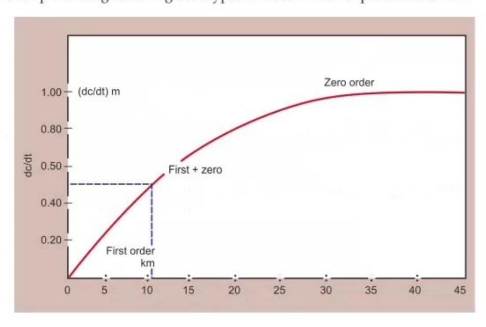
- What does the graph suggest?
- Define zero-order and first-order kinetics.
- Give an example of a drug following both types of kinetics in a dose-dependent manner (Michaelis-Menten kinetics).
Answer
- Graph: Michaelis-Menten / Capacity-limited saturation pharmacokinetics (transition from first-order to zero-order elimination kinetics as drug concentration rises).
- Definitions:
- First-order kinetics: The rate of drug elimination is directly proportional to plasma drug concentration (constant fraction or percentage of drug eliminated per unit time; constant half-life).
- Zero-order kinetics: The rate of drug elimination is constant and independent of plasma drug concentration (constant amount of drug eliminated per unit time; enzyme pathways are saturated).
- Drug example: Phenytoin (also Aspirin/Salicylates, Alcohol/Ethanol).
MNJ09-012
A 4.5-year-old child on therapy for ALL has relapsed and presents with a WBC count of
Following the first cycle of reinduction, the child has the following parameters:
- What is the diagnosis?
- Mention the doses of hyperhydration fluid,
, and Allopurinol. - What clinical and laboratory parameters will you monitor?
- What is the choice of dialysis if needed?
- Mention a newer modality of treatment with its mechanism of action.
Answer
- Diagnosis: Acute Tumor Lysis Syndrome (TLS).
- Initial therapeutic doses:
- Hydration:
(or maintenance) without potassium. - Allopurinol:
(or ) divided tid. : (routine alkalization is controversial and generally avoided if hyperphosphatemia/hypocalcemia is present).
- Hydration:
- Monitoring: Urine output (
or ), urine specific gravity/pH, daily weights, serum electrolytes ( ), uric acid, BUN/Creatinine every 4–6 hours, and ECG for hyperkalemia. - Choice of dialysis: Hemodialysis (or Continuous Renal Replacement Therapy / CRRT).
- Newer modality: Rasburicase (recombinant urate oxidase) — directly converts insoluble uric acid to highly soluble allantoin.
MNJ09-013
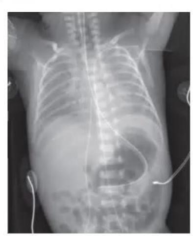
- What is abnormal in this radiograph?
- What is the ideal position of placement for an umbilical arterial catheter (UAC) and an umbilical venous catheter (UVC)?
- After placing a UAC, the right lower limb appears pale/blanched. What would you do?
- What is the anatomical level of the renal arteries?
- How do you maintain a UAC line?
Answer
- Abnormality: Malpositioned umbilical arterial catheter (e.g., tip coursing abnormally into the subclavian artery or carotid artery).
- Ideal Positions:
- UAC (Umbilical Arterial Catheter):
- High line: T6 to T9 (thoracic aorta, above diaphragm, between ductus arteriosus and celiac axis).
- Low line: L3 to L4/L5 (abdominal aorta, between inferior mesenteric artery and aortic bifurcation).
- UVC (Umbilical Venous Catheter): Inferior vena cava (IVC) at the junction with the right atrium (just above diaphragm, level T8–T9).
- UAC (Umbilical Arterial Catheter):
- Management of pale lower limb (vasospasm/thrombosis):
- Reflex vasodilation: Warm the contralateral (opposite) limb.
- If pallor persists
, immediately remove the umbilical arterial catheter.
- Level of renal arteries: L1–L2 vertebral level.
- Maintenance of UAC: Continuous flush with heparinized saline (
) at , strict asepsis, and avoid administering vasoactive drugs/blood products through UAC.
MNJ09-014
A 5-year-old child is caught in a house fire and brought to the ER with 60% BSA burns. His weight is around 16 kg:
- What is the immediate life-threatening complication likely to occur in this case and how will you identify it?
- What are the 4 most important initial steps in management?
- Calculate the fluid therapy for the first 24 hours for this child.
Answer
- Immediate complication: Upper airway obstruction and inhalational injury.
- Identification: Singed facial/nasal hairs, soot/carbonaceous sputum in oropharynx, hoarseness of voice, stridor, tachypnea, severe burns to the face and neck.
- Key management steps:
- Airway maintenance with early endotracheal intubation (before airway edema worsens).
- High-flow 100% humidified oxygen.
- Establish large-bore IV access (avoid burnt skin if possible) and start prompt fluid resuscitation.
- Analgesia/pain control and prevention of hypothermia.
- Fluid resuscitation (Parkland Formula):
- Burn Resuscitation Fluid:
- First 8 hours:
- Next 16 hours:
- First 8 hours:
- Plus Maintenance Fluid:
- For
- For
- Total 24-hr fluid:
.
- Burn Resuscitation Fluid:
MNJ09-015
(May 2013)
A mother in the pediatric emergency room states that her 1.5-month-old infant has been vomiting after every feed for 2 weeks. The vomit is non-bilious and non-bloody. Birth history was normal. Physical examination reveals a hungry, dehydrated infant.
Vital signs:
- What is your diagnosis?
- What will be the classic ABG and electrolyte findings in this child?
Answer
- Diagnosis: Infantile Hypertrophic Pyloric Stenosis (IHPS).
- ABG and Electrolytes: Hypochloremic, Hypokalemic Metabolic Alkalosis with elevated
and compensatory elevated (paradoxical aciduria may be seen).
MNJ09-016

- Define a reactive Non-Stress Test (NST).
- What is the definition of Chronic Lung Disease (CLD) / Bronchopulmonary Dysplasia in neonates?
- What is permissive hypercapnia?
- Enumerate Bell's staging criteria for Necrotizing Enterocolitis (NEC).
- What is threshold Retinopathy of Prematurity (ROP)?
Answer
- Reactive NST: Baseline heart rate 120–160 bpm with normal baseline variability and
fetal heart rate accelerations of lasting over a 20-minute recording window. - Chronic Lung Disease (CLD / BPD): Requirement for supplemental oxygen
in infants born weeks PMA assessed at 36 weeks post-menstrual age (or days of life for infants weeks). - Permissive Hypercapnia: A ventilatory strategy where mild-to-moderate respiratory acidosis (
, ) is accepted to minimize ventilator-induced lung injury (barotrauma/volutrauma). - Modified Bell's Staging for NEC:
- Stage I (Suspect NEC): Temperature instability, lethargy, abdominal distension, heme-positive stools, normal/ileus X-ray.
- Stage II (Definite NEC): Absent bowel sounds, abdominal tenderness, metabolic acidosis, thrombocytopenia, pneumatosis intestinalis / portal venous gas on X-ray.
- Stage III (Advanced NEC): Shock, DIC, peritonitis, impending/proven perforation (pneumoperitoneum).
- Threshold ROP:
contiguous or cumulative clock hours of Stage 3 ROP in Zone I or Zone II in the presence of "plus" disease.