THG25-001
Match the hearing milestones with their appearance age:
| Hearing Milestone | Code | Appearance Age |
|---|---|---|
| i. Turns head to one side and then upward if sound is made above ear level | a | 7 months |
| ii. Turns head diagonally and directly toward the sound source | b | 5 months |
| iii. Turns head to the side of sound | c | 6 months |
| iv. Turns head to one side and then downward when sound is made below level of ears | d | 9 months |
| v. Turns head in a curving arc toward the sound source | e | 3 months |
Answer
- i — c (6 months: Turns head to one side and then upward if sound is made above ear level)
- ii — d (9 months: Turns head diagonally and directly toward the sound source)
- iii — e (3 months: Turns head to the side of sound)
- iv — b (5 months: Turns head to one side and then downward when sound is made below level of ears)
- v — a (7 months: Turns head in a curving arc toward the sound source)
THG25-002
An anterior rhinoscopy finding is shown:
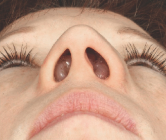
a. What is the diagnosis?
b. What is the most common cause?
c. What are the other causes/associations?
d. Explain common symptoms and signs.
e. Name symptomatic treatment.
f. What is the surgical modality of choice?
g. What are the indications for surgery?
Answer
- a. Bilateral nasal polyposis (nasal polyps in both nostrils)
- b. Cystic fibrosis (CFTR mutation)
- c. Allergic rhinitis, chronic rhinosinusitis with nasal polyps (CRSwNP), aspirin-exacerbated respiratory disease (AERD / Samter triad), allergic fungal rhinosinusitis (AFRS), and Kartagener syndrome / primary ciliary dyskinesia.
- d. Chronic bilateral nasal obstruction, mouth breathing, hyponasal speech (rhinolalia clausa), profuse mucoid/mucopurulent anterior and posterior rhinorrhea, anosmia/hyposmia, and widening of the nasal bridge (frog-face deformity).
- e. Intranasal corticosteroid sprays (e.g., Fluticasone, Mometasone), oral corticosteroids for acute shrink, nasal saline irrigations, and oral antihistamines/antileukotrienes.
- f. Functional Endoscopic Sinus Surgery (FESS)
- g. Complete mechanical airway obstruction unresponsive to medical therapy, recurrent refractory sinusitis, severe sleep apnea/hypopnea, or craniofacial deformity.
THG25-003
Tuning fork tests are shown:
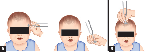
a. Name of two tests given in images A and B.
b. Name the frequency of tuning fork used.
c. What is the purpose of test?
d. What is the interpretation if test A is positive or negative?
e. What is the interpretation if test B is central or lateral?
Answer
- a. Image A: Rinne test; Image B: Weber test
- b. 512 Hz tuning fork (ideal balance between tactile vibration and pure tone frequency; 256 Hz may also be used).
- c. To clinically differentiate between conductive and sensorineural hearing loss.
- d. Rinne Test Interpretation:
- Rinne positive: Air conduction (AC) > Bone conduction (BC)
Normal hearing OR Sensorineural hearing loss. - Rinne negative: Bone conduction (BC)
Air conduction (AC) Conductive hearing loss in that ear.
- Rinne positive: Air conduction (AC) > Bone conduction (BC)
- e. Weber Test Interpretation:
- Weber central: Sound heard equally in midline/both ears
Normal hearing OR symmetrical hearing loss. - Weber lateralized to affected/worse ear
Conductive hearing loss in that ear. - Weber lateralized to normal/better ear
Sensorineural hearing loss in the contralateral ear.
- Weber central: Sound heard equally in midline/both ears
THG25-004
Interpret the following tuning fork test combinations to determine whether the hearing loss is sensorineural or conductive:
| Test Pattern | Right Ear (Rinne) | Left Ear (Rinne) | Weber Test | Clinical Interpretation |
|---|---|---|---|---|
| a | Rinne positive | Rinne positive | Weber central | |
| b | Rinne negative | Rinne positive | Weber right | |
| c | Rinne positive | Rinne positive | Weber left | |
| d | Rinne negative | Rinne negative | Weber central |
Answer
| Test Pattern | Right Ear (Rinne) | Left Ear (Rinne) | Weber Test | Clinical Interpretation |
|---|---|---|---|---|
| a | Rinne positive | Rinne positive | Weber central | Normal hearing OR Bilateral symmetrical sensorineural hearing loss |
| b | Rinne negative | Rinne positive | Weber right | Right conductive hearing loss |
| c | Rinne positive | Rinne positive | Weber left | Right sensorineural hearing loss (sound lateralizes to better left ear) |
| d | Rinne negative | Rinne negative | Weber central | Bilateral symmetrical conductive hearing loss |
THG25-005
An adolescent presented with history of fever, sore throat, trismus, and dysphagia.
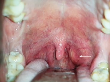
a. What do you see? What is the diagnosis?
b. What is the etiology?
c. Name the most common pathogens.
d. What are the complications?
e. What is the management?
Answer
- a. Unilateral erythematous peritonsillar bulging with contralateral uvular deviation
Acute peritonsillar abscess (Quinsy). - b. Suppurative complication of acute tonsillitis spreading into the loose peritonsillar space (Weber gland suppuration).
- c. Streptococcus pyogenes (Group A beta-hemolytic Streptococcus) and mixed oral anaerobes (Fusobacterium, Bacteroides, Peptostreptococcus).
- d. Parapharyngeal abscess, retropharyngeal abscess, airway obstruction, erosion into internal carotid artery, and mediastinitis.
- e. Needle aspiration or incision and drainage (I&D), intravenous broad-spectrum antibiotics (Ampicillin-sulbactam or Ceftriaxone + Metronidazole), analgesia, hydration, and interval tonsillectomy in recurrent cases.
THG25-006
A 7-month-old infant with history of excessive crying, irritability, fever, tugging of left ear—otoscopic examination reveals in given image.
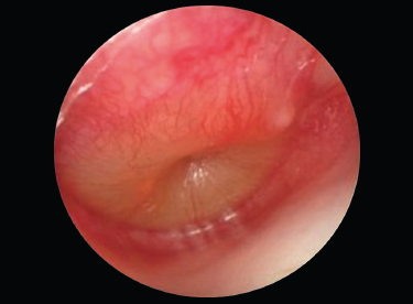
a. What are the otoscopic findings?
b. What is the diagnosis?
c. Describe the prevention strategies (any four).
d. Name the most common pathogens.
e. Mention antibiotics recommended for first-line treatment and duration.
f. What are the complications (at least eight)?
Answer
- a. Marked bulging of the tympanic membrane with diffuse erythema, loss of light reflex, and distortion/obscuration of normal ossicular bony landmarks.
- b. Acute Otitis Media (AOM)
- c. Prevention Strategies:
- Exclusive breastfeeding for at least the first 6 months
- Complete immunization against Streptococcus pneumoniae (PCV) and Haemophilus influenzae
- Avoidance of exposure to second-hand tobacco smoke
- Avoidance of supine bottle-feeding / propped bottles
- Limiting pacifier use after 6 months of age
- d. Streptococcus pneumoniae, non-typeable Haemophilus influenzae, and Moraxella catarrhalis.
- e. High-dose oral Amoxicillin (80–90 mg/kg/day divided 12-hourly) or Amoxicillin-clavulanate for 10 days in infants
years. - f. Complications:
- Intratemporal: Tympanic membrane perforation, acute mastoiditis, labyrinthitis, facial nerve palsy, conductive hearing loss, and chronic suppurative otitis media (CSOM).
- Intracranial: Meningitis, epidural abscess, subdural empyema, brain abscess, lateral/sigmoid sinus thrombosis, and otitic hydrocephalus.
THG25-007
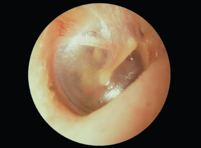
Identify this image:
Answer
- Identification: Normal tympanic membrane (otoscopic view of right ear).
- Anatomical Landmarks:
- Translucent, pearly-gray circular membrane
- Handle (manubrium) of malleus pointing posterosuperiorly
- Lateral process of malleus and umbo
- Cone of light (light reflex) radiating anteroinferiorly from the umbo
- Incus long process visible in the posterosuperior quadrant
- Intact pars tensa and pars flaccida without erythema, effusion, or perforation.
THG25-008
A 7-year-old boy developed severe ear pain 1 day after swimming in a community pool. Pain is accentuated on manipulation of pinna, there is tenderness over tragus and pinna. On examination, there is erythema, edema of ear canal, and otorrhea.
a. What is the diagnosis?
b. Name the most common causative organisms (any three).
c. What are the otoscopic findings?
d. What is the mainstay of treatment?
e. If the same boy develops facial paralysis, vertigo, sensorineural hearing loss, lateral displacement of pinna what will you suspect?
Answer
- a. Acute otitis externa ("Swimmer's ear")
- b. Pseudomonas aeruginosa (most common), Staphylococcus aureus, Enterobacteriaceae (Proteus, Klebsiella), and fungal species (Aspergillus, Candida).
- c. Diffuse erythema and edema of the external auditory canal with purulent debris/otorrhea; tympanic membrane is intact though often partially obscured.
- d. Gentle ear canal aural toilet/cleansing, topical antibiotic-corticosteroid otic drops (e.g., Ciprofloxacin-dexamethasone or Polymyxin B-neomycin-hydrocortisone)
placement of an ear wick if canal is severely edematous, and keeping ear strictly dry. - e. Necrotizing (malignant) otitis externa / Skull base osteomyelitis (or acute mastoiditis).
THG25-009
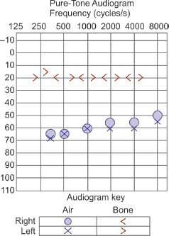
A 6-year-old boy presents with audiogram as given in below image. A history of recurrent upper respiratory tract infections treated with recurrent courses of antibiotics. His speech is limited, but he has no other medical problems. His father and grandfather who are with him are smokers.
a. What does the audiogram show?
b. At what age is pure tone audiometry possible?
c. What is the most likely cause for the speech defect?
d. Give three steps to help him with his hearing and speech defect.
Answer
- a. Bilateral conductive hearing loss (air conduction thresholds elevated to 30–40 dB HL with normal bone conduction thresholds
, demonstrating an air-bone gap). - b. 4–5 years of age (conditioned play audiometry from 2.5–5 years, standard pure tone audiometry
). - c. Chronic otitis media with effusion (OME / "glue ear") causing persistent fluctuating conductive hearing loss during critical speech acquisition years.
- d. Management Steps:
- Bilateral myringotomy with ventilation tube (grommet) insertion
- Elimination of environmental passive tobacco smoke exposure
- Speech and language therapy, and classroom preferential seating in the front row.
THG25-010
Parents of a 6-year-old boy reports that he has restless sleep, loud, frequent, and disruptive snoring, breathing pauses, choking arousals, and nocturnal diaphoresis. During daytime, he has mouth breathing and dry mouth, chronic rhinorrhea, drowsiness, hyponasal speech, and poor appetite.
a. What is your diagnosis?
b. Enumerate anatomical factors (at least six) that predispose to this condition.
c. What is the gold standard test for diagnosing this condition?
d. Name most commonly used parameter in this test for evaluation.
e. If gold standard test is not available what alternate tests you can perform?
f. ________ is the first-line treatment advocated in a child with ________?
Answer
- a. Pediatric Obstructive Sleep Apnea Syndrome (OSAS)
- b. Predisposing Anatomical Factors:
- Adenotonsillar hypertrophy (most common)
- Craniofacial anomalies (micrognathia, retrognathia, midface hypoplasia)
- Deviated nasal septum / turbinate hypertrophy
- Macroglossia (e.g., Down syndrome, Beckwith–Wiedemann)
- Laryngomalacia
- Obesity / neck adiposity
- Achondroplasia (skull base narrowing)
- c. Overnight in-laboratory attended Polysomnography (PSG)
- d. Apnea–Hypopnea Index (AHI) or Obstructive AHI (
is abnormal in children). - e. Overnight continuous pulse oximetry, nocturnal video recording, ambulatory home sleep apnea testing, or daytime nap PSG.
- f. Adenotonsillectomy is the first-line treatment advocated in a child with adenotonsillar hypertrophy.
THG25-011
A 28-day-old well thriving term infant is brought to outpatient department with complaints of persistent stridor, noticed since birth. This is associated with increased work of breathing as in inspiratory obstruction like suprasternal and subcostal retractions. He is able to feed well and is growing well but frequently regurgitates.
a. What is most likely diagnosis?
b. How you will confirm your diagnosis?
c. What is the treatment?
Answer
- a. Laryngomalacia (congenital flaccid supraglottic collapse)
- b. Flexible awake fiberoptic laryngoscopy (demonstrating omega-shaped epiglottis, shortened aryepiglottic folds, and inward prolapse of arytenoid cartilages into airway on inspiration).
- c. Expectant observation and reassurance (resolves spontaneously by 12–18 months in >90%); anti-reflux medical therapy (PPI/H2-blocker) for associated GERD; surgical supraglottoplasty for severe cases with failure to thrive or cyanotic spells.
THG25-012
A 2.5-month-old infant has been experiencing worsening biphasic stridor. Her parents give history of two brief episodes of croup. There is a 1-cm-diameter hemangioma on the infants’ right thigh.
a. What is the most likely diagnosis?
b. What will be radiographic finding?
c. How will you manage?
Answer
- a. Congenital subglottic hemangioma
- b. Lateral and AP neck radiograph showing asymmetric, smooth, eccentric subglottic soft-tissue narrowing / stenosis below the vocal cords.
- c. Oral Propranolol (
divided 2–3 times daily for 6–12 months as first-line pharmacotherapy), oral corticosteroids (prednisolone) in acute airway compromise, and endoscopic surgical laser ablation or tracheostomy for refractory life-threatening obstruction.
THG25-013
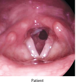
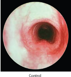
A 3-month-old infant developed biphasic stridor and barking cough during an episode of upper respiratory tract infection; Past history of recurrent such episodes once/twice a month. Bronchoscopy was done.
a. What is your diagnosis?
b. How will you grade the severity of this condition?
c. How will you treat this child?
Answer
- a. Congenital subglottic stenosis (circumferential narrowing of cricoid ring lumen).
- b. Myer–Cotton Grading System:
- Grade I:
luminal obstruction - Grade II: 51%–70% obstruction
- Grade III: 71%–99% obstruction (with identifiable lumen)
- Grade IV: Complete obstruction / no detectable lumen (100% stenosis)
- Grade I:
- c. Treatment:
- Grades I and II: Conservative observation and medical management during viral infections (humidified oxygen, nebulized adrenaline, and systemic steroids); most widen spontaneously as child grows.
- Grades III and IV / symptomatic: Endoscopic balloon dilation, anterior cricoid split, laryngotracheal reconstruction (LTR) with cartilage grafting, or tracheostomy until definitive surgical repair at 3–4 years of age.